
Conclusion Heredity material was packaged in discrete transferable units came up with law of segregation and law of independent assortment Thomas Morgan early 1900s Discovered that fruit flies genes were associated with chromosome inheritance ID: 658403
Download Presentation The PPT/PDF document "DNA Gregor Mendel – 1840’s" is the property of its rightful owner. Permission is granted to download and print the materials on this web site for personal, non-commercial use only, and to display it on your personal computer provided you do not modify the materials and that you retain all copyright notices contained in the materials. By downloading content from our website, you accept the terms of this agreement.


Slide1
DNASlide2
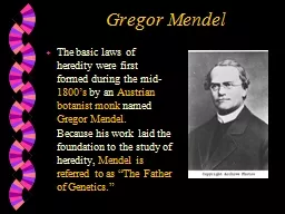
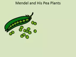
Gregor Mendel – 1840’s
Conclusion:
Heredity material was packaged in discrete transferable units; came up with law of segregation and law of independent assortment.Slide3
Thomas Morgan – early 1900’sDiscovered that fruit flies’ genes were associated with chromosome inheritance.
Conclusion:
Chromosomes were known to be composed of proteins and DNA, so genes must be one of these two macromoleculesSlide4
Experiment
If dead (heat-killed) pathogenic bacteria was mixed in a culture with living harmless bacteria, the harmless bacteria would become deadly.
Conclusion
Transformation
in bacteria allows a change in genotype and phenotype due to the assimilation of external DNA by the cell.
FREDRICK GRIFFITH -1928Slide5
He separated the components of the heat-killed, deadly bacteria & divided it into smaller samples (proteins, lipids, carbohydrates, or nucleic acids) & left the other molecules intact. He then mixed each sample of the treated lethal strain with living samples of the non-lethal strain.
AVERY’S EXPERIMENT -1944
Only the DNA extract from the deadly bacteria would allow the live harmless bacteria to become deadly.
AVERY’S CONCLUSIONSlide6
HERSHEY AND CHASE - 1952
Conclusion:
The DNA molecule entered the bacteria cell & not the protein. This showed that DNA and not protein controls traits that are passed on.Slide7
CHARGAFF - 1947
Experiment:
Studied the composition of DNA and the concentration of each of the nitrogenous bases.
Conclusion:
DNA base composition varies from one species to another; bases are not present in equal amounts in any one species but they are found in a predictable ratio; concentration of T=A and concentration of C=G.Slide8
Franklin and Wilkins
Experiment:
Took x-ray diffraction pictures of DNA in its different forms
Conclusion:Discovered that the B form of DNA was double helix in structure.Slide9
Watson and Crick
Experiment:
Built a 3-D model that reflected the base pairing rules determined by
Chargraff & the distance between bases suggested by Franklin’s X-ray photos.
Conclusion:DNA was a double helix that made one full turn every 3.4nm with bases 0.34nm apart & sugar/phosphate molecules on the outside of the ladder.Slide10
So what are the various parts of DNA?Nitrogen basesAdenineCytosine
Guanine
Thymine
PhosphatesDeoxyribose
Hydrogen bondsSlide11
DNA Molecule
Sugar and phosphate backbones are
antiparallel
(their subunits run in opposite directions)
Adenine and guanine are purines
(both have 2 organic rings)Cytosine and thymine are pyrimidines (both have 1 organic ring)Adenine forms 2 hydrogen bonds with thymineCytosine forms 3 hydrogen bonds with guanineSlide12
WHY 5’ AND 3’?Slide13
Let’s make some DNA Red = phosphateWhite =
deoxyribose
Yellow = adenine
Blue = thymineOrange = cytosine
Green = guaninePink = uracilPlastic connectors = hydrogen bonds
Minimum of 10 pairsSlide14
DNA ReplicationSlide15
What is the purpose of DNA Replication?To produce a copy of DNA identical to the original in preparation for mitosis or meiosis.Slide16Slide17
Meselson and Stahl experiment
Conclusion:
DNA replication follows the semiconservative model.
1
st replication in the 14 N medium produced a band of hybrid. This eliminated the conservative model.2nd replication produced both light and hybrid DNA this eliminated the dispersive model & supported the semiconservative model.Slide18
E. coli vs Human DNA ReplicationE. coli
Has a single chromosome
4.6 million nucleotide pairs
Can replicate its chromosome in less than an hour.
Human46 DNA molecules; each in a chromosome6 billion nucleotide pairsCan replicate all chromosomes in a few hours
Replication process is similar in prokaryotes and eukaryotes.Slide19
Some of the “players” involved…
DNA Polymerase-
adds nucleotides to a preexisting chain.
Ligase
- joins the sugar phosphate backbones of all Okazaki fragments.Primase- synthesizes the primer that’s 5-10 nucleotides long.
Helicase- unzips the DNA Topoisomerase-relieves the strain of overtwisting DNA by braking, swiveling, & rejoining DNA strands.Slide20
DNA unwinds & unzips with the help of helicaseSlide21
In order for replication to begin a primer is needed & it is an RNA primer
The primer is about 5-10 nucleotides long
The new DNA strand starts from the 3’ end of the RNA primer
DNA Polymerase adds nucleotides to the preexisting strandSlide22
Replication occurs from 5’ to 3’
In eukaryotes Okazaki fragments are 100-200 nucleotides long.
Leading strand- is made going towards the replication fork and is continuous
Lagging strand- is made going away from the replication fork and is synthesized discontinuously, as a series of segments called Okazaki fragments.
Leading strand needs only one primer & lagging strand needs a primer for each Okazaki fragment.Slide23
Ligase
-
joins sugar-phosphate backbone.
DNA polymerase I –
removes the primer and replaces it with DNA nucleotides; one by oneDNA polymerase III-Continuously synthesizes the leading strand
LET’S WATCH A VIDEOSlide24
How is replication of one side of each double strand different than the other?Because bases can only be added in the 5’ to 3’ direction, the 3’ to 5’ strand must be assembled in fragments that are later annealed together by a ligase protein.Slide25
Proofreading and Repairing DNA
Errors amount to 1 in 10 billion nucleotides in the final DNA product
Initial pairing error amount to 1 in 100,000 more common.Slide26
How is the new strand ensured to be identical?The bases are matched in a consistent patternThe daughter strands are half new, half old
There is a proofreading mechanism that checks for errors in both strands.Slide27
Why are mistakes made?Spontaneous chemical changes under normal conditions.Exposure to mutagens
EX: cigarette smoke and X-rays
There are many different DNA repair enzymes.
-E. coli has 100 known repair enzymes
-Humans have 130 identified repair enzymesSlide28
Teams of enzymes detect & repair damaged DNA, such as this thymine
dimer
(often caused by UV radiation), which distorts the DNA molecule.
A nuclease enzyme cuts the damaged DNA strand at 2 points, & the damaged section is removed.
Repair synthesis by a DNA polymerase fills in the missing nucleotides.
DNA
ligase
seals the free end of the new DNA to the old DNA, making the strand complete.Slide29
If your body is unable to repair the thymine dimer…Slide30
Replicating the ends of DNA molecules
This kind of thing does not occur prokaryotes with a circular chromosome
The primer on the end is removed but can’t be replaced with DNA because DNA polymerase can only add nucleotides to the 3’ end of a preexisting polynucleotide
The strand will get shorterSlide31
What is done to compensate for this problem?Telomeres (located at the ends of DNA molecules) are made of repeated units that are non-coding so that, as they get shorter, no genes are lost.
The enzyme
telomerase
lengthens telomeres in germ cells
Cells can only go through a limited number of replications before they are put to death.plus
alsoSlide32Slide33
Let’s Replicate!!!
with narrative Slide34
How Does a Gene Become a Protein?With a lot of help, I’ll tell you that!!!Slide35
Let’s start with the 2 nucleic acids involvedDNA and RNA
These molecules have structural similarities and differences that define function.Slide36
Compare DNA to RNASlide37
Made of nucleotides
Connected by covalent bonds to form a linear molecule from 5’ to 3’
Contains
deoxyribose
Nitrogen bases A,T,G, & CDouble strandedRestricted to the nucleus (eukaryotes)Made of nucleotidesConnected by covalent bonds to form a linear molecule from 5’ to 3’Contains riboseNitrogen bases A,U,C, & GSingle strandedAble to travel out of the nucleus (eukaryotes)
Comparison of DNA to RNADNA
RNASlide38
Is Uracil a purine or a pyrimidine?Since thymine is a
pyrimidine
and in essence
uracil replaces thymine in RNA it would make
sence that uracil is also a pyrimidine. Wouldn’t it?Slide39
The sequence of the RNA bases, together with the structure of the RNA molecule, determines RNA function.
mRNA:
carries information from the DNA to the ribosome.
tRNA:
are molecules that bind specific amino acids and allow information in the mRNA to be translated to a linear peptide sequence.rRNA: are molecules that are functional building blocks of ribosomes.RNAi: plays a role in regulation of gene expression at the level of mRNA transcription.To be discussed laterSlide40
Genetic information flows from a sequence of nucleotides in a gene to a sequence of amino acids in a protein
This occurs in two partsSlide41
What signals the cell to make a specific protein?Cell signalingCell receptorCell hormonesSlide42
Transcription
Is the synthesis of RNA using one side of a segment of a DNA strand.
The DNA segment serves as a template.
The RNA made is mRNA.
It is antiparallel to the DNA templatemRNA is made from 5’ to 3’ (reading the DNA in a 3’ to 5’ direction).This is all completed in the nucleus.Slide43
RNA Polymerase opens the DNA strands & joins the RNA nucleotides that are complimentary to the DNA template.Needs promoter to begin
Bacteria have a single type of RNA polymerase that synthesizes all types of RNA.
Eukaryotes have at least 3 types of RNA polymerase.Slide44Slide45
An example of a promoter is the TATA boxSlide46
Termination of Transcription
Differs between bacteria and eukaryotes
In bacteria
Go through a terminator sequence in DNA.
Once RNA polymerase hits the terminator signal it releases from the DNA and the mRNA that was being made.In eukaryotesRNA Polymerase II transcribes a sequence on the DNA which codes for a polyadenylation signal (AAUAAA) in the pre-mRNA.About 10-35 nucleotides later proteins cut the pre-mRNA from the polymerase & undergoes processing…Slide47
Modifying of the pre-mRNA in EukaryotesBoth ends of the mRNA transcript are altered.
In most cases, certain interior sections are cut out and the remaining pieces are spliced together.
These actions produce a mRNA molecule that is ready for action!!!Slide48
RNA Processing
The cap and tail:
help facilitate the mRNA leaving the nucleus & help protect the
mRNA strand from degradation by hydrolytic enzymes.
help the ribosomes attach to the 5’ end of the mRNA
For ribosome bindingSlide49
More RNA ProcessingRNA splicing:
Removing portions of the RNA molecule & putting the other ends together.
The parts to be “edited out” are interspersed between the coding segments.
The segments that intervene with the coding segments are called:
intronsThe segments that will eventually be expressed & exit the nucleus are called: exonsAverage length of transcription unit = 8000 nucleotides, but average size protein is 400 amino acids so only about 1,200 nucleotides long. This indicates long noncoding stretches of nucleotides which happen to be interspersed in the coding segments.Slide50
HOW IS PRE-mRNA SPLICING CARRIED OUT?
A small nuclear
ribonucleoproteins
(
snRNPs) recognize the splice sites. These are located at the end of introns
.Composed of RNA & proteinSeveral snRNPs and additional proteins form a larger assembly: spliceosome
These molecules release introns & join the exonsSlide51
Why have introns?One idea is that introns play regulatory roles in the cell.
Splicing process is necessary for mRNA to leave the nucleus.
Consequence for having introns & exons
Genes are known to give rise to 2 or more different polypeptides, depending on which segments are treated as exons during RNA processing =
alternative RNA splicing.Slide52
TranslationDivided into 3 stages: initiation, elongation, & terminationSlide53
TranslationMolecular components of translationmRNA: has nucleotide triplets called:
codons
Written in the 5’ to 3’ direction
tRNA
: has nucleotide triplets called: anticodonsThey are complimentary to the codonsMain function is to transport amino acids to the ribosome & drop off the amino acid to add to the polypeptide chain.Ribosomes: made of rRNA and proteinsAdds each amino acid brought by the tRNA
to the growing end of the polypeptide chain.Slide54
How do you know the code from the mRNA to the amino acid?Slide55
Stage 1: InitiationmRNA interacts with the rRNA of the ribosome to initiate translation at the (start) codon & travels from the 5’ to 3’ end.
The sequence of nucleotides on the mRNA is read in triplets called
codons
.Each
codon encodes a specific amino acid, which can be deduced by using a genetic code chart. Many amino acids have more than one codon.Slide56
tRNA
brings the correct amino acid to the correct place on the mRNA.
The amino acid is transferred to the growing peptide chain, with the help of ATP.
The process continues along the mRNA until a “stop” codon is reached
Stage 2 : ElongationSlide57
Stage 3: TerminationA release factor (protein) binds directly to the stop codon.This causes an addition of a water molecule instead of an amino acid to the polypeptide chain.
This breaks the bond between the chain & the
tRNA
.
The process terminates by the ribosome falling off the mRNA strand releasing the newly synthesized peptide chain.Slide58
The polypeptide chain folds and becomes activeSlide59
What happens to the mRNA strand?They eventually degrade in the cytoplasm and become free floating nucleotides once again.Slide60
Transcription and translation can happen simultaneously.
In other words, translation of an mRNA molecule begins while still being transcribed.Slide61
Picture summary of transcription and translation in a eukaryotic cell.
Each amino acid attaches to its proper
tRNA
with the help of a specific enzyme & ATP.Slide62
Questions
Transcription
Translation
Where?
NucleusCytosol/CytoplasmWhat is used as a template?DNA
mRNAWhat is used to synthesize the new strand?RNA PolymeraseRibosomes
What is the new strand made
of?
RNA
Amino acidsSlide63
LET’S MAKE SOME PROTEIN!
I need 4 volunteers
Slide64
Which parts played what in transcription & translation?
Master Chef
= RNA Polymerase
Prep-chef
= ribosome
Cookbook
=DNA
Scrap paper
= mRNA
Ingredients
= amino acids
Customer 1
= receptor mediated cell signal
Customer 2
= cell signal- hormone
Tabs on cookbook
= TATA boxes (telling chef where to find the
recipe
=geneSlide65
Now it’s your turn You will get together with 4-5 people in class.
You will now create a completely different analogy for the transcription & translation process.
You have 20 minutes to come up with your analogy & then we will present themSlide66
How Genes Influence Traits
Genes specify the amino acid sequence of proteins
The amino acid sequence determines the shape and activity of proteins
Proteins determine a majority of what the body looks like and how it functionsSlide67
Fig. 8.11 The journey from DNA to phenotypeSlide68
Fig. 8.11 The journey from DNA to phenotypeSlide69
HIV
- HIV is a unique virus in that its genetic material is a single-stranded RNA.
The flow of genetic information travels from RNA to DNA.
Once the HIV virus enters the white blood cells, it activates an enzyme called reverse transcriptase.
This enzyme uses the RNA of the virus to synthesize complimentary double stranded DNA. The process of reverse transcription is very error-prone, hence there is a large degree of mutation which is why finding a cure for AIDS is so difficult
.This new DNA integrates itself into the host genome & becomes transcribed and translated for the assembly of new viral progeny.
RETROVIRUSSlide70
Targeting polypeptides to specific locationsTwo populations of ribosomes
Free and bound
Free are in the
cytosol
Make proteins that stay and function hereBound ribosomesUsually attached to the cytosolic side of the endoplasmic reticulum (ER) or the nuclear envelope.Make proteins of the endomembrane system as well as proteins secreted from the cell (ex: insulin)Slide71
One gene/one polypeptide hypothesisIn the 1940’s experimental work led to the hypothesis:
Every one gene of DNA produce one enzyme
This was amended to include all proteins.
It was later discovered that many proteins are actually composed of more than one polypeptide.
This led to the proposal that each individual polypeptide required one gene.Slide72
In the last few years…Researchers have discovered that at least some genes aren’t quite that straightforward.For Example:
One gene may lead to a single mRNA molecule, but the mRNA molecule may then be modified in many different ways.
Each modification may result in a different polypeptide.Slide73
So…what is a gene?A region of DNA that can be expressed to produce a final functional product that is either a polypeptide or an RNA molecule.Slide74
Point MutationsWhat can affect protein structure and function?Slide75
Types of Small-Scale MutationsSubstitutions:Nucleotide-pair substitutionReplacement of one nucleotide & its partner with another pair of nucleotides.
Results in one of the following: silent mutation,
missense
mutation, or nonsense mutation.
Insertions & deletionsAdditions or loses of nucleotide pairs in a geneDisastrous effect on the resulting proteinWhenever the number of insertions or deletions aren’t a multiple of three = frameshift mutation. Slide76Slide77
EX: Point mutation: Sickle-cell disease