
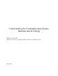
This is a cartoon of the heart and the bypass machine as seen from the head of the bed by the anesthesiologist Note the 4 chambers ofLVOT then to the aortic valve beyond which the coronary On the ID: 959681
Download Pdf The PPT/PDF document "Understanding the Cardiopulmonary Bypass..." is the property of its rightful owner. Permission is granted to download and print the materials on this web site for personal, non-commercial use only, and to display it on your personal computer provided you do not modify the materials and that you retain all copyright notices contained in the materials. By downloading content from our website, you accept the terms of this agreement.


Understanding the Cardiopulmonary Bypass Machine and Its Tubing Robert S. Leckie, MD Division of Cardiac Anesthesia, Beth Israel Deaconess Medical Center This is a cartoon of the heart and the bypass machine as seen from the head of the bed by the anesthesiologist. Note the 4 chambers ofLVOT, then to the aortic valve, beyond which the coronary On the CPB machine, there is a reservoir bucket at the left side, and then 4 roller-pumps, labeled A,B,C and D. After heparinization, the first cannula that is almost always put in is the aortic. It takes oxygenated blood from roller pump A, and It is put in first because if there is a problem, arrhythmia, or ischemia, and you need to it is best to have that one already in place. You do not want to hurry when putting this ght tear/dissect. In additio
n, it serves as a conduit for rapid fluid resuscitation. Keep the systolic BP in Hg range during placement to lessen the chance of a tear, while this is being placed. The second cannula that is placed is the venous. Usually the appendage of the right atrium is used, since tying it off at the end of the case doesn't matter. Atrial arrhythmias are very common during its placement, so you need to have checked the defibrillator in case cardioversion is needed. Remember to synchronize the cardioverter/defibrillator for atrial dysrhythmias. always low to the ground. It has to be lower than the patient, as much of the drainage into the venous cannula is gravity-dependent siphoning. Vacuum It drains into the reservoir bucket, where the oxygenator resides. Then it goes around roller pump A and back into the p
atient by way of the the aortic cannula. With adequate heparinization and these two cannulae, you can go on bypass and rescue patients from cardiac arrest, severe hypothermia, bupivicaine injection, etc. achieved using the femoral artery and vein, without opening the chest. If cardiac surgery is planned, you need cardioplegia and a crossclamp. The Plege The cardioplegia has to get into the meat of the ventricles. It does no good in the chambers. There are 2 ways to get it in. One is called antegradethe normal course of blood down the coronary arteries. The other is , meaning it goes backward through the veins via the coronary sinus to feed Cardioplegia, or plege, is pushed by roller pump goes to a cannula stabbed into the proximal ascending aorta. The cross-clamp (X-C) is placed between the aortic cannula
and the plegline. The X-C's job is to keep the plege from keep the regular bypass blood from the aortic cannula from chasing the plege down the coronaries and flushing it out which would allothe heart to wake up from its period of intended potassium- and cold-induced hibernation. The closed space between the X-C and the pressure there is via the coronaries. If you are using plege, you need a X-C. When you are done with th, the X-C can plege doesn't get flushed out come off. The Retrograde line goes to the coronary sinus, rium. The retrograde line looks like a Swan in the sense that it is a long tubwith a balloon near the end and an infusion port beyond that. When the balloon is wedged up into the sinus, the plege is given under pressure and can only go back up the coronary the myocardium. You still n Cannul
a #4 is called the vent. It has nothing to do with ventilation of the lungs. It is a line CPB. If your CPB is good, all the blood coming to the RA is being diverted to the CPB machine and will not go through the RV, PA, and PV's to the LA and LV. But there is still "stuff" that “stuff” is made up of the drainage from the Thebesian veins which come out of the LV chamber, some trickle of plege that finds its way through the aortic valve if it is not 100% perfectly competent, and drainage from the bronchial circulation which drains into the pulmonary veins into the LA and LV. The more COPD someone has, the more bronchial blood flow they will have. In this situation, ented properly. r "flavent is flat and he will turn it off. There are a few ways to do this. Nobody wants to something bad and leave a
bleeding hole afterwards. You could put a cannula thrutimes done, especially in aortic valve surgery, to keep the field decompressed. In a CABG case, we simply turn a stopcock on the Plege line, after some plege has been given and allowed to set in the myocardium, rollepump C can be used to geallowed out thru the aortic ted by the vent line. The pulled into the aorta around the cannula site andf, these bubbles will go out the aorta. When he sees that the vent linis being sucked enough to go from a round Cannula #5 is called the pump sucker. There are 2 cell-saver lines in the OR. One is like for any other ortho or trauma case, patient's right foot. The problem with it is that when blood is salvaged from the of RBC's which we can re-the platelets and plasma are thrown in the trash! Heparinized Blood !! °
d cell-saver, which is on the machine. It will take blood from the field and put it back into the reserplasma and platelets "in play". The only problem being taken into the CPB tubes or it will clot off something called the pumpsucker. It can come on has to be turned off as soon as the protamine starts room must agree and be aware that the protamine is OK to start, otherwise going back on CPB to fix something is a big problem. At the end of bypass, the tubes come out in the reprotamine. The plege line/vent can come out either just before or just after separation from bypass. as much blood as possible to the patient, and we sometimes use reverse Trendelenberg and touches of NTG to make the patient accommodate this volume so they become temporarily hypervolemic. We would like to leave the aortic cannula in
for as long as possible in case there is a problem and you have to crash back on CPB. It is a good safety net, and as stated earlier, it is not good to hurry it in. Therefore, we leave it in as long as we can but once about ½ or so of the protamine is in, there is too great a chance of getting clots on the end of it, so it has to be pulled. Again, a pressure in the 90 to 100 range is a good idea during decannulation to prevent the aorta can become tense again after the knots are all finished. Keep in mind being held together by these knots, so from this point on systolic pressures of 130 and higher might be dangerous, so keep a lid on it. Once all the cannulae are out, the chest will be closed. Watch for sudden drops in BP load. Also, there is a chance that a graft wischemia with hypotension or dysrhythmi