
Project Background A fundus camera is used to photograph the retina to provide information about a patients eye Diabetic retinopathy is a complication associated with diabetes eventually leading to blindness ID: 779356
Download The PPT/PDF document "Low-Cost Fundus Camera Detailed Design R..." is the property of its rightful owner. Permission is granted to download and print the materials on this web site for personal, non-commercial use only, and to display it on your personal computer provided you do not modify the materials and that you retain all copyright notices contained in the materials. By downloading content from our website, you accept the terms of this agreement.


![[Insert Project Name] Detailed Design Review (DDR)](https://thumbs.docslides.com/672195/insert-project-name-detailed-design-review-ddr.jpg)


Slide1
Low-Cost Fundus Camera
Detailed Design Review
Slide2Project Background
A fundus camera is used to photograph the retina to provide information about a patient's eye.
Diabetic retinopathy is a complication associated with diabetes, eventually leading to blindness.
Most people will only see a specialist after damage to the retina has occurred
Used exclusively in an Opthamologist's office
With proper care, patients with retinopathy can reduce their risk of blindness by 95%.
Typically very expensive and heavy
Slide3Problem Statement
Due to the need for pupil dilation, specialized camera equipment, staff, and facilities, diabetic patients often do not receive regular screenings.
By creating a low-cost, portable, and easy to use fundus camera, screening for retinopathy in diabetic patients can be performed by a general practitioner.
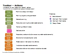
Slide4Software
Linux distribution - Ubuntu
Embedded program written in Python and Java
Python for opening communication ports, and control
Java for implementing active video filters and noise reduction.
60Hz flicker filter can be implemented in both
GUI written in Java
Slide5Hardware
ODROID XU-4
ODROID-VU7 (7 inch screen)
See3CAM
Solid State Drive
8Gb eMMC
Slide6Hardware - ODROID-VU7
Slide7Hardware - ODROID-VU7
Slide8Mechanical Base
Preconstructed Base
provided by Prof. Sisson
FDA - approved
Allows for large and fine movements
Commercially available
Slide9Controls
The X and Z axis are controlled using a joystick for fine movements and a loose base for large movements.
The Y axis is controlled by a dial connected to a screw allowing for fine movements.
The X axis can be locked by tightening a thumb screws.
Slide10Additional Mechanical Design (Central housing)
The central stand will hold a detachable component which will hold:
ODROID-C processor (Green)
ODROID-VU7 7in Screen (Black)
See3CAM (Yellow)
Solid State Drive (Blue)
Lens housing (Yellow)
Slide11Additional Mechanical Design (Lens housing)
By modifying a film based fundus camera we achieve:
Has a proven lens structure.
Has a dated but optically sound lighting system.
This will require:
Integrate sensor at correct distance.
Replace dated electrical lighting components with LED system.
Slide12Mechanical Design Risk analysis
Stand is unsecured and is unstable while in transport causing difficulties in portability.
Will make Central housing separable allowing them to be transported separately.
Team does not own design for stand
Central housing attachment to stand will be made adaptable.
Lens housing was recovered from an old device.
Will make CAD diagrams of the housing to insure ability to duplicate final design.
Slide13Illumination and Flash
Kowa RC-2
Xenon bulb
Infrared lighting required to align camera
with pupil while avoiding dilation
Slide14Kowa RC-2 Illumination
Diagram of Kowa RC-R retinal camera produced to image a rat’s eye.
Shows use of a hole mirror (HM), xenon light source (Xe LS), liquid light guide (LLG), source ring (SR), and green filter (GF).
Xe light source requires 10W
Replace with LEDs?
https://www.osapublishing.org/boe/fulltext.cfm?uri=boe-2-11-3094
Slide15FDA Requirements
Must conform to:
ISO 10940: 2009 Ophthalmic instruments - Fundus cameras
IEC 60601-1:2005 Medical Electrical Equipment – Part 1: General Requirements for Basic Safety and Essential Performance
IEC 60601-1-2 Medical Electrical Equipment – Part 1-2: General Requirements for Basic Safety and Essential Performance: Collateral Standard: Electromagnetic Capability – Requirements and Tests;
ISO 15004-1:2006 Ophthalmic instruments – Fundamental requirements and test methods – Part 1: General requirements applicable to all ophthalmic instruments
ISO 15004-2:2007 Ophthalmic Instruments – Fundamental requirements and test methods – Part 2: Light hazard protection
Slide16Form FDA 3654
Form used to ensure compliance standards have been met.
Checks that standards match regulations
Slide17Form
FDA
3654
Slide18Current BOM
Slide19BOM of Previous Team’s Materials
Slide20Additional Items
Slide21Budget Overview
P16590- MSD I
P15590
Prototype Approximate Part Cost
Budget
Purchased
% Budget Spent
$2000
$445.63
22.3%
Purchased
$2087.99
Item Cost
$973.19
Slide22End of Semester Overview
Task
Assigned To
Due Date
Go over design review notes, create new action items if necessary
Team
12/10/2015
Put out any extra purchase requests
Team
12/10/2015
Gate review
Team
12/11/2015
Go over gate review notes
Team
12/11/2015
Design central mechanical housing
Matt
TBD
Load kernel
Cecelia
TBD