

Bhajneesh Singh Bedi Objectives Approach to Adenopathy Who to investigate When to investigate How to define risk for underlying malignancy Lymph Nodes Anatomy Collection of lymphoid cells attached to both vascular and lymphatic systems ID: 1033185
Download Presentation The PPT/PDF document "Lymphadenopathy Presented by :" is the property of its rightful owner. Permission is granted to download and print the materials on this web site for personal, non-commercial use only, and to display it on your personal computer provided you do not modify the materials and that you retain all copyright notices contained in the materials. By downloading content from our website, you accept the terms of this agreement.


1. LymphadenopathyPresented by : Bhajneesh Singh Bedi
2. ObjectivesApproach to AdenopathyWho to investigateWhen to investigateHow to define risk for underlying malignancy
3.
4. Lymph NodesAnatomyCollection of lymphoid cells attached to both vascular and lymphatic systemsOver 600 lymph nodes in the bodyFunctionTo provide optimal sites for the concentration of free or cell-associated antigens and recirculating lymphocytes – “sensitization of the immune response”To allow contact between B-cells, T-cells and macrophagesLymphadenopathy - node greater than 1cm in size
5. Why do lymph nodes enlarge?Increase in the number of benign lymphocytes and macrophages in response to antigensInfiltration of inflammatory cells in infection (lymphadenitis)In situ proliferation of malignant lymphocytes or macrophagesInfiltration by metastatic malignant cellsInfiltration of lymph nodes by metabolite laden macrophages (lipid storage diseases)
6. Epidemiology0.6% annual incidence of unexplained adenopathy in the general population10% were referred to a subspecialist and 3.2 % required a biopsy and 1.1% had a malignancy
7. When to worry?AgeCharacteristics of the nodeLocation of the nodeClinical setting associated with lymphadenopathy
8. AgeChildren/young adults – more likely to respond to minor stimuli with lymphoid hyperplasiaLymph nodes in patients less than the age of 30 are clinically benign in 80% of cases whereas in patients over the age of 50 only 40% are benignBiopsies done in patients less than 25 yrs have a incidence of malignancy of <20% vs the over-50 age group has an incidence of malignancy of 55-80%
9.
10.
11. Characteristics of the nodeNodes lasting less than 2 weeks or greater than one year with no progression of size have a low likelihood of being neoplastic – excludes low grade lymphomaCervical nodes – up to 56% of young adults have adenopathy on clinical examInguinal adenopathy is common – up to 1-2 cm in size and often benign reactive nodes
12. Characteristics of the nodeConsistency – Hard/Firm vs Soft/Shotty; FluctuantMobile vs Fixed/MattedTender vs PainlessClearly demarcatedSizeWhen to worry – 1.5-2cm in sizeEpitroclear nodes over 0.5cm; Inguinal over 1.5cmDuration and Rate of Growth
13. Location of the nodeSupraclavicular lymphadenopathy Highest risk of malignancy – estimated as 90% in patients older than 40 years vs 25% in those younger than 40 yrsRight sided node – cancer in mediastinum, lungs, esophagusLeft sided node (Virchow’s) – testes, ovaries, kidneys, pancreas, stomach, gallbladder or prostateParaumbilical node (Sister Joseph’s)Abdominal or pelvic neoplasm
14. Location of the nodeEpitroclear nodesUnlikely to be reactiveIsolated inguinal adenopathyLess likely to be associated with malignancy
15. Clinical SettingB symptoms – fever, night sweats, weight lossFatiguePruritisEvidence of other medical conditions – connective tissue diseaseYoung patient – mononucleosis type of syndrome
16. Physical ExamFull nodal examination – nodal characteristicsOrganomegalyLocalized – examine area drained by the nodes for evidence of infection, skin lesions or tumours
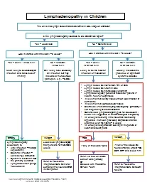
17. Approach to LymphadenopathyLocalized – one area involvedGeneralized – two or more non-contiguous areas
18.
19. Generalized LymphadenopathyMalignancy – lymphoma, leukemia, Kaposi’s sarcoma, metastasesAutoimmune – SLE, RA, Sjogren’s syndrome, Still’s disease, DermatomyositisInfectious – Brucellosis, Cat-scratch disease, CMV, HIV, EBV, Rubella, Tuberculosis, Tularemia, Typhoid Fever, Syphilis, viral hepatitis, PharyngitisOther – Kawasaki’s disease, sarcoidosis, amyloidosis, lipid storage diseases, hyperthyroidism, necrotizing lymphadenitis, histiocytosis X, Castlemen’s disease
20.
21. Laboratory InvestigationCBCSerologyCXRCT and MRIUltrasound
22. DrugsAllopurinolAtenololCaptoprilCarbamazepineGoldHydralazinePenicillinsPhenytoinPrimidonePyrimethamineQuinidineTrimethoprim/SulfamethozoleSuldinac
23. ManagementIdentify underlying cause and treat as appropriate – confirmatory testsGeneralized adenopathy – usually has identifiable causeLocalized adenopathy3-4 week observation period for resolution if not high clinical suspicion for malignancyBiopsy if risk for malignancy - excisional
24. Fine Needle AspirateConvenient, less invasive, quicker turn-around timeMost patients with a benign diagnosis on FNA biopsy do not undergo a surgical biopsy
25. Follow-up and Treatment Follow-up at 2-4 weeks interval for benign causes. Antibiotics are given only if there is strong evidence of bacterial infection. DO NOT USE GLUCOCORTICOIDS-might obscure diagnosis or delay healing in cases of infection (EXCEPTION: life-threatening pharyngeal obstruction by enlarged lymph tissue in Waldeyer’s ring caused by IM.)
26. ConclusionsLymphadenopathy – initial presenting symptomReactive vs MalignantProbabilityHistoryPhysical ExamBiopsy if not resolved in 3-4 weeks for low risk patientsBiopsy all high risk patients – excisional biopsy
27. Rosai-Dorfman Lymphadenopathy
28. Thanks forYour Attention