
Dr Ayisha Qureshi Assistant Professor MBBS MPhil Location of the Heart The Pericardium amp the Pericardial Sac The heart is enclosed in the doublewalled membranous pericardial sac ID: 408998
Download Presentation The PPT/PDF document "CARDIAC MUSCLE" is the property of its rightful owner. Permission is granted to download and print the materials on this web site for personal, non-commercial use only, and to display it on your personal computer provided you do not modify the materials and that you retain all copyright notices contained in the materials. By downloading content from our website, you accept the terms of this agreement.


Slide1
CARDIAC MUSCLE
Dr. Ayisha Qureshi
Assistant Professor,
MBBS, MPhilSlide2Slide3Slide4
Location of the Heart:Slide5Slide6Slide7
The Pericardium & the Pericardial Sac:
The
heart is enclosed in the double-walled, membranous
pericardial sac
(
peri
means “around”).
The
sac consists of
two layers—a
tough, fibrous covering and a secretory lining. The outer fibrous covering of the sac attaches to the connective tissue partition that separates the lungs. This attachment anchors the heart so that it remains properly positioned within the chest. The sac’s secretory lining secretes a thin pericardial fluid, which provides lubrication to prevent friction between the pericardial layers as they glide over each other with every beat of the heart.Pericarditis, an inflammation of the pericardial sac that results in a painful friction rub between the two pericardiallayers, occurs occasionally because of viral or bacterial infection.Slide8
Heart walls are composed of 3 distinct layers:
ENDOCARDIUM (inner):
thin layer of endothelium that lines the entire circulatory system (
endo
means “within”
)
MYOCARDIUM (middle):
composed of cardiac muscle that forms the bulk of heart wall (
myo
means “muscle”
)EPICARDIUM (outer): thin external membrane covering the heart (epi means “on”)Slide9
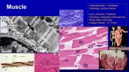
The Cardiac Muscle
Branched muscle cells (myofibrils)
Centrally
located
nucleus
Outside
membrane
is called
sarcolemma.
Cardiac
muscles have the same arrangement of actin and myosin, and the same bands, zones and Z discs as skeletal muscles forming sarcomeres
.They do have a poorly developed sarcoplasmic reticulum than skeletal muscles and require Calcium from ECF for contraction as T-tubules are well organized.Slide10
Arrangement of the heart muscles
The myocardium consists of interlacing bundles of cardiac muscle fibers arranged spirally around the circumference of the
heart.
What
is the
advantage of the spiral arrangement?
When the cardiac muscle contracts & shortens, a wringing effect is produced, efficiently pushing blood upwards towards the exit of the major arteries of the heart.Slide11
Intercalated Discs
Although the cardiac muscles interdigitate & branch, there is
NO
anatomical continuity b/w the individual muscle fibers.
Cardiac muscles are branched, have a single nucleus and are interconnected to each other, end to end by specialized structures called
INTERCALATED DISCS.
The intercalated discs are further composed of:
Gap Junctions
DesmosomesSlide12Slide13
HEART AS A DUAL PUMP:
Even thought the heart is a single organ, the left and the right side of the heart is anatomically and functionally separate.
This is done with the help of the
interventricular
spetum
.
It ensures that the blood from the left and right side of the heart does not mix
.
Although the left and the right sides are separated, the heart contracts in a co-ordinated fashion: the atria contract together and the ventricles contract together….Slide14Slide15
O
2
poor blood returns from the body thru the Superior & Inferior Vena Cava
↓
Enters the Right atrium
↓
Right ventricle
↓
Pulmonary artery
↓
Lungs
↓Blood is Oxygenated↓Pulmonary Veins↓Left Atrium↓Left Ventricle ↓Aorta ↓Circulated to the body Slide16