/
Confirming Pages Confirming Pages

Confirming Pages - PDF document

danika-pritchard
danika-pritchard . @danika-pritchard
Follow
421 views
Uploaded On 2016-08-14

Confirming Pages - PPT Presentation

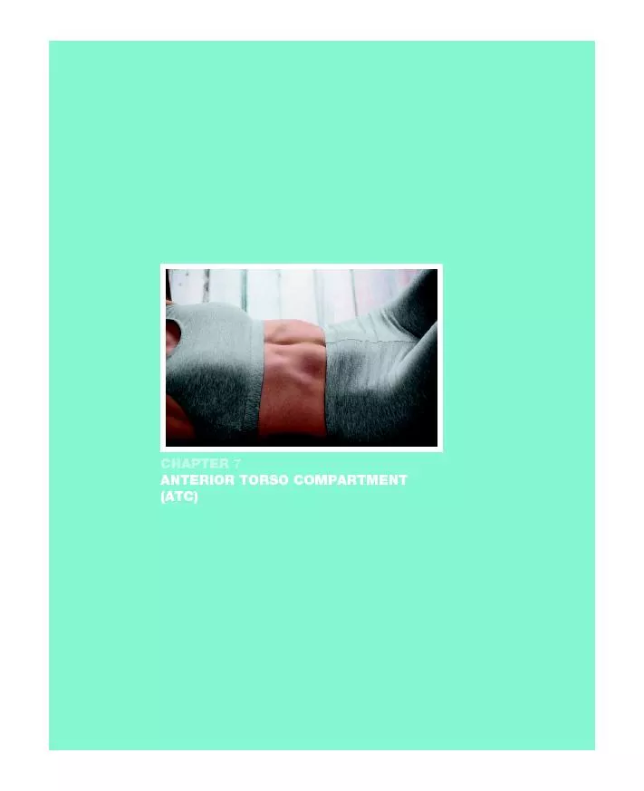
ANTERIOR TORSO COMPARTMENT wri73923ch07164217indd 164 Confirming Pages LEARNING OUTCOMES After completing this chapter you will be able to 71 Define the origins and insertions of the ID: 446492

ANTERIOR TORSO COMPARTMENT wri73923_ch07_164-217.indd

Share:

Link:

Embed:

Download Presentation from below link

Download Pdf The PPT/PDF document "Confirming Pages" is the property of its rightful owner. Permission is granted to download and print the materials on this web site for personal, non-commercial use only, and to display it on your personal computer provided you do not modify the materials and that you retain all copyright notices contained in the materials. By downloading content from our website, you accept the terms of this agreement.


Presentation Transcript

Confirming Pages ANTERIOR TORSO COMPARTMENT wri73923_ch07_164-217.indd 164 Confirming Pages LEARNING OUTCOMES After completing this chapter, you will be able to: 7.1 Define the origins and insertions of the muscles of the ATC. 7.2 Describe the actions of the ATC. 7.3 Recognize the pain patterns of the ATC. 7.4 Discuss the clinical notes for and the importance of treating the muscles of the ATC. 7.5 Demonstrate the client positioning and treatment routines related to the ATC 7.6 Demonstrate safe and effective stretching techniques for the muscles of the ATC. OVERVIEW Like the posterior lower extremity compartment (PLEC), the anterior torso compartment (ATC) Shortened abdominal muscles will pull the rib cage down and cause a forward neck and head quently decreasing cervical range of motion and many back, shoulder, and upper-extremity pain terior compartment muscles that countertighten and resist the forward and downward pulls of tension from the shortened trunk flexors and hip extensors. (An analogy: A tree is held up with guide wires and the tree bends to one side from sustained windsÑit is the lengthened wires that nals, psoas, serratus anterior, pectorals, and anterior cervicals are a kinetic chain that, when short, often causes chronic pain of the low back, neck, and shoulders. Often, chronic back, neck, and shoulder pain associated with muscular imbalances that effect stooped posture can be reduced and ful ones often experienced in the posterior torso. Lengthening the abdominals will create space between the pelvis and the rib cage. Lengthening the serratus anterior and pectorals will create space between the shoulder girdles and the pelvis, thereby allowing the scapulae to drift up and back, restoring the body to a more upright posture. Treatment for stooped posture and forward neck and head positioning should consist of combining both the abdominals and the hamstrings within each session. Lengthening the hamstrings will cause the pelvis to rotate up and forward, increasing the lumbar curve and thereby greatly reducing the compression effect on the lumbar spinal disks. The combined treatments (ATC and PLEC) will restore the body to an upright posture, thereby decreasing the chronic pain that is often experienced in the posterior torso. wri73923_ch07_164-217.indd 165 Confirming Pages The anterior torso is the emotional cylinder of the body. Playing the edge with precision Precise hand placements and treatment routines of the abdominal, chest, and anterior cervical muscular imbalances and stooped posture. CLINICAL SUCCESS In 2001, a professional golfer on the PGA tour went to a therapist. He complained of a Òslight During a soft-look evaluation of the clientÕs body, the therapist observed his projected forward The client was placed in the supine position on the table and a hard-look evaluation of his body was conducted. After thoroughly examining for lower-body deviations, the therapist concluded that the clientÕs leg lengths and pelvic bones were symmetrical and that his primary complaint of bilateral upper-extremity numbness and weakness was most likely isolated in the neck and the tho-racic outlet. The client was moved into a side-lying position aided by a cushioned body support sys-tem. The treatment plan consisted of releasing the anterior cervical muscles, primarily the longus colli, the longus capitis, and the scalene muscles. Releasing these muscles would help restore much of his cervical lordosis, thereby reducing the compression effect on his spinal nerves and at the base of the occiput. The muscles attached on the anterior tubercles were isolated, and the therapist proceeded to Slow, focused, and isolated work in the appropriate areas produced remarkable results. These apy sessions and no residual discomfort during his tournament play. wri73923_ch07_164-217.indd 166 Confirming Pages ABDOMINALS RECTUS ABDOMINIS, FIGURES 7.1 AND 7.2 ORIGIN Costal cartilages 5, 6, and 7 INSERTION Anterior pubic bone ACTION Flexes vertebral column; compresses abdominal contents MYOTOME T7–T12 FIGURE 7.1 Rectus Abdominis Muscles EXTERNAL OBLIQUE, FIGURES 7.3 AND 7.4 ORIGIN Lower eight ribs INSERTION Abdominal fascia, iliac crest, and linea alba ACTION flexes trunk; compresses abdominal contents laterally flexes trunk; rotates trunk to opposite side MYOTOME T8–T12 RectusabdominisPyramidalis wri73923_ch07_164-217.indd 167 Confirming Pages FIGURE 7.2 Rectus Abdominis Muscle and Referred Pain Patterns FIGURE 7.3 External and Internal Obliques and Transverse Abdominis Muscles FIGURE 7.4 External Oblique Muscle and Referred Pain Patterns Rectusabdominis Rectus Transverse External External wri73923_ch07_164-217.indd 168 Confirming Pages CLINICAL MASSAGE THERAPY AND STRUCTURAL BODYWORK169 INTERNAL OBLIQUE, FIGURES 7.3 AND 7.5 ORIGIN Inguinal ligament and iliac crest INSERTION Costal cartilages of last four ribs, abdominal fascia, and linea alba ACTION laterally flexes trunk; rotates trunk to same side MYOTOME T8–T12 TRANSVERSE ABDOMINIS, FIGURES 7.3 AND 7.6 ORIGIN Inguinal ligament, iliac crest, and thoracolumbar fascia INSERTION Abdominal fascia, linea alba, and pubis ACTION Compresses abdominal contents MYOTOME T7–T12 FIGURE 7.5 Internal Oblique Muscle and Referred Pain Patterns Internaloblique Internal wri73923_ch07_164-217.indd 169 Confirming Pages FIGURE 7.6 Transverse Abdominis Referred Pain Pattern CLINICAL NOTES: Abdominals The abdominal muscles lie in the following order from superficial to deep: rectus abdominis, which The abdominals work together to compress the abdominal contents and hold them in place. The Any sustained pain (acute or chronic) of the abdomen should be referred to a primary care physi- Transverse wri73923_ch07_164-217.indd 170 Confirming Pages The primary referred pain pattern of the oblique muscles is in the upper abdominal quadrants just Increased tension in the abdominal musculature can be the result of physical activities that require A Òseated-chair clientÓ is a person who spends most of the time sitting. Inadequate lumbar support chair client to slouch. Sitting slouched causes posterior rotation of the pelvis and loss of lumbar vis from slouched posture, the transverse abdominis is often left slack and often loses its ability to function properly. As the space between the rib cage and the pelvis decreases, so does the space for the abdominal contents. Loss of space increases the abdominal pressure and forces the abdominal contents to Òpooch outÓ; this further inhibits the function, maintaining lumbar lordosis. Slouched mary protractor of the scapula, and its fibers interdigitate with those of the external oblique along the anterior-lateral rib cage. Protraction from the serratus anterior will cause the scapula to slide forward around the rib cage, further shortening the pectoral muscle and bringing the arm toward the chest; this is viewed as a rounded shoulder. It must be emphasized again that short trunk ologically efficient posture. Pain is a signal of abnormal physiology; and the signaling of pain is often a sign of neuromuscular exhaustion from chronic muscle and nerve tension and is frequently experienced in the lengthened posterior torso muscles of the lower back, neck, and shoulders. These frequently experienced pain symptoms are the bodyÕs way of communicating that itÕs tired of holding the body upright from the forward collapsing that is associated with stooped posture. The muscles of the posterior torso, primarily the extensors and retractors, countertighten in an effort to support the upper body and resist its falling forward off the gravitational line. Forward neck and head positioning resulting from stooped posture causes posterior neck pain, especially occipital joints, as this is the bodyÕs last-ditch effort to rock the head back, leveling the eyes to the horizon. These compressed joints often cause headaches and restrict cervical motion. Postoperative scar tissue in the abdominal area can be treated with massage therapy. Interweaving will assist in the softening of the scar. Superficial work, including fascial stretching along the Langer wri73923_ch07_164-217.indd 171 Confirming Pages 172 CHAPTER 7 are lines of tension in often follow the skinÕs natural wrinkles. ROUTINE: Abdominals supine. Any sustained pain (acute or chronic) of the abdomen should be referred to the primary physician for proper medical and diagnostic evaluation. 1. Treat the anterior torso compartment with compression, separating the muscle divisions from the pubic bone to the rib cage. (See Figure 7.7 .) Continue superiorly through the sternal and pectoral regions in a fan-shaped 2. This is a bilateral treatment; if the leg lengths are even, choose one side to treat first. If the clientÕs leg lengththe rib cage, using compression and slow gliding to treat the rectus abdominis muscle ( Figure 7.8 ). Move inferi- 3. Flex the trunk laterally, away from the side being treated. Move laterally into the obliques, and continue treat-allowing you, the therapist, to proceed further into the deeper layers of the abdominal wall. (See Figure 7.9 .) In FIGURE 7.7 FIGURE 7.8 wri73923_ch07_164-217.indd 172 Confirming Pages FIGURE 7.10 FIGURE 7.11 FIGURE 7.12 Figure 7.9 d, the use of the elbow gives a broader contact point and a deeper compression into the muscles and 4. Have your client turn to a side-lying position, arm extended over the head. Place a bolster in the space between with both hands moving horizontally down toward the table. (See Figure 7.10 .) 5. Using the fingertips of one hand while stretching the myofascia with the other, treat the obliques with com-posterior, further treating the abdominals with compression and stretching. (See Figure 7.11 .) Treat slowly, fol- 6. Using the thumbs, treat the muscles attaching to the rib cage with slow compression and friction. Opening up the spaces between the ribs will allow a fuller expansion of the rib cage on deeper inhalation. (See Figure 7.12 .) Coach the client in deep breathing, and encourage the client to focus on full relaxation. Maintain the clientÕs edge to avoid any pain or increased discomfort while continuing treatment throughout the anterior and lateral rib cage. FIGURE 7.9a FIGURE 7.9b FIGURE 7.9c FIGURE 7.9d wri73923_ch07_164-217.indd 173 Confirming Pages ILIOPSOAS PSOAS MAJOR, FIGURES 7.13 AND 7.14 ORIGIN T12–L5 INSERTION Lesser trochanter of femur ACTION proximates lumbar vertebrae toward femur MYOTOME L1–L4 FIGURE 7.13 Iliopsoas Muscles: Psoas Major, Psoas Minor, and Iliacus FIGURES 7.13 AND 7.14 ORIGIN Inner surface of iliac fossa INSERTION Lesser trochanter of femur ACTION Flexes hip MYOTOME L2, L3 IliacusPsoas majorPsoas minor wri73923_ch07_164-217.indd 174 Confirming Pages CLINICAL MASSAGE THERAPY AND STRUCTURAL BODYWORK175 CLINICAL NOTES: Iliopsoas The iliopsoas is composed of two distinct muscles: the psoas major and the iliacusÑwith one common attachment. The psoas major arises from the lumbar vertebrae. The iliacus originates from the inner surface of the iliac fossa and is well established as the primary hip flexor. They both insert on the lesser trochanter of the femur. Psoas major is the only muscle that attaches the lumbar spine to the femur. Many people think of the psoas as a primary postural stabilizer of the spine. Although it is often Functions of Psoas There is no debate that the psoas major is a hip flexor. Yet there is much debate among researchers Part of the confusion is that the psoas major is a hip flexor, and hip flexion is usually associated (For clarification, it should be noted that the lifting of the femur toward the pelvis and the tilting vis toward the front of the joint. Conversely, a posterior tilt of the pelvis is hip extension. Learning to think about joint movement in this fashion increases the studentÕs ability to analyze posture and A number of factors influence the various actions of the psoas muscles. When the psoas contracts, FIGURE 7.14 Iliopsoas Muscles and Referred Pain Patterns Iliopsoaspain pattern Iliopsoas wri73923_ch07_164-217.indd 175 Confirming Pages An increase in lumbar lordosis is by definition an increase in the curve of the lumbar spine. In a true When viewed from the side, the psoas major lies in an almost vertical line along the lumbar spine and must pull each vertebral body downward toward the pelvis and femur. When this occurs, the psoas is actually decreasing the space between the vertebral bodies; flattening the lumbar arc. If there is a bar vertebrae do not fan open to create lumbar extensionÑthey close, creating lumbar flexion. The psoas major muscle is, therefore, with a minor but significant exception at the L5-S1 joint (dis-bar extension or contribute to an actual lumbar lordosis; it actually resists it. Psoas Controls Lumbar Extension If the torso starts to lean backward, the psoas major will generate an eccentric contraction (a con-ing too far and too fast. Psoas Reacts to Other Muscles If the body is slouching forward and the abdominal wall and hip extensors are chronically short, In response to the lumbar spine being pushed posteriorly (rearward) and the pelvis moving into a If indeed it is determined that psoas is contracted by way of reacting to overshortening of the the long run. Compression of Lumbar Spinal Disks To reiterate, the psoas major muscle is perfectly placed to apply direct powerful downward forces wri73923_ch07_164-217.indd 176 Confirming Pages Much back pain is caused by muscles irritating and sometimes entrapping sensory nerves. Many M uscle tension also produces vertebral dysfunction and disk pathology often from muscles distant from the posterior torso, such as the hamstrings, gluteals, and abdominal muscles. The Pars Vertebralis of Psoas The pars vertebralis portion of the psoas consists of small slips of muscle, which are the deepest lay-tebral column and compressor of the intervertebral disks. (See Figure 7.15a .) The pars vertebralis portion of the psoas major and the deepest fibers of the main body of the psoas too traumatized. A short ened or traumatized psoas requires the approach of the minimum-edge While it is true that the psoas major is, along with the iliacus, a primary hip flexor, it simultaneously “Mini-” or “Pseudo-” Lordosis The minor but significant exception referred to earlier is produced by the structure of the L5-S1 joint. Because the 5th lumbar, as viewed from the side, appears as if it were positioned on a small, downward-sloping shelf projecting forward from the top of the sacrum, the psoas major can also be a powerful force pulling the 5th lumbar vertebra, and the whole lumbar column, forward and down. It is common to have muscles pulling so hard in opposite directions that while the hip extensors attempt to rotate the pelvis posteriorly, the psoas attempts to rotate the lumbar spine anteriorly. This causes a forward and downward shearing action of the L5 vertebrae that creates a ÒkinkÓ at the posterior face of the lumbosacral joint. The ÒkinkÓ contributes to the illusion of the Òdeep curveÓ in the lumbar area. terior spine (which is restricted to the L5-S1, and possibly L5-L4, area) and other common postural illusions for a true lordosis of the entire lumbar spine. (See Figure 7.15b .) Posterior Fibers of Psoas Also complicating lower-back pain, though not all of them are directly attached to the lumbar wri73923_ch07_164-217.indd 177 Rev. Confirming Pages pain-sensitive than muscle fibers) could be a significant source of pain. Lumbar Pain Pattern The iliopsoas, abdominals, hamstrings and the erector spinae, multifidus, rotatores and quadratus lum-borum muscles are often the culprits of a failed low back and/or chronic pain because they can generate opposing forces on the pelvis, sacrum and lumbar vertebrae, and intervertebral disks. Prolonged sitting with the hips acutely flexed will cause the iliopsoas to adaptively shorten. This, plus reactive forces from opposing muscles, creates compression, stress, and vertical pain along the spine (lamina groove) and sacroiliac joints. Secondary pain of the iliopsoas muscle will likely be present at the insertion sites on the lesser trochanter of the femur and on the anterior thigh and groin. A hypertense psoas major also contributes to the so-called hip-click phenomena as well. Asymmetrical imbalances of the iliopsoas may contribute to a compensatory lumbar scoliosis. Both FIGURE 7.15a Pars vertebralis C Pars vertebralis: Deep, superior portion(in green), small bridges between bodiesof T-12 thru L-5. Fully covered by portionA of psoas major.T-12B Deep, posterior portioniginating at transverseA Superficial, anteriorportion (in purple) ofpsoas major, originatingat vertebral bodies &intravertebral discs ofT-12 through L-4 or L-5Femur wri73923_ch07_164-217.indd 17811/14/09 12:07:22 PM Rev. Confirming Pages FIGURE 7.15b T-12 Gluteus maximus Psoas Major,vertebral bodies, flattening the curve from T–12 through L–4 or L–5The greater the ThoracicHamstrings, and Adductor Magnusof joint, TENDING toward pushing pelvisposterior TILT of pelvis. However, highby hip extensors. ... The resultinganterior SHIFT is often mistaken for an anterior TILT.creating lumbar flexion of T-12A pseudo-lordosis appears as a deep curve (exaggerated lordosis) of the lumbar spine. Yet often, this is a false illusionof the actual lumbar vertebrae fromT–12 to L– 4. The overlying flesh often masks the true picture of what is actually occurring structurally(relationship between the bones). Treating such cases by tucking the pelvis or sacrum, flattening the low back, prematurely releasing wri73923_ch07_164-217.indd 17911/14/09 12:07:24 PM Confirming Pages 180 CHAPTER 7 lumbar spine, pelvis, and/or lower extremities. For example, unless countered by an opposing muscle, a hypertonic iliopsoas muscle may cause a client to lean toward the hypertonic side, further involving the obliques, quadratus lumborum, erector spinea and latissimus dorsi muscles on the same side. ROUTINE: Iliopsoas Treating the abdominal muscles before treating the iliopsoas is recommended, as the tension in a hypertonic abdominal muscle can prevent lengthening of the iliopsoas. supine. 1. If pain is present in the lumbar and sacroiliac region, bend the knee and laterally rotate and abduct the thigh. the abdominal wall separating the muscular divisions. (See Figure 7.16 .) 2. Place the fingertips just lateral to the rectus abdominis. Use circular pressure to penetrate slowly, gradually to help avert her natural tendency to guard against abdominal penetration. (See Figure 7.17 .) When the client 3. To isolate the psoas major, keep the hands in the same place and have the client contract the psoas muscle by moving superior toward the rib cage. (See Figures 7.18 and 7.19 .) Using the edge technique, treat with com- 4. Using the fingertips of both hands, treat the iliacus muscle where it merges with the psoas major muscle, along Periodically have the client contract the iliopsoas group to verify the location. (See Figure 7.20 .) 5. Outline the length of the psoas with both hands. Using fingertips and/or contoured hands, apply compression FIGURE 7.16 FIGURE 7.17 FIGURE 7.18 wri73923_ch07_164-217.indd 180 Confirming Pages FIGURE 7.19 deep-gliding effleurage up to and as far under the inguinal ligament as the client will allow. (See Figure 7.21 .) FIGURE 7.21 FIGURE 7.20 PECTORALIS MAJOR, FIGURES 7.22 AND 7.23 ORIGIN medial half of clavicle sternum from manubrium to zyphoid process first six costal cartilage surfaces INSERTION Lateral lip of bicipital groove of humerus ACTION Adduction, flexion, medial rotation of humerus; depression of arm and shoulder MYOTOME C5–T1 PECTORALIS MINOR, FIGURES 7.24 AND 7.25 ORIGIN Anterior 3rd, 4th, and 5th ribs near costal cartilage INSERTION Coracoid process of scapula ACTION Protracts, depresses, downwardly rotates scapula MYOTOME C8–T1 wri73923_ch07_164-217.indd 181 Confirming Pages FIGURE 7.22 Pectoralis Major Muscles FIGURE 7.23 Pectoralis Major Muscle and Referred Pain Patterns FIGURE 7.24 Pectoralis Minor and Subclavius Muscles Pectoralismajor muscle Pectoralis Pectoralis PectoralisSubclavius wri73923_ch07_164-217.indd 182 Confirming Pages FIGURE 7.25 FIGURES 7.24 AND 7.26 ORIGIN 1st rib costocartilage junction INSERTION Middle third of inferior surface of clavicle ACTION Depression of shoulder; stabilization of clavicle during shoulder movement MYOTOME C5, C6 FIGURE 7.26 Subclavius Muscle and Referred Pain Patterns Pectoralisminor pain Subclavius Subclaviuspain pattern wri73923_ch07_164-217.indd 183 Confirming Pages 184 CHAPTER 7 CLINICAL NOTES: Pectoralis Major, Pectoralis Minor, and Subclavius The pectoralis minor originates on the 3rd, 4th, and 5th ribs and attaches to the coracoid process Because of the varied directions that its three divisions travel and their attachment on the humerus, the pectoralis major influences many movements of the humerus, including flexion, adduction, and medial rotation of the arm, as well as depression of the arm and shoulder. The pectoralis minor assists the serratus anterior in forced inspiration and contributes to protraction, depression, and downward rotation of the scapula. The subclavius stabilizes the clavicle and assists in depression of the shoulder. Anterior shoulder pain, breast pain, and diffuse soreness are symptoms of trigger point pain referred by the pectoralis muscles. Pain in the shoulder that traces down the arm and into the hand could be a sign of thoracic outlet syndrome: a condition in which the cervical spine compresses toward the rib cage because of anterior cervical musclesÕ and the pectoralis minorÕs pulling the shoulder down from the front, thus narrowing the space between the cervical spine and the 1st rib. Also contrib-uting to thoracic outlet syndrome is the latissimus dorsiÕs pulling the shoulder down from the back and, therefore, pulling the clavicle down and back against the 1st rib. These muscular imbalances can compress and entrap the neurovascular structures exiting and entering the neck, chest, and arm; if left untreated, they can lead to pain, motor weakness, immobility, and atrophy of the shoulder and upper extremity. Dr. Janet Travell, coauthor of refers to the pectoralis Travell has a second nickname for the pectoralis major: the Òpoor posture muscle.Ó A stooped pos- wri73923_ch07_164-217.indd 184 Confirming Pages CLINICAL MASSAGE THERAPY AND STRUCTURAL BODYWORK185 The three divisions of the pectoralis major should be treated independently with treatment directed The chest chest, where there can be metabolic waste accumulation, causing hypersensitivity. ROUTINE: Pectoralis Major, Pectoralis Minor, and Subclavius supine, with the clientÕs arm extended away from the body. Support the arm with an extended bol-ster if the arm does not fit on the table, but allow the clientÕs hand to hang off the table. (See Figure 7.27 .) 1. Treat the anterior torso compartment with slow, deep effleurage from the pubic bone, over the rib cage, and superiorly in a fan-shaped pattern through the sternal and pectoral region, ending at the clavicles. 2. Using the fingertips, apply compression and friction along the muscle fibers, separating the three muscular divi-sions of the pectoralis major. 3. Using the fingertips, slowly compress downward and apply slow, rhythmical friction to the pectoralis minor by treating the pectoralis major. (See Figure 7.28 .) 4. Isolate the pectoralis major by picking it up, off the rib cage. Treat with compression. Using thumbs and fingers, friction in 1-inch segments. (See Figure 7.29 .) 5. Using a collapsed fist, treat the pectoralis major from the sternum to the humerus with deep, slow effleurage. 6. Contour one hand along the lateral rib cage, and slowly apply inward compression into the pectoralis minor while the other hand lightly lifts the pectoralis major. (See Figure 7.30 .) FIGURE 7.28 FIGURE 7.27 wri73923_ch07_164-217.indd 185 Confirming Pages FIGURE 7.29 FIGURE 7.30 FIGURE 7.31 FIGURE 7.32 7. Maintain the same contoured hand position. Glide superiorly toward the coracoid process while lifting the arm comfort. (See Figure 7.31 .) Have the client move to the side-lying position to ensure that the pectoralis major is off the rib cage, exposing it and the pec-toralis minor for treatment. Sit at the side of the table at the clientÕs waist. Place the clientÕs arm out in front of his or her 8. Place one hand around the clientÕs scapula. Place the other hand underneath the clientÕs arm, and grasp the pectoralis major. Treat with slow, firm petrissage, exploring the muscular divisions. (See Figure 7.32 .) Face the friction. Use the edge technique for maintaining the clientÕs comfort. (See Figure 7.33 .) 9. Allow the clientÕs arm to roll forward so that the clavicle lifts off the upper ribs. Using the fingertips of one hand friction in 1-inch segments. (See Figure 7.34 .) 10. Using the fingertips, glide along the inferior border of the clavicle and re-treat the pectoralis major around the attachment and the subclavius muscle, as shown in Figure 7.34 . wri73923_ch07_164-217.indd 186 Confirming Pages CLINICAL MASSAGE THERAPY AND STRUCTURAL BODYWORK187 SERRATUS ANTERIOR, FIGURES 7.35 AND 7.36 ORIGIN Upper nine ribs INSERTION Anterior vertebral border of scapula ACTION Protracts scapula; rotates scapula upward MYOTOME C5–C7 FIGURE 7.35 Serratus Anterior Muscle FIGURE 7.33 FIGURE 7.34 Serratusanterior wri73923_ch07_164-217.indd 187 Confirming Pages FIGURE 7.36 Serratus Anterior Muscle and Referred Pain Patterns CLINICAL NOTES: Serratus Anterior The serratus anterior anchors the scapula to the thorax, originating on the upper nine ribs and attaching on the entire anterior vertebral border of the scapula from the inferior angle to the supe-rior angle. The lower fibers of the serratus anterior interdigitate with the upper fibers of the exter-nal oblique where they attach to the lower ribs. These fibers can pull the inferior angle of the scapula forward, causing the scapula to rotate upward elevating the shoulder girdle. From a posterior view, the vertebral border of the scapula will appear to protrude from the body. The primary function of the serratus anterior is protraction of the scapula, assisted by the pectoralis Shortening of the pectoralis major, pectoralis minor and the serratus anterior is often the result of protraction of the shoulder, it is likely that the serratus anterior is involved. Serratusanterior Serratuspain pattern Serratusanterior Serratus wri73923_ch07_164-217.indd 188 Rev. Confirming Pages ROUTINE: Serratus Anterior side-lying. 1. Using the fingertips, treat the lateral torso with slow, deep effleurage; stretching the myofascia from the iliac crest to the axilla, continuing to the olecranon processes of the ulna. (See Figure 7.37 .) 2. With a contoured hand and the fingertips, glide along the rib cage, simultaneously compressing the serratus anterior onto the rib cage. (See Figure 7.38 .) Position the other hand under the vertebral border of the scapula. 3. Using flexed fingertips, treat the underlying surface of the vertebral border of the scapula with friction in 1-inch segments. (See Figure 7.39 .) 4. Using fingertips, treat along the upper nine ribs with compression and slow, deep effleurage. Then apply a broad, slow and isolated friction in 1-inch segments. (See Figure 7.40 .) 5. Using the fingertips, treat the muscles along the lamina groove with friction while retracting the shoulder girdle for easier access. (See Figure 7.41 .) FIGURE 7.37 FIGURE 7.38 FIGURE 7.39 FIGURE 7.40 FIGURE 7.41 wri73923_ch07_164-217.indd 18911/14/09 12:08:56 PM Confirming Pages CERVICAL MUSCLES Prior to treating the cervical muscles, treat the compartment distortions from the originating By now, a complete postural assessment and palpatory examination of the clientÕs muscles that Chasing a clientÕs pain (i.e., treating only where it hurts) often produces only temporary results. tal joints, and decreasing the intensity and frequency of the neck pain. Quite often, lengthening the FIGURES 7.42 AND 7.43 ORIGIN 1. Manubrium of the sternum; 2. medial clavicle INSERTION Mastoid process ACTION flexion of neck; elevation of sternum on deep inspiration lateral flexion of neck; rotation of head to opposite side MYOTOME C3, C4 wri73923_ch07_164-217.indd 190 Confirming Pages FIGURE 7.42 Sternocleidomastoid Muscle FIGURE 7.43 Sternocleidomastoid Referred Pain Patterns CLINICAL NOTES: Sternocleidomastoid The sternocleidomastoid muscle (SCM) is the largest and most superficial muscle of the antero- The SCM muscles are synergistic and antagonistic to themselves. Bilaterally, they flex the neck and Sternocleidomastoid Sternocleidomastoid(sternal head) Sternocleidomastoid wri73923_ch07_164-217.indd 191 Confirming Pages 192 CHAPTER 7 Pain patterns of the SCM muscle frequently include multiple referral zones to the face and head. Refer-ral pain from the clavicular section may occur independently of referral pain from the sternal section. Trigger points in the sternal section of the SCM typically cause migraine arc pain above the eyes that travels laterally through the sinuses and spills downward to the face, cheek, and temporomandibular joint region. A person with these symptoms may also be medically diagnosed with atypical facial neu-ralgia or tension headache. Trigger points of the clavicular section of the SCM typically refer pain deep within the ear and may cause dizziness, disturbed equilibrium, and vertigo. Treatment of the SCM should include restoring an upright body posture and treating any and taining stability and strength of the neck muscles. ROUTINE: Sternocleidomastoid supine. Stand or sit at the head of the therapy table. 1. Using the fingers, glide in a superior direction and explore the intermuscular septum between the sternal and the medial and lateral borders of the SCM. (See Figure 7.44 .) 2. Using flexed fingers of both hands, penetrate under the lateral borders of the SCMÕs and apply light, static com-revealed, will determine the amount of time needed for treatment. (See Figure 7.45 .) 3. Isolate the sternal head of the SCM. Pick up and compress the fibers, moving superiorly in 1-inch segments. (See Figure 7.46 .) FIGURE 7.44 FIGURE 7.45 wri73923_ch07_164-217.indd 192 Confirming Pages FIGURE 7.46 FIGURE 7.47 FIGURE 7.48a FIGURE 7.49 4. Isolate the clavicular head of the SCM. Pick up and compress the fibers, moving superiorly in 1-inch segments. 45-degree angle. (See Figure 7.47 .) 5. Using the fingertips of the other hand, rake into the SCM fibers beginning at the merge of the two heads, isolated using standard compression. (See Figure 7.48 .) Glide 6 to 10 times. 6. Using a contoured index finger, friction the mastoid process in all directions. (See Figure 7.49 .) wri73923_ch07_164-217.indd 193 Confirming Pages ANTERIOR CERVICALS—SUPERFICIAL: SUPRAHYOIDS FIGURES 7.50 AND 7.51 ORIGIN Inside surface of mandible INSERTION Hyoid bone ACTION Elevates hyoid bone; raises floor of mouth and tongue MYOTOME C1–C3, facial nerves FIGURES 7.50 AND 7.51 ORIGIN Inside surface of mandible; mastoid process of temporal bone INSERTION Intermediate tendon attached to hyoid bone and inside surface of mandible ACTION Raises hyoid bone; assists in opening jaw; moves hyoid bone forward or backward MYOTOME C1–C3, facial nerves FIGURE 7.50 Anterior Cervicals—Suprahyoid Muscles: Mylohyoid and Digastric Hyoid boneMylohyoidDigastric wri73923_ch07_164-217.indd 194 Confirming Pages ANTERIOR CERVICALS—SUPERFICIAL: INFRAHYOIDS FIGURE 7.52 ORIGIN Inside surface of mandible INSERTION Hyoid bone ACTION Protracts hyoid bone and tongue MYOTOME C1–C3, facial nerves FIGURE 7.51 Suprahyoid Referred Pain Patterns FIGURE 7.52 Anterior Cervicals—Infrahyoid Muscles: Geniohyoid, Stylohyoid, Thyrohyoid, Sternohyoid, Sternothyroid, and Omohyoid Suprahyoidspain patterns GeniohyoidThyrohyoidSternothyroidStylohyoidThyroid cartilageSternohyoidOmohyoid wri73923_ch07_164-217.indd 195 Confirming Pages 196 CHAPTER 7 FIGURE 7.52 ORIGIN Styloid process of temporal bone INSERTION Hyoid bone ACTION Retracts hyoid bone; elevates tongue MYOTOME C1–C3, facial nerves FIGURE 7.52 ORIGIN Lamina of thyroid cartilage INSERTION Hyoid bone ACTION Depresses hyoid or elevates thyroid cartilage MYOTOME C1 FIGURE 7.52 ORIGIN Medial end of clavicle, manubrium of sternum INSERTION Hyoid bone ACTION Depresses hyoid MYOTOME C1–C3 FIGURE 7.52 ORIGIN Manubrium of sternum INSERTION Lamina of thyroid cartilage ACTION Depresses hyoid or depresses thyroid cartilage MYOTOME C1–C3 wri73923_ch07_164-217.indd 196 Confirming Pages CLINICAL MASSAGE THERAPY AND STRUCTURAL BODYWORK197 ANTERIOR CERVICALS—DEEP FIGURE 7.52 ORIGIN Superior border of scapula near scapular notch INSERTION Hyoid bone via central tendon on clavicle ACTION Depresses hyoid bone MYOTOME C2, C3 LONGUS COLLI, FIGURES 7.53 AND 7.54 ORIGIN Multiple attachments from C3–T2 vertebral bodies and transverse tubercles INSERTION Multiple attachments from C3–T2 vertebral bodies and transverse tubercles ACTION Flexes cervical spine MYOTOME C2–C7 FIGURE 7.53 Anterior Cervicals—Longus Colli Muscles Longuscolli wri73923_ch07_164-217.indd 197 Confirming Pages FIGURE 7.54 Anterior Cervicals—Longus Colli Referred Pain Pattern FIGURE 7.55 Anterior Cervicals—Longus Capitis Muscles LONGUS CAPITIS, FIGURES 7.55 AND 7.56 ORIGIN Transverse tubercles of C3–C6 INSERTION Occipital bone, anterior to foramen magnum ACTION Flexes head MYOTOME C1–C3 Longuscapitis Longus colli wri73923_ch07_164-217.indd 198 Confirming Pages FIGURE 7.56 Anterior Cervicals—Longus Capitis Referred Pain Pattern CLINICAL NOTES: Anterior Cervicals The superficial anterior cervical muscles include the suprahyoids and the infrahyoids, which attach Throat pain, difficulty swallowing, and the feeling of a ÒknotÓ in the throat are some symptoms of pain pattern wri73923_ch07_164-217.indd 199 Confirming Pages 200 CHAPTER 7 Include the suprahyoids and infrahyoids in the treatment plan. It is necessary to thoroughly release these muscles before attempting to displace the trachea and treat the longus colli and longus capitis. ROUTINE: Anterior Cervicals supine. 1. Treat the fascia of the anterior and lateral neck for 5 minutes. Use fascial stretching and rolling techniques.(See Figure 7.57 .) (The musculature thickens as multiple tendons merge in a 1-inch diameter in the area of the 2. Assess the tension of the suprahyoids and infrahyoids by gently moving the trachea and hyoid bone back and forth. (See Figure 7.58 .) 3. Isolate the sternohyoid and sternothyroid by picking up these superficial muscles at midline. Using the finger-tips and thumbs, treat the muscles from superior to inferior with compression and friction. (See Figure 7.59 .) FIGURE 7.57 FIGURE 7.58 FIGURE 7.59 wri73923_ch07_164-217.indd 200 Rev. Confirming Pages FIGURE 7.60 FIGURE 7.61 FIGURE 7.62 4. Stabilize the trachea and hyoid bone with one hand. Using the index finger of the other hand, treat the infrahy-treat underneath the hyoid bone with friction. (See Figure 7.60 .) 5. Using the fingertips, treat the attachments of the suprahyoids directly above the hyoid bone from lateral to medial with cross-fiber friction. (See Figure 7.61 .) Compressions should be made slowly and precisely for A slight cervical extension will allow easier access to and better treatment of the longus colli and longus capitis muscles. (See Figure 7.62 .) 6. To treat the longus capitis, it is necessary to displace the trachea. This is a bilateral treatment. To treat the nective tissue toward the side being treated before displacing the trachea. (See Figure 7.63 .) 7. Stabilize the trachea in its displaced position with the left thumb. Using the right thumb or index finger, compress the longus colli and longus capitis muscles against the anterior vertebral bodies and cervical disks. (See Figure 7.64 .) 8. Using both index fingers, effleurage the longus colli and longus capitis muscles just lateral to the midline. (See Figure 7.65 .) 9. Using the right thumb or index finger, treat the anterior longitudinal ligament, longus colli, and longus capitis with cross-fiber friction. (See Figure 7.66 .) This is done with a focused intent and should not be painful. Change direction and stand at the head of the table. 10. Lift the clientÕs head, and flex the neck and head forward. With the left thumb or index finger, compress and increasing the cervical extension with each compression. (See Figure 7.67 .) 11. After treating the superficial and deep anterior cervical muscles, stretch the muscles by cupping the hands under wri73923_ch07_164-217.indd 20111/14/09 12:09:49 PM Rev. Confirming Pages pression effect on the cervical disks. (See Figure 7.68 .) This is a slow, non-forceful stretch. The client should 12. Repeat steps 6 to 11 on the clientÕs right side. FIGURE 7.63 FIGURE 7.64 FIGURE 7.65 FIGURE 7.66 FIGURE 7.67 FIGURE 7.68 wri73923_ch07_164-217.indd 20211/14/09 12:09:58 PM Confirming Pages SCALENES SCALENUS ANTERIOR, FIGURES 7.69 AND 7.70 ORIGIN Anterior tubercles of cervical vertebrae C3–C6 INSERTION 1st rib ACTION inspiration MYOTOME C3–C5 FIGURE 7.69 Scalene Muscles: Scalenus Anterior, Scalenus Medius, SCALENUS MEDIUS, FIGURES 7.69 AND 7.70 ORIGIN Anterior tubercles of cervical vertebrae C2–C7 INSERTION 1st rib ACTION inspiration MYOTOME C3–C5 ScalenusposteriorScalenusScalenusOblique view wri73923_ch07_164-217.indd 203 Confirming Pages FIGURE 7.70 Scalene Referred Pain Patterns SCALENUS POSTERIOR, FIGURES 7.69 AND 7.70 ORIGIN Posterior tubercles of cervical vertebrae C5–C7 INSERTION 2nd rib ACTION raises 2nd rib on deep inspiration MYOTOME C3–C5 CLINICAL NOTES: Scalenes The scalene muscle group originates on the cervical vertebrae and inserts on the 1st and 2nd ribs. In most conventional medical practices, the scalene muscle group is often overlooked as a source of Scalenespain patterns pain patterns wri73923_ch07_164-217.indd 204 Confirming Pages Chronic unilateral contraction of the scalene muscles is primarily due to asymmetry elsewhere in ROUTINE: Scalenes Note: If the client experiences an electriclike shock during this treatment, reposition the flexed fingers on the posterior tuber-exit the spine between the anterior and posterior tubercles. side-lying, with the clientÕs arm extended. (See Figure 7.71 .) This position ensures that the clavicle 1. Using one hand, grasp the SCM muscle and move it toward the therapy table. Using the other hand, penetrate C3 to C7. (See Figure 7.72 .) 2. Using the fingertips, continue the compressions and friction, moving posteriorly approximately Å inch to ½ inch to treat the medial and posterior scalene attachments on the anterior and posterior tubercles. 3. Using the fingertips, penetrate under the clavicle as far inferior as possible to influence the deeper fibers of the scalenes toward the 1st and 2nd ribs. (See Figure 7.73 .) Utilize the ÒminimumÓ edge to ensure clientÕs comfort. FIGURE 7.71 FIGURE 7.72 FIGURE 7.73 wri73923_ch07_164-217.indd 205 Confirming Pages Ensure the clientÕs comfort by treating slowly and precisely. If a pulse is palpated, reposition the fingers.cular structures of the body. supine. Pick up the clientÕs head and cup it in the hand. Rotate the head away from the side being treated, and elevate it approxi-mately 45 degrees. 4. Lightly place the thumb of the other hand between the sternal and clavicular tendons of the sternocleidomas-(See Figure 7.74 .) 5. Continue with thumb and glide superiorly along the anterior tubercles up to the point where the heads of the SCM merge. (See Figure 7.75 .) 6. Using flexed fingers, continue to treat the scalenes where they lie under the SCM muscle with compressions and 1-inch glides. (See Figure 7.76 .) Release the compression, move, then reenter with compression, and con-tinue 1-inch glides until the entire scalene attachments and bellies have been treated. FIGURE 7.74 FIGURE 7.75 TEMPOROMANDIBULAR JOINT FIGURES 7.77 AND 7.78 ORIGIN Zygomatic arch INSERTION Angle of mandible (outer surface) ACTION Closes lower jaw; clenches teeth MYOTOME 5th cranial nerve (trigeminal) FIGURE 7.76 wri73923_ch07_164-217.indd 206 Confirming Pages FIGURE 7.77 Masseter Muscle FIGURE 7.78 Masseter Referred Pain Patterns Masseter Masseterpain pattern Masseter Masseterpain pattern wri73923_ch07_164-217.indd 207 Confirming Pages 208 CHAPTER 7 FIGURES 7.79 AND 7.80 ORIGIN Temporal bone INSERTION Coronoid process of mandible ACTION Closes jaw; clenches teeth MYOTOME 5th cranial nerve (trigeminal) FIGURE 7.79 Temporalis Muscle FIGURE 7.80 Temporalis Referred Pain Patterns Temporalis Temporalispain patterns wri73923_ch07_164-217.indd 208 Confirming Pages CLINICAL MASSAGE THERAPY AND STRUCTURAL BODYWORK209 MEDIAL PTERYGOID, FIGURES 7.81 AND 7.82 ORIGIN Medial pterygoid plate of sphenoid bone INSERTION Angle of mandible (inner surface) ACTION Closes lower jaw; clenches teeth; deviates jaw to opposite side MYOTOME 5th cranial nerve (trigeminal) FIGURE 7.81 Pterygoid Muscles: Medial and Lateral FIGURE 7.82 Pterygoid Referred Pain Pattern Lateralpterygoidpterygoid Pterygoidpain patterns wri73923_ch07_164-217.indd 209 Confirming Pages 210 CHAPTER 7 LATERAL PTERYGOID, FIGURES 7.81 AND 7.82 ORIGIN Lateral pterygoid plate of sphenoid bone INSERTION Mandibular condyle, temporomandibular joint capsule, and articular disk ACTION Opens jaw; protracts mandible; deviates jaw to opposite side MYOTOME 5th cranial nerve (trigeminal) CLINICAL NOTES: Temporomandibular Joint The temporomandibular joint (TMJ) is the articulation between the condyle of the mandible and All of these muscles attach to the mandible and influence its movement. The masseter, medial pter- Symptoms of TMJ dysfunction include ear pain, sore jaw muscles, temple and/or cheek pain, jaw A therapist and a dentist should work together to address TMJ dysfunction so that the dental and When the mouth is opened, the mandibular condyles should translate forward and downward; gery may be indicated to correct this kind of TMJ dysfunction. wri73923_ch07_164-217.indd 210 Confirming Pages ROUTINE: Temporomandibular Joint supine. 1. Treat the cranial fasciae for approximately 10 minutes. Use a hair-pulling technique utilizing traction and/or Figures 7.83 and 7.84 .) 2. With one hand, rotate the clientÕs head away from the side of treatment. Using flexed fingertips, treat the entire temporalis muscle with circular friction. Friction above and below the zygomatic arch. (See Figure 7.85 .) 3. Isolate the temporalis tendon where it inserts on the coronoid process. To do this, have the client open her ensure the clientÕs comfort (See Figure 7.86 .) 4. Using a contoured index finger, treat the mastoid process with slow, deep friction. (See Figure 7.87 .) FIGURE 7.83 FIGURE 7.84 FIGURE 7.85 FIGURE 7.86 FIGURE 7.87 wri73923_ch07_164-217.indd 211 Confirming Pages FIGURE 7.88 FIGURE 7.89 5. Using the thumb, effleurage the entire masseter muscle from the zygomatic arch to the angle of the mandible. 6. Using the thumb, friction the attachments of the masseter on the zygomatic arch and the angle of the mandible. (See Figure 7.88 .) 7. Using the thumb, again effleurage the entire masseter muscle from the zygomatic arch to the angle of Figure 7.89 .) Note: Before performing intraoral treatments to muscles in the mouth, check your stateÕs regulations to ensure that these before administering intraoral treatments. 8. Using the index finger, penetrate laterally and inward until reaching the masseter. To confirm location of the ½-inch sections. (See Figure 7.90 .) 9. Using the pad of the thumb, apply pressure straight down toward the therapy table. Use the edge technique to penetrate the deep section of the muscle once the superficial section has been released. (See Figure 7.91 .) 10. Support the clientÕs head with the nontreating hand for stabilization. To isolate the medial pterygoid, first pal-behind the upper back molars. Using a flexed index finger, treat the medial pterygoid with compression into the upper fibers of the muscle and hold the pressure until the muscle releases. (See Figure 7.92 .) Using a flexed 11. Isolate the lateral pterygoid. To do this, have the client deviate the jaw toward the side of treatment so that it is (See Figure 7.93 .) FIGURE 7.90 wri73923_ch07_164-217.indd 212 Confirming Pages FIGURE 7.91 FIGURE 7.93 FIGURE 7.92 STRETCHING FIGURE 7.94 Using a body ball, have the client relax and hyperextend Using a body ball, have the client relax and hyperextend FIGURE 7.95 stretch (over body ball) Using a body ball, have the client relax and hyperextend wri73923_ch07_164-217.indd 213 Confirming Pages FIGURE 7.96 body ball) FIGURE 7.97 body ball) Using a body ball, have the client relax and hyperextend Assisted hip flexor stretch (with rope) This is the most effective stretch when an assistant is Two-person assisted hip flexor stretch With the client in the side-lying position and the leg Stand behind the client, and stabilize one shoulder while wri73923_ch07_164-217.indd 214 Confirming Pages With the client in the standing position, instruct the cli-extend and stretch the anterior cervical muscles. FIGURE 7.100 Assisted medial rotator stretch FIGURE 7.101 Anterior cervical stretch BRIEF SUMMARY The anterior torso compartment (ATC) is a chain of connecting muscles and fasciae that arise The Clinical Notes boxes in this chapter cover muscular imbalances that affect posture and niques that are safe, effective, and easy to do. REVIEW QUESTIONS 1. Name the primary muscle that compresses the abdominal contents. wri73923_ch07_164-217.indd 215 Confirming Pages 216 CHAPTER 7 2. What is the main action of the rectus abdominis muscle? 3. The abdominal routine is one of the most important treatments for people suffering from 4. Name the abdominal muscle that rotates the trunk to the opposite side. 5. If a client experiences sustained pain (acute or chronic) of the abdomen, what should the 6. The iliopsoas muscle group comprises what two muscles? 7. List the three divisions of the pectoralis muscle. wri73923_ch07_164-217.indd 216 Confirming Pages CLINICAL MASSAGE THERAPY AND STRUCTURAL BODYWORK217 8. Acting unilaterally, the sternocleidomastoid (SCM) rotates the head to which side? 9. Name the two primary flexors of the neck that are located deep within the anterior cervical 10. List the three primary muscles that close the jaw and clench the teeth. CRITICAL-THINKING QUESTIONS A client complains of occasional tingling and numbness of the fingers and a painful, weak grip. 1. What side-lying routine should the therapist consider treating first, and why? 2. Releasing and lengthening which muscles would help to restore the cervical lordosis? wri73923_ch07_164-217.indd 217