
Dr Zaki Bettamer Zikoechoyahoocom February 15 2019 50 years old male KC DM HTN And smoker CO central ch Pain 3 wks duration radiate to Lt arm with Exercion amp by rest staying less than 15 min no sweating no vomiting ID: 1006644
Download Presentation The PPT/PDF document "CORONARY ARTERY DISEASE" is the property of its rightful owner. Permission is granted to download and print the materials on this web site for personal, non-commercial use only, and to display it on your personal computer provided you do not modify the materials and that you retain all copyright notices contained in the materials. By downloading content from our website, you accept the terms of this agreement.


1. CORONARY ARTERY DISEASEDr. Zaki BettamerZikoecho@yahoo.comFebruary 15, 2019
2. 50 years old male K/C DM , HTN And smokerC/O central ch. Pain , 3 wks duration, radiate to Lt. arm ↑ with Exercion & ↓ by rest, staying less than 15 min. no sweating , no vomiting.O/E NormalHis ECG normalCE: normal
3. Normal Myocardial Balanced Oxygen Supply and DemandCoronaryBlood FlowOxygen SupplyOxygen DemandContractilityHeart RatePreloadAfterload
4. Reduced CoronaryBlood FlowOxygen SupplyOxygen DemandContractilityHeart RatePreloadAfterloadMyocardial Ischemia Unbalanced Oxygen Supply and Demand
5. Causes:A) Decrease coronary blood flow by mechanical obstruction.I-Atherosclerosis.2-Coronary spasm.3-Coronary thrombosis or embolism.4- vasculitis Young female (SLE) & Young male ( Polyarteritis nodosa)5- Syphilitic coronary osteal obstruction.B) Decrease the flow of oxygenated blood to the myocardium. 1-Hypoxia / anaemia.2-Hypotension and decreased COP.C) Increased oxygen demand-e.g left ventricular hypertrophy Hypertension or Aortic stenosis
6. CORONARY ARTERY DISEASECoronary artery disease is a chronic disease caused by the gradual narrowing or/& occlusion of coronary arteries usually due to atherosclerosis Narrowed artery reduces oxygen supply to the myocardium. Ischemia (reduced blood & O2 supply) often causes chest pain or discomfort known as angina pectoris or Myocardial Infarction due to complete blockage.
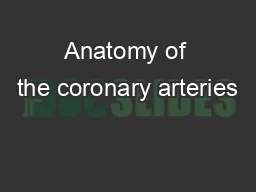
7. Coronary arterial system
8. AtherosclerosisDeposition of lipids under the intima of arteries. Chronic process that develops in following stages over 30-40 years Fatty streak Fibrous plaque Plaque inflammation , instability & Rupture
9. Atherosclerosis
10.
11.
12.
13.
14.
15.
16.
17. Fatty streak:Injury to intimal endotheliumLipid depositsNo obstruction
18. Fibrous plaque:Plaque and thrombus formation Protrusion into arterial lumen Proliferation of smooth muscle cells Irreversible progression
19. Complicate lesion:Total occlusionPlaque calcificationPlaque ruptureThrombus formation
20.
21. C.A.D.• Is the most common form of heart disease.• The single most important cause of prematuredeath in Europe.• In UK 1 in 3 men and 1 in 4 women die fromCAD.
22. Clinical manifestations & pathology PathologyFixed atheroma stenosis of Coronary artery.Dynamic obstruction of coronary artery due to plaque rupture with thrombosis.acute occlusion of coronary artery leads to myocardial necrosis.Clinical problemStable angina:Unstable angina:Myocardial infarction:
23. Heart failure: Arrhythmia:Sudden death: Myocardial dysfunction due to infarction/ ischemia.Altered conduction due to ischemia.Ventricular arrhythmia, asystole, or massive infarction.
24. ATHEROSCLEROSIS: HARMFUL CONSEQUENCESAtherosclerosis causes decrease in lumen of arteries and thus decreases blood supply
25. Artery involvedOrganConditionSymptomsEventsCoronary Cerebral carotid Peripheral HeartBrain Peripheral tissue (sk muscle)CADCVDPVD/PADAnginaMI weakness strokeIntermittent claudication gangren
26. Risk factors for Atherosclerosis &CADModifiableC. smokingDyslipidemiaDMHTNPhysical inactivityObesityNon modifiableAgeGenderFamily historyRecent associations: High Plasma homocysteine, High fibrinogen, High CRP Chlamydia pneumoniae.
27. The Cardiovascular ContinuumRisk factorsDiabetes, hypertension, Smoking, hyperlipedimiaAtherosclerosisAngina pectorisMyocardialinfarctionMyocardial scaring RemodelingVentriculardilationHeart failure End-stageheart diseaseDeath
28. Ischemic heart disease spectrumAcute coronary syndrome ACS Stableangina Unstable anginaNon STEMISTEMISuddenischemicdeathIncreasing disease severity
29. Presentation of IHD1. Asymptomatic.2. Sudden death.3. Angina pectoris 4. Myocardial Infarction.S. Heart failure. 6. Arrhythmia.
30. SYMPTOMS OF CADChest pain (angina) : Patient may feel pressure or tightness in chest, as if someone is standing on chest. Shortness of breath.
31. Location of Chest Pain
32. ANGINA PECTORISPrinciple symptom of CAD (IHD, CHD)Sudden, severe & temporary chest pain caused by an inadequate supply of oxygen to the myocardium due to narrowing of the coronary arteryPain may radiate to left arm or shoulder, back, neck or jawThe symptoms usually last for few minutes and pain subsides on rest.No permanent damage occurs Angina may be is precipitated by exercise, cold, stress, heavy meal
33. MYOCARDIAL INFARCTIONMyocardium : heart muscle Infarction : death of the tissue (necrosis)MI is death of the myocardial tissue due to complete blockade of coronary artery
34. ACUTE CORONARY SYNDROMEIt is an umbrella term used to describe symptoms and signs occuring because of acute atherothrombosis in coronary artery due to unstable plaque with critical stenosis or complete occlusionIs characterized by prolonged chest pain at restIt is an emergency condition and patient has to be hospitalized
35. Classification of ACS• STEMI of ACSC.P + ST elevation/ LBBB + high Troponin (TNI)• NSTEMI of ACSC.P + ST depression/T inversion +high TNI• Unstable anginaC.P + Abnormal /normal ECG +normal TNI
36. ACS Algorithm
37. Diagnosis of ACS• Typical chest pain: Severe prolonged pain, not relieved by GTN.• ECG changes• Cardiac enzymes
38. Investigations in CADECGRecording of electrical activity of the heart via electrodes attached to the skinNet sum of depolarisation and repolarisation potentials of all myocardial cellsP-QRS-T patternP - atrial depolarisationQRS - ventricular depolarisationT - ventricular repolarisation
39. ECGST elevation M.I.: Early changes- ST elevation. Late changes – Q waves.Non ST elevation M.I (NSTEMI): ST /T wave depression.
40. ECG ABNORMALITIES IN ISCHEMIA & MIECG (Electrocardiogram)
41.
42. Acute STEMI (Anterior)
43. Acute STEMI (Inferior)
44. Acute STEMI (anteriorlateral)
45. Acute Non-STEMI (anteriorlateral)
46. Exercise (stress) ECG
47. Exercise (stress) ECGMaximum HRMale= 220-AgeFemale = 210-AgeTarget HR 80-85% of MHRHigh risk findings : Fall in BP on exercise Horizontal ST depression Exercise induced arrhythmias Low threshold for ischaemia
48. Cardiac scanA (Thallium, Technetium)Thallium IV Uptake by the heart reflect coronary perfusion. Thallium with exercise is more accurate.The heart also can be stressed with dobutamine in patients unable to do exertion.
49. Echocardiography & dobutamine echoTo assess ventricular function (ejection fraction). Also to detect wall motion abnormalities reflecting ventricular damage. Dobutamine echo is very useful in diagnosis of IHD .
50. Cardiac Catheterization
51.
52. Coronary angiography• Gold standard test.• Invasive procedure with cannulation of Rt.femoral artery.• Gives detailed anatomical information ofcoronary arteries.
53. Coronary angiographyA diagnostic procedureCatheter’ refers to a long narrow rubber tube inserted through artery of leg & advanced upto the opening of coronary artery in aorta‘A small amount of contrast material (dye) is injected through the catheter into coronary arteries
54. Coronary AngiographyLeft Main TrunkLeft Anterior DescendingLeft CircumflexDiagonal branch
55. RCANORMAL RCA
56. RCAStenosis RCA
57. Cardiac enzymeCreatinine kinase isoenzyme MB (CK-MB).• Start to raise 4 – 6 hours after start of chest pain. raise for 3-4 days.• False +ve results trauma, I.M.injections,D.C.shock.• Serum troponin I & T most sensitive test.• TNI continue raised for upto 2 weeks.• False +ve results in: renal failure, Myocarditis, pulmonary embolism.
58. TLC, ESR. Normal/No tissue damage.Lipid profile, S. Homocysteine, CRP & B. sugar
59. TREATMENT OF IHDLife style modification Regular physical exercise Stop smoking Stop alcohol Dietary controls : weight controlRestrict saturated fats
60. Pharmacological Antiplatelets Aspirin, clopidogrilStatines Nitrates : short & long acting Beta Blockers CCBsInterventionalPCICABG
61. Management of ACSManagementASABeta blockerNitratesST Elevation MIPrimary interventionThrombolytic therapyNon ST Elevation MI and USALMWHPlatelet inhibitorsRole of catheterization
62. Early management of ACSImmediate: Oxygen. I.V. access. ECG monitor & 12 lead ECG. Aspirin. I.V. analgesia. nitrate
63. Early TT. of STEMI• STEMI need reperfusion:Primary angioplasty.Thrombolysis• NSTEMI & unstable angina:Low molecular weight heparin.Antiplatelet TT. Aspirin & clopidogrel.Coronary angiography.
64. NITRATESDilates veins & large arteries Dilation of veins lead to reduction of the preload Dilation of the arteries leads to reduction of the afterload Coronary artery dilation
65. BETA BLOCKERSReduces Myocardial Oxygen Demand↓ Heart Rate ↓ Force of contraction Increases Coronary filling↑ Diastolic time
66. CCBsBlock Calcium channelsPrevent entry of Ca++ ionsMyocardial CellDecreased force of contraction Vascular smooth muscle Coronary ArteryCoronary blood flow Peripheral arteriolePVR
67. Primary angioplasty• More effective than thrombolysis in TT. ofSTEMI.• Death, reinfarction, & stroke reduced from 14to 8%.• Need specialised centers with experiencedoperators to perform it within 90 minutes ofpresentation.
68. Coronary angiography (PCI)
69. PCI with STENTINGAlso known as.Balloon angioplastyAngioplasty.PCI (percutaneous coronary intervention) Done in Cardiac Catheterization Laboratory (Cath Lab) by an interventional cardiologist
70.
71.
72. PTCA WITH STENTINGThe arrow on the angiogram shows block in the artery.Opening of the artery after stenting Opening of the artery after stenting
73. Thrombolytic therapy• Prompt thrombolytic TT.(within 12 hours ofthe onset of chest pain) reduce mortality inpatients with ST elevation MI.• Not effective in NSTEMI & unstable angina.• Complications of TT.Hemorrhagic CVA.Bleeding.
74. Contraindications or cautions:absolute contraindicationPrevious ICH Ischemic stroke within the last 3 months except acute ischemic stroke within 3 hours Malignant intracranial neoplasm Aortic dissection active bleeding excluding menses relative contraindicationMajor surgery within 3 weeks BP >180/110 Pregnancy Concurrent warfarin therapy Active peptic ulcer disease
75. CABG(Coronary Artery Bypass Graft Surgery)(Done by CVTS in Cardiac OT)
76.