

M tashvighi Pediatric Hematology oncologist MAHAK Pediatric Cancer Treatment and Research Center 2018 Primary Hepatic tumor is Rare only 12 of all childhood cancers Hepatoblastoma ID: 918634
Download Presentation The PPT/PDF document "Primary liver tumor Hepatoblastoma" is the property of its rightful owner. Permission is granted to download and print the materials on this web site for personal, non-commercial use only, and to display it on your personal computer provided you do not modify the materials and that you retain all copyright notices contained in the materials. By downloading content from our website, you accept the terms of this agreement.

![Brain Tumor 101 Presented by [NAME]](https://thumbs.docslides.com/775177/brain-tumor-101-presented-by-name.jpg)


Slide1
Primary liver tumor Hepatoblastoma
M. tashvighiPediatric Hematology oncologistMAHAK Pediatric Cancer Treatment and Research Center2018
Slide2Primary Hepatic tumor is Rare - only 1-2% of all childhood cancers
Hepatoblastoma & hepatocellular carcinoma are the two most common malignancies that arise de novo in the liverHepatic tumors have a wide geographic variation in incidence: They are seen more frequently in Asian and African childre They are the third most common abdominal cancer in Japan.
Slide3The geographic variation is thought to reflect the etiologic role of environmental conditionsWhite males /
females 1.4/0.5 per million African American males/females 0.9/ 0.0 per million
Slide4BenignHemangioendotheliomaMesenchymal
HamartomaMesenchymal (mixed) AdenomaAngiomyolipomaEmbryonal sarcoma HamartomaMalignantHepatoblastoma
Hepatocellular carcinoma
Rhabdomyosarcoma
Undifferentiated
Angiosarcoma
Biliary cyst
Sarcoma
Teratoma
Rhabdoid tumor Myofibroblastic tumorYolk sac tumorLeiomyosarcomaLangerhans Cell HistocytosisAML-M7
Different
Diagnosis
Slide5Hepatoblastoma is Approximately two-thirds of liver tumors in childrenMedian age 1 year Male : female ratio
1.7:1.0
Slide6EtiologyUnknownCongenital anomalies which have been reported with
hepatoblastoma & HCC
Slide7Disorders Associated with Increased Risk of HepatoblastomaLow-birth-weight infantCongenital
cystathioninuria and hemihyperplasiaMaternal use of hormonal therapyExposure to metals such as in welding and soldering fumesBeckwith -Wiedemann syndromeHemihypertrophy syndromes Li.Fraumeni syndromeVon Gierke diseaseFAB
Trisomy 18
Fetal alcohol syndrome
Gardner
syndromea
Type I glycogen storage disease(Von
Gierke
)
Prader
Willi – syndromewilm’s tumor (WT1)Meckel’s diverticulumCongenital absence of adrenal gland
Congenital absence of kidney
Umbilical hernia
Slide8Conditions associated with a high risk for HCC development are ;Antitrypsin deficiency
Wilson’s diseaseHemochromatosisHereditary tyrosinemiaFanconi’s anemiaFamilial adenomatous polyposis Gardener’s syndrome
Slide9Familial Adenomatous PolyposisFAPAD
Mean age 16 yr.Mutation in APC gene (5q21)Other manifestation;Congenital hypertrophy of retinal epithelium(CHRPE)Supernumerary teethSkull & jaw osteomasEpidermoid cysts,fibromas, lipomas, skin, hyperpigmentation,keloids
Slide10Neoplasia in FAPColon carcinomaAdrenal carcinoma
Thyroid papillary carcinomaPeriampullary carcinomaFibrosarcomaGastric adenocarcinomaMedulloblastomaHepatoblastomaAstrocytomaETC
GENETIC TEST FOR
FAB
Protein
Truncation test(PTT)
80-90% of FAB families
100% accurate if know
Direct
sequenceing
Difficult due to large gene100% accurate if know
1/3 new mutation
Slide11Most common sign of primary liver malignancy;
Upper abdominal mass May have abd. Pain ,wt. loss ,anorexia ,N/V50-70% Unresectable mass at diagnosisGeneralized abdominal enlargementAbdominal distention Pallor Jaundice Fever Diarrhea Constipation
Slide12Children with a ruptured tumor usually present with;VomitingS
ymptoms of peritoneal irritationSevere anemiaRare cases manifest precocious puberty/virilization due to β-human chorionic gonadotropin (hCG) secretion by the tumorPediatric hepatoblastoma: diagnosis and treatmentTransl Pediatr 2014;3(4):293-29
Slide13DIAGNOSTIC EVALUATIONHistoryPhysical examination
Diagnostic test:Complete blood count Anemia Moderate leukocytosis is common Thrombocytosis >500,000/mm3 UrinalysisLiver profile and electrolytes
(
Bilirubin and liver enzymes are usually
normal )
Fibrinogen
,
PT & PTT
HBsAg
,
HBcAg and core antibody(positive hepatitis B e Ag., higher HBV-DNA level, HBV genotype C infection, core promoter mutation are associated with a higher risk of HCC)AFP (Low AFP levels are associated with anaplastic histology and poor outcome )
β-
hCG
CEAg
Slide14High AFPHepatoblastomaHepatocellular Carcinoma
Germ cell tumorsTerato CarcinomaViral Hepatitis & other Active liver diseaseCirrhosisInflammatory Bowel DiseaseYolk Sac tumors
Pregnancy
Gastrointestinal
tumors
Slide1590% of children with hepatoblastomas 50% -70%
of children with HCC have elevated AFP some variants of both HBL ,HCC that have low or normal AFP levelsRhabdoid tumor, have low AFP levels and worse prognosis
half-life
of AFP is
4-9 days
levels fall to within reference range within
4-6 weeks
following resection
Reference range AFP levels are high
at birth
and higher in premature infantsPediatric hepatoblastoma: diagnosis and treatmentTransl Pediatr 2014;3(4):293-29
Slide16Radiographic evaluation of intrahepatic disease;• Sonogram• Abdominal
CT-scan• Abdominal -MRI• MRI angiogram• MRI cholangiogramRadiographic evaluation of extrahepatic disease;CXR-AP, latChest CT-scanBrain CT-scanBone
scan
Bone
marrow
aspirate/biopsy
Slide17Abdominal ultrasonography ;large mass in liver, sometimes with satellite lesions &
areas of hemorrhage within the tumorUltrasound,may not be as sensitive in the evaluation of the postoperative bed due to the presence of either omental flap or gas-filled loops of bowelPercutaneous biopsy, either ultrasound or CT guidance can be used
Pediatric
hepatoblastoma
: diagnosis and treatment
Transl
Pediatr
2014;3(4):293-29
Slide18The most useful diagnostic modality is multiphase CT or MRI;Focal / Multifocal solid tumor
Hypervascular lesions in the liver with delayed contrast excretion are highly suggestive of a malignant liver tumorStippled or chunky calcifications detect in 40%-50% of patients, is significantly higher than in patients with benign lesions such as hemangiomas and
hemangioendotheliomas
MRI
is more sensitive than
CT
in discriminating between;
Disease
recurrence
Postoperative abnormalities(fibrosis and post-treatment necrosis in the liver Angelina Cistaro Editor Atlas of PET/CT in Pediatric Patients
Slide19Positron emission tomography (PET) scanning: Studies support a potential role for PET scanning at diagnosis &
follow-up in hepatoblastomaSeveral articles on the impact of PET/CT in adult HCC published, but there are no reports on the use of this imaging technique in pediatric patients Uptake of the 18F -FDG tracer reflect the ability of HB cells to store large amounts of glycogen granules in their cytoplasm
Slide20lung metastases by 18 F-FDG– PET/CT was
lower than that obtained with CT & for lesions below 1 cm, because of the limited resolution18 F-FDG– PET/CT was significantly superior to bone scan in patients with bone metastasesFDG–PET/CT in the assessment of tumor response and tumor viability after interventional therapy
(
transcatheter
arterial chemoembolization & radiofrequency ablation
)
Slide21Slide22Conventional arteriographyNewer modalities for extent of hepatic involvement by tumor and its
proximity to the portal vein Transarterial chemoembolization is a therapeutic consideration. Ultrasound in conjunction with color Doppler, a noninvasive modality, is especially useful in young infants
Slide23hepatoblastoma usually affects one or more contiguous liver segments. Occasionally, the tumor can be multifocal.
This presents a surgical challenge & liver transplantation may be required.
Slide24MetastasesHepatoblastoma, Right lobe of the liver is more commonly
involved than LeftTumor involves both lobes in 30% of patientsMetastases at diagnoses occur in 10%-20% of patients lung being the predominant site 10-20% of patients with hepatic tumors have pulmonary metastases
D
istant
metastases, including
brain
and
bone
, are
rare
Higher incidence of nonpulmonary metastases in congenital hepatoblastoma Childhood Cancers: Hepatoblastoma-The Oncologist 2000;5:445-453
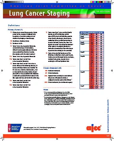
Slide25StagingStage I Complete resection of tumor by wedge resection lobectomy
or by extended lobectomy as initial treatmentStage II GTR with microscopic residual Stage III A. Gross residual tumor involving both lobes of liver B. Regional lymph node involvement
Stage IV
Metastatic disease
with complete or incomplete resection
Slide26In Europe, the Childhood Liver Tumor Study Group of the International Society of Pediatric Oncology (SIOPEL) has developed the preoperative evaluation of the tumor extent (PRETEXT) staging system
Segmental assessment of the extent of the tumor main hepatic vessels Pediatric hepatoblastoma: diagnosis and treatmentTransl Pediatr 2014;3(4):293-29
Slide27Hepatic segmental anatomy according to Couinaud. This method of hepatic segmentation is based on portal venous supply &
hepatic venous drainage
Slide28PRETEXT SIOP(based on presurgical findings) Staging for Hepatoblastoma
Stage 1 Tumor involves one quadrant Three adjoining quadrants are free of diseaseStage 2 Tumor involves two adjoining quadrants with remaining two free of diseaseStage 3 Tumor involves three adjoining quadrants or two nonadjoining quadrants.One quadrant or two
nonadjoining
quadrants are free of disease
Stage
4
Tumor involves
all four
quadrants
Slide29The pathologic classifications for hepatoblastoma and HCC are :
Hepatoblastoma Epithelial type (56%) Pure fetal pattern Embryonal pattern -Macrotrabecular type CholangioblasticSmall cell undifferentiated type or anaplastic
Mixed
epithelial and mesenchymal
type (
44%)
HCC
Fibrolamellar
HCC
(
Histologic variant of HCC - most frequent &is commonly observed in children & adolescents- has a similar prognosis when adjusted for stage)
Slide30Slide31Some histological types are associated with prognosis so;Pre-chemotherapy specimens for the initial diagnoses and tumor classification.
tissue banking for biological studiesWell differentiated fetal histology composed only of cells resembling fetal hepatocytes with minimal mitotic activity (COG);pure fetal histologyand low mitotic activity may be treated exclusively with surgery, and
no chemotherapy
is
necessary
7%
of the total number of
patients & showed
100%
(
EFS)Most HBs are extremely heterogeneous, often with closely intermixed histological components, and only rarely composed of a single histological type
Slide32Small cell undifferentiated (SCU, component intermixed with other histologies ,associated with
low serum AFP levels, and poor response to chemotherapysome, but not all, of these tumors may represent INI1 negative neoplasms within the spectrum of primary rhabdoid tumorsshould be submitted for molecular testing, and patients and family members referred to a genetics counselor to possible be screened for germline mutations
significance of small cell component when ad
mixed with other epithelial types
, and whether th
ese
small foci are sometimes
INI1expressing
Hepatoblastoma
State of the Art ;Pathology, Genetics, Risk Stratification, and Chemotherapy
Piotr Czauderna, Dolores Lopez
Terrada
Slide33The most common mesenchymal elements are osteoid and cartilage The presence of
mesenchymal elements associated with improved prognosis in patients with advanced disease
Slide34Cytogenetic AbnormalitiesGain of chromosome 20 ,most common
,Gain of chromosome 2 or 8Associated with FAP and trisomy 20 is a common finding in colon adenomas Chromosomal aberration, der(4)t(1q;4q) t(1;4)(q12;q34) Gain
of material on
1q
Studies of DNA content have shown that
diploid tumors with low proliferation index
have a
better
prognosis than aneuploid tumors & high proliferative index
Slide35Changes in the expression of H19 and IGF2 have also been implicated in the etiology of hepatoblastoma
Slide36Prognostic factorGood prognostic factor
(COG) ;Complete resection(stage I) completely resected tumors pure fetal histologyRate of fall alpha- fetoproteinLarge early response (>2 log decline in AFP)strongest dependent predictor of outcome ( P<0001)Poor prognostic factor (SIOPEL and the COG) ;
AFP<100
ng/ml or
>
1/000/000
ng/ml at
diagnosis
and
/ or with small cell undifferentiated (SCUD) histology Regardless of the PRETEXT staging
systemVascular invasionPediatric hepatoblastoma: diagnosis and treatmentTransl Pediatr 2014;3(4):293-29
Slide37Potential Prognostic Factors in Hepatoblastoma
PRE- TREATMENTRESPONSE TO TREATMENTPRETEXT StagePositive Surgical MarginsMetastasis at diagnosisSurgical ResectabilityUnresectable
Vessel involvement
Tumor Relapse
Extrahepatic tumor extension
Response to
Chemotherapy
Lymph Nodes
Tumor Rupture at diagnosis
AFP level (<100,100-1000, >1 million)
Pathologic subtype (Pure Fetal
, Small Cell Undifferentiated)
Age (<1 year - >6 year)
Birth weight
Platelet Count
Co-Morbidity
Slide38TREATMENTOveral survival IN stage 3 & 4 is 20-30%
Surgery; complete resection can be chance of cure Initial surgical resection-most prognostic factorDelayed surgical resection after chemotherapyOrthotopic liver transplantationChemotherapy ;
plays
an important role, not only in
eradicating subclinical
metastases
in
completely resected disease
,
but
also may allow unresectable disease to become resectableTranscatheter arterial chemoembolization
Slide39Timing of surgeryIn the United States;
Initial surgical resection in appropriate patients is preferred The European approach is different; Patients are staged by PRETEXT and neoadjuvant chemotherapy is administered prior to surgery to all patients except PRETEXT stage 1Surgery is recommended
for
limited metastatic disease
(especially in
lung
)
This surgery is often done at time of liver
tumor resection
Slide40Classic reasons for unresectable ;Extremely large tumor that may lead to excessive
bleeding Involvement of both the right and left lobesInvolvement of major hepatic veins or the inferior vena cava (IVC) Diffuse multifocal diseaseOnly 30% have been
considered
resectable
at
diagnosis
In the
unresectable
patient
Biopsy should be performed
Slide41Surgical BiopsyDiagnostic surgical biopsy is strongly recommended ;
Children <6 months ,Wide range of possible tumours presenting at this age Possible confounding effect of a physiologically elevated AFP level Children older > 3 years of age, to distinguish hepatoblastoma from hepatocellular carcinomaAll patients with a normal serum AFP
Slide42BiopsyBiopsy may not be necessary for young children (6 months to 3 years) with a very high AFP level
Avoiding a biopsy theoretically reduces the risks of tumor seeding or disseminationThe Japanese Study Group for Pediatric Liver Tumors (JPLT) strongly recommends that liver tumors of children should be treated after definitive diagnosis of a biopsy specimen, except in urgent life threatening circumstances such as tumor invasion of the right atrium or tumor rupture Pediatric hepatoblastoma: diagnosis and treatmentTransl
Pediatr
2014;3(4):293-29
Slide43Most common intraoperative complication ;Hemorrhage
The risk of bleeding is increased ;Extended hepatectomy Tumor proximity to the IVC or hepatic vessels Air embolus
D
amage
to
the portal vein
,
hepatic artery
, or
hepatic
duct
Slide44Postoperative complications;Sub phrenic abscessBile leak
Postoperative bleedingSmall bowel obstruction
Slide45Radical hepatic resection results in many potential postoperative complications:• Hypovolemia• Hypoglycemia• Hypo albuminemia
• Hypo fibrinogenemia and deficiency of coagulation proteins• Hyper bilirubinemia persists for 24 weeks after resectionHepatic regeneration is complete by 13 months post surgery
Slide46Transcatheter arterial chemoembolization Hepatic arterial chemoembolization involves giving chemotherapy and
vascular occlusive agents via catheter into the artery supplying the tumorCryoablation, and more recently radiofrequency ablation, have also been used in the treatment of liver tumors in adults with little experience in children
Slide47ABLATION THERAPY Tumors are destroyed using
;Heat (Radiofrequency ablation)Cold (Cryoablation) Chemical agents (Percutaneous ethanol instillation)Ablative therapy is for tumors involving the liver,
kidney
,
lung
and
painful tumors of
bone
The
goal of ablative therapy is
complete tumor destructionAblative therapy is an alternative to surgical resection and appropriate primarily for patients with four or fewer tumors limited to the liver
Slide48RADIOFREQUENCY ABLATIONGeneration of heat to destroy the
tumorExposure of both normal and cancer cells to heat above 122 F° - 9 up to 14 min. )causes the cells to die, resulting in cellular destructionSound waves to interact with molecules in the tumor, causing them to vibrate and generate heatEnergy is delivered through a
needle
G
uidance
provided by
ultrasound
,
CT
or
MRI
Slide49Slide50ADVERSE REACTIONnot uncommon ;Fatigue
Muscle ache Low grade fever Rarely Liver injury in the form of bleeding leakage of liver fluid (bile) Heat damage to surrounding organs ,the gall bladder or bowelLung surrounds the liver may be lung injury or collapse
Slide51Success is influenced ;Tumor size, as larger tumors are more difficult to completely eradicate than smaller
onesTumor location adjacent to flowing blood initial treatment did not destroy all of the tumor, the procedure may be repeatedDestroys very little normal liver Radiofrequency ablation
does not interfere
with
future surgical
procedures or
other types of
therapy
Continued
monitoring
with CT or MRI at 3 mo.- 6 mo.
Slide52CRYOABLATIONIce ball with subzero temperatures is created by circulating liquid nitrogen in a probe that is directly inserted into the tumor
Tumor in ,liver, kidney, prostate, lung , bone
Slide53PERCUTANEOUS ETHANOL INSTILLATIONAlcohol destroys cells on contact through destruction of their lining membranes
More treatment sessions Slightly less effectiveoften employed in conjunction with radiofrequency ablation to enhance the success of the procedure. Vascular Interventional Radiology of the Robert Wood Johnson University Hospital and the UMDNJ- Robert Wood Johnson Medical School
Slide54Combination chemotherapy plays several roles in the management:Adjuvant therapy for patients who have undergone complete resection, its use
improves disease-free survivalPreoperative therapy for patients who have initially unresectable disease to shrink the primary tumor Palliative therapy for patients with metastatic disease at diagnosisThe outcome of high risk hepatoblastomas with multi focally disseminating growth in the liver, invasion of large vessels, extrahepatic extension and metastases is still poor,
especially since these tumors often rapidly develop resistance against cytotoxic
drugs
Chemotherapy
Slide55Patients are assigned to the following risk groups(COG):
Very low-risk:Grossly resected tumors (stage I) with PFH & an elevated AFP level > 100 ng/mL Low-risk: Grossly resected tumors (stage I-II) & lacking any unfavorable biologic feature ( any SCU elements or a low diagnostic AFP level < 100 ng/mL)
Intermediate-risk;
G
ross
residual disease/
unresectable
disease OR grossly resected disease with any SCU elements but no metastatic disease and no low diagnostic AFP level < 100 ng/mL
High-risk:
M
etastatic disease OR low diagnostic AFP level < 100 ng/mL regardless of stage
COG AHEP0731: Phase III Study of Combination Chemotherapy in Pediatric Patients With Newly Diagnosed
Hepatoblastoma
Slide56Slide57Children’s Hepatic Tumors International Collaboration (CHIC)Hepatoblastoma Risk Stratification;
PRETEXT Standard risk High risk (HR) Very high risk (VHR)Low risk
(primary resection at diagnosis)
Intermediate risk
Any M+
–
–
–
M+
I M–
VPERF—(any AFP, any age) –
VPERF + AND age <8 years (any AFP)
VPERF + AND age ≥8 years
II M–
VPERF—AND Age <3 AND AFP >1,000 ng/mL
VPERF—AND Age <3 AND AFP 100-1,000 ng/mL
Age 3-7 AND/OR VPERF +
AFP <100 ng/mL, AND/OR age ≥8 year
III M–
–
VPERF—AND Age<3 AND AFP >1,000 ng/mL
Age 3-7 AND/OR VPERF + AND/OR AFP 100-1,000 ng/mL
AFP <100 ng/mL, AND/OR age ≥8 year
IV M–
–
–
AFP >100 ng/mL
AFP <100 ng/mL, AND/OR age ≥8 years
Slide58Multicenter groups in CHIC are JPLT, SIOPEL, GPOH and COG;Standard low-risk patients;
PRETEXT I, II, and III tumorsno extrahepatic features [hepatic vein/cava involvement (V), portal vein involvement (P), contiguous extrahepatic tumor (E), rupture at diagnosis (R), and multifocality (F)] or distant metastasis (M)JPLT and COG -primary hepatectomy for PRETEXT I and II tumorsSIOPEL -preoperative chemotherapy for every patient followed by tumor resectionor
liver transplantation
and a short course of postoperative chemotherapy for most
cases
CIHC
; initial
resection
for
PRETEXT I or II tumors if the tumor is located at least 1 cm from the middle hepatic vein &bifurcation of the portal veinPreoperative chemotherapy performed for other situationsCisplatin monotherapy recently achieved similar rates of complete resection and survival among children with resectable tumors
Slide59High-risk (HR) patients Unresectable tumor at diagnosis and/or associated with so-called “combi
factors” without distant metastasisCombi factor is a combination of the cross sectional imaging componentsMacrovascular involvement retrohepatic vena cava or all three hepatic veins (V)Macrovascular involvement portal bifurcation or both right and left portal veins (P)Contiguous extrahepatic tumor (E)M
ultifocal
disease (
F)
S
pontaneous
rupture (R) at
diagnosis
Pediatric
hepatoblastoma: diagnosis and treatmentTransl Pediatr 2014;3(4):293-29
Slide60High-risk (HR) patients CHIC ;these patients were included as
high-risk patients, even if their tumor was resectable Conventional preoperative chemotherapy used in the PLADO,and C5V trials for patients with PRETEXT IV unresectable tumors resulted in tumors that could be resected by hepatectomy in some patients; but the outcome of patients with unresectable tumors at diagnosis remained unsatisfactory. Pediatric hepatoblastoma: diagnosis and treatmentTransl Pediatr 2014;3(4):293-29
Slide61Regimen 1-CCG823F (every 3-4 weeks)-(six cycles of chemotherapy)
Doxorubicin (25 mg/m2/day×3 days - > 10 kg 0.83 mg/kg/day - continuous infusion by central line)Cisplatin( 20 mg/m2/day- ×5 days - > 10 kg ,0.66 mg/kg/day - continuous infusion)Regimen 2- (C5V)(every 3-4 weeks)
Cisplatin
(100 mg/m2 IV infused over 6 h
,
>1 yr. 3.3 mg/kg
- day
1)
Vincristine
(1.5 mg/m2 IV (max 2 mg), >1 yr. 0.05 mg/kg - day 3, 10, 17)Fluorouracil (600 mg/m2 IV - day 3)
Slide62Regimen 3 (every 3-4 weeks)Cisplatin
(90 mg/m2 - >1 yr. - 3 mg/kg - IV infused over 6 h ), day 0Doxorubicin( 20 mg/m2/day - >1 yr. 0.66 mg/kg - continuous infusion × 4 days, days 0-3)Toxicity of this regimen
was more
severe
All
patients will receive
G-CSF
and
bactrim
to reduce toxicity
After four cycles, surgical resection followed by four additional courses of chemotherapy
Slide63Treatments for HBLCOG and JPLT studies have approved
primary resection for children with resectable tumors, especially PRETEXT I or II Patients with stage I PFH treated with surgery alone. Stage I
hepatoblastoma
(non-pure fetal histology [PFH])
,
non-small cell undifferentiated [SCU])
and
Stage II
(non-SCU)
is a highly curable disease with 2 cycles of adjuvant cisplatin, 5-fluorouracil, and vincristine (C5V) SIOPEL studies have not permitted the used of primary resectionPediatric hepatoblastoma: diagnosis and treatmentTransl Pediatr 2014;3(4):293-29
COG AHEP0731: Phase III Study of Combination Chemotherapy in Pediatric Patients With Newly Diagnosed
Hepatoblastoma
Slide64Intermediate-risk group : Receive C5VD chemotherapy comprising;
Cisplatin (100 mg/m2 IV infused over 6 h, >1 yr. 3.3 mg/kg- over 6 hours on day 1)Fluorouracil (600 mg/m2 IV - on day 2)Vincristine (1.5 mg/m2 IV (max 2 mg), >1 yr. 0.05 mg/kg - IV on days 2, 9, and 16) Doxorubicin
(
20 mg/m2/day - >1 yr. 0.66 mg/kg
-
IV over 15 minutes on days 1-2)
Repeats
every 21 days for 6 courses in the absence of disease progression or unacceptable
toxicity
Surgical
resection after course 2 OR surgical resection or liver transplantation after course 4 of C5VD COG AHEP0731: Phase III Study of Combination Chemotherapy in Pediatric Patients With Newly Diagnosed
Hepatoblastoma
Slide65Vincristine/irinotecan upfront window treatment of high-risk hepatoblastoma:
Vincristine (1.5 mg/m2/day -IV on days 1 & 8) Irinotecan (50 mg/m2/day -IV over 90 minutes on days 1-5) Repeats every 21 days for 2 courses in the absence of disease progression or unacceptable toxicityResponders ;
30
% decrease in tumor burden according to RECIST criteria
90
% (> 1 log
10
) decline in the AFP
level
Reevaluation every
2 cycle/Until second surgerySignificant antitumor activity & acceptable toxicity in relapse hepatoblastomapediatr hematol oncol 2015 feb;32
Slide66Responders were VI , receive 2 additional cycles of VI intermixed with 6 cycles of
cisplatin/doxorubicin/5-fluorouracil/vincristine (C5VD)- VI in between each 2-course (C5VD)Patients undergo tumor resection or liver transplantation after course 4 of C5VD followed by 2 courses of adjuvant C5VD. Referral for orthotopic liver transplant (OTL) is as potentially unresectable following central surgical review and staging according to the PRE TEXT (Pretreatment Extent of Disease) grouping system
(COG) AHEP0731
Upfront VI treatment of high risk
hepatoblastoma
(COG) AHEP0731- CANCER 2017 JUN15
Slide67' High risk' HB SIOPEL group;
Intensive multi-agent regimen including CARBO, CDDP and DOXO, preceded by a single dose of CDDP Pre-operative phase alternating administration ;Cisplatin at days 1, 29, 57 & 85 80 mg/m2 over 24 hours as a continuous i.v- ( >5kg-1.7 mg/kg: if well tolerated increase to 2.6 mg/Kg )Administered regardless of the blood cell count
CARBO
/DOXO at
days 15, 43 &71
CARBO
-
500
mg/m2 as an
i.v.
infusion from hours 0-1 (>5kg -11.5 mg/kg: if well tolerated increase 16.6 mg/Kg)DOXO- 60 mg/m2 /48 hours continuous - starting soon after the end of the CARBO (>5kg -1.34 mg/Kg in 48 hours continuous infusion: if well tolerated increase to 2.0 mg/Kg/48 hours)Tumour response evaluate before days 29, 57 and 85, and just before surgery
Childhoodliver
tumor
stratege
group-HR
HB Guidelines 11.01.2010
Slide68Slide69German Society for Pediatric Oncology two courses- (IPA) ;
Ifosfamide (3 g/m2/ over 72 h (days 1-3)Cisplatin ( 20 mg/m2/d for 5 days (days 4-9)Doxorubicin ( 60 mg/m2 /over 48 h (days 9-10) two
courses of IPA, followed by a tumor resection and a 4th course of IPA
2003 May-Jun;215(3):159-65.
[Differentiated treatment protocols for high- and standard-risk
hepatoblastoma
--an interim report of the German Liver Tumor Study HB99].
' High risk' HB
Slide70German group has given carboplatin and etoposide to patients who have failed IPA ;Carboplatin
(800 mg/m2) Etoposide (400 mg/m2) In case of tumor response, they received one or two courses of HD-chemotherapy with carboplatin (2000 mg/m2) and etoposide (2000 mg/m2) after sampling of peripheral stem cells, followed by resection of the primary tumor and metastases, whenever possible.2003 May-Jun;215(3):159-65.
[Differentiated treatment protocols for high- and standard-risk
hepatoblastoma
--an interim report of the German Liver Tumor Study HB99].
Slide71Topotecan may prove useful in preparative regimens for autologous stem cell transplant in the treatment of hepatoblastoma. Stem
cell transplant following; thiotepa, melphalan and busulfan has been used in the treatment of microscopic residual disease with good results ifosfamide, carboplatin, and etoposide and achieved a very good partial response prior to liver transplant.
Slide72Orthotropic liver transplantation has improved the outcome for some patients with unresectable tumors (PRETEXT IV tumors or tumors with portal or hepatic vein involvement)
Pediatric hepatoblastoma: diagnosis and treatmentTransl Pediatr 2014;3(4):293-29
Slide73Very high-risk patients (metastatic HBL)with lung metastases have a poor prognosisOlder patients (≥8 years old at diagnosis)
Patients with low AFP levels (<100 ng/mL) have unfavorable outcomes Pediatric hepatoblastoma: diagnosis and treatmentTransl Pediatr 2014;3(4):293-29
Slide74Very high-risk patients (metastatic HBL)Resection of lung metastases has been effective for some patients with metastatic tumorsCISPLAIN intensification therapy such as that used in the SIOPEL-4 protocol seems to be effective
new molecular targeting therapy using vincristine and irinotecan will be investigated by COGliver transplantationTransarterial embolization (TAE) is used to control peritoneal hemorrhage in patients with ruptured tumorsPediatric hepatoblastoma: diagnosis and treatmentTransl Pediatr 2014;3(4):293-29
Slide75Very high-risk patients (metastatic HBL)Malignant liver tumors, including HBL, are mainly fed by the hepatic arteryTrans arterial
chemoembolization (TACE) Cisplatin & Anthracycline are used for embolization Effect of TACE equivalent to systemic chemotherapy less toxic in comparison with systemic chemotherapyAdministering TACE to children is somewhat difficult and requires general
anesthesia
Pediatric
hepatoblastoma
: diagnosis and treatment
Transl
Pediatr
2014;3(4):293-29
Slide76SIOPEL-1 ; four triweekly preoperative ,two postoperative cycles
Cisplatin Doxorubicin JPLT ; four preoperative & two postoperative cycles Cisplatin Pirarubicin
Pediatric
hepatoblastoma
: diagnosis and treatment
Transl
Pediatr
2014;3(4):293-29
Slide77Radiotherapyis not curative
useful in shrinking unresectable disease or microscopic residual diseaseRadiation dosages 1200 to 2000 cGyOccasionally, higher doses to localized areas of tumor Radiotherapy immediately after hepatic resection
will limit hepatic
regeneration
Slide78Criteria for judging tumor response Complete R
esponse ; no evidence of disease and normal serum AFP value (for age) Partial Response :Any tumor volume shrinkage associated with a decreasing serum AFP value, > 1 log below the original measurement Stable Disease ;no tumour volume change and no change, or <l log fall of the serum AFP concentration. Progressive Disease;
unequivocal
increase in 1 or more dimensions and/or any unequivocal increase of the serum AFP concentration
Childhoodliver
tumor
stratege
group-HR
HB Guidelines 11.01.2010
Slide79toxicity Pure tone audiometry is the method of choice in children older than 3 years of age
If a child starts to show signs of high frequency hearing loss he/she should be followed carefully If grade 3 or 4 ototoxicity is documented, CDDP should be withdrawnBILATERAL HEARING LOSS GRADE Designation
< 40 dB at all frequencies
0
None
> 40 dB at 8,000 Hz only
1
Mild
> 40 dB at 4,000 Hz and above
2
Moderate > 40 dB at 2,000 Hz and above 3 Marked
> 40 dB at 1,000 Hz and above 4
Severe
a
possible increase of ototoxicity in patients treated with CARBO and
CDDP
Slide80Renal toxicity Glomerular toxicity Nephrotoxicity of CDDP in children (as in adults) is
dose-related and Plasma creatinine measurements and creatinine clearances are not reliable in childrenCareful measurement of GFR by isotope clearance is essential for accurate monitoring of renal statusTubular toxicity Renal loss of Magnesium and consequent Hypomagnesemia Renal tubulopathy
(careful
monitoring of
electrolytes)
Hypomagnesemia
may
persist years
after stopping therapy
Slide81Bone marrow toxicity; ANC >1000/mm3 platelet count
>100.000/mm3 are necessary before starting chemotherapy with Carboplatin and DoxorubicinIt is better to delay slightly the institution of chemotherapy until these criteria are met, rather than decreasing the doseIf a delay of one or more additional weeks is required, decrease dosage by 25% for the next course only, and use GCSF ANC <
500/mm3
associated with
fever and sepsis
or severe infection and/or severe thrombocytopenia
(< 10.000/mm3 lasting
more than 5 days) associated with bleeding, decrease dosage by 25%
Slide82Cardiotoxicity; Echocardiogram should be performed when the patient is;n
ormothermicnormal haemoglobinis not being hyperhydratedprior to administration of DOXO
Slide83Hepatic toxicity;No standardised criteria exist for the adjustment of DOXO dosage in the presence of hepatic dysfunction
50% reduction of DOXO dosage if Total Bili. > 3 mg/100 ml, +/-AST, ALT) are >5 times the normal value. Neither CARBO nor CDDP doses need to be modified.
Slide84Abdominal tumorImaging-tumor marker(AFP)
TUMOR RUPTURETAE/TACEBIOPSYBIOPSY
Primary resection
Pre –op CX
Pre –op CX
Post
–op CX
Resection/LTX
R
esection
M
etastectomy
Post –op CX
CXs
CXs
Metastectomy
Resection/LTX
CXs
Recurrence /
Progression
Rescue CXs
Diagnosis and treatment algorithm for
hepatoblastoma
M(+):VHR
M(-)
IR or
HR
L
R
Slide85Hepatoblastoma report
at MPCTRC
Slide86sex
frequencyPercentFemale1043.5%Male1356.5%
total
23
100%
Slide87Age & Sex
FemaleMaleAge31<1Y391-3Y
1
2
3-5Y
3
1
5-10Y
10(43.5%)
13(56.5%)
Total
Slide88Nationality
Frequency21Iranian2Afghan23Total
Frequency
Iranian
10
Tehran
2
Alborz
1
Markazi
2
Gilan
2
Kerman
1
Fars
1
kashan
1
Golestan
1
Bandar Abbas
21
Total
Slide89Consanguinity
FrequencyConsanguinity13No9Yes1Missing(child adopt)
23
Total
Slide90Family History Of CancerOne pa. has 3 Family History Of Cancer(Liver-Skin-Uterus)
Five pa. have GI Family History Of Cancer(Pancreas-Intestine-Colon-GI)One pa. has Family History Of (brain Cancer)two pa. has Family History Of Leukemia
Slide91Signs & symptom
Slide92Staging
PercentFrequencyStaging17.4%4 Stage10
0
Stage2
46.8%
12
Stage3
34.8%
7
Stage4
100%23Total
1 child in Stage1 after relapse was
in stage 4
Slide93Metastasis
relapseFrequencyMetastasis316No-7Yes
-
23
Total
Slide94Total
Uni FocalMulti Focal16412Rt. Lobe11
0
Lt. Lobe
6
0
6
Both Lobes
23
5
18Total
Slide95After Chemotherapy
Before ChemotherapyMaxMinMaxMin19.81
21.7
5.2
WBC
36.4
6.7
16.4
8.1
Hb
947391. 754.9
MCV
529
26
998
71.2
Plt
8950
5
201
24
SGOT
1469
11
93
10
SGPT
2133
177
798
159
ALK
15.2
.2
1.5
.3
Bill
.5
.5
17
.3
HBsAg
249
1
810
.6
HBsAb
.1
.1
49
.1
HCVAb
Lab Data
Slide96AFP
Minimum =12Maximum =120000Mean=17464
Frequency
Percent
<100
9
39.0%
100-1000
6
26.3%
>1000
8
34.7%
Slide97First Chemotherapy
Slide98Second Chemotherapy
Slide99status
FrequencyPercentUnder treatment313.1%
Off treatment/Follow
up/alive
9
39.1%
death
11
47.8%
Slide100Relapse
percentFrequencyRelapse74%17No26%
6
Yes
100%
23
Total
One pa. has two relapse
Slide101Pathology Type
Type14Epithelial4Mixed Epithelial& Mesenchymal5Unknown type23
Total
Slide102age
ConsanguinityHistory of cancer in family
AFP
biopsy
Second look surgery
Patology
First chemo
Second chemo.
RELAPSE.
status
Cause death
Stage 1
<1
+
-
100-1000
+
+
Epithelial
Cis/VCR
-
-
Alive
1-3
+
+/GI
100-1000
+
+
Epithelial
Doxo
/Cis
-
-
Alive
1-3
-
+/
GI
<100
+
-
hepatoblastoma
C5V
IPA
+
lung
DC
relapse
3-5
?
-
>1000
_
+
Epithelial
Doxo
/Cis
-
-
Alive
Stage 3
<1
?
+/GI
>1000
-
+
Mixed Epithelial& Mesenchymal
Doxo
/Cis
_
-
Alive
<1
+
/IVF
-
100-1000
+
-
hepatoblastoma
C5V
-
-
Chemo
<1
-
-
<100
+
hepatoblastoma
C5V
-
-
Alive
-
1-3
-
+/
Leukemia
>1000
+
-
hepatoblastoma
Doxo
/Cis
-
-
DC
pneumonia
1-3
+
-
>1000
-
+
Epithelial
Doxo
/Cis
-
-
Alive
1-3
+
-
100-1000
-
+
Epithelial
C5V
IPA
+
Liver/
peritoneum
Chemo
1-3
-
-
>1000
-
+
Epithelial
C5V
-
-
Chemo
1-3
+
-
>1000
+
+
Epithelial
Cis/VCR
C5V
-
-
Alive
1-3
adopt
?
>1000
+
+
Epithelial
Doxo
/Cis
_
-
Alive
5-10
-
-
100-1000
+
-
Mixed Epithelial& Mesenchymal
Doxo
/Cis
-
-
DC
Electrolyte imbalance
Slide103age
ConsanguinityHistory of cancer in family
AFP
biopsy
Second look surgery
Patology
First chemo
Second chemo.
RELAPSE.
status
metastasis
Cause Death
stage3
5-10
-
-
100-1000
-
+
Epithelial
Cis/VCR
C5V
IPA
+/
liver
DC
-
DIC/ sepsis
5-10
?
+/
leukemia
<100
-
+
Mixed
Epithelial& Mesenchymal
C5V
-
-
Alive
-
-
stage4
1-3
+
+/GI
<100
-
+
Epithelial
C5V
-
-
DC
IVC /mass in
Rt.A
1-3
+
-
>1000
+
-
Epithelial
C5V
DOX/CIS
+
DC
Lung/
adenopathy
Relapse
DIC
1-3
-
-
<100
+
+
Mixed Epithelial& Mesenchymal
C5V
DOX/CIS
CIS/carbo.
+
DC
Heart
Relapse
1-3
-
+/liver
<100
+
-
hepatoblastoma
C5V
-
-
DC
lung
Respiratory
insuf
.
3-5
-
-
<100
+
-
Epithelial
C5V
-
-
DC
Lung/
adenopathy
Respiratory
insuf
.
3-5
-
+/GI
<100
+
+
Epithelial
DOX/CIS
C5V
rinotecan
+
DC
Lung
Brain
Adenopathy
L.Transplant
Relapse
DIC
5-10
+
+/brain
<100
+
-
Epithelial
C5V
-
-
DC
lung
Sepsis/
Electrolyte imbalance
Slide1043&5
year survival=37.4%±.13Global 5-year survival rates for hepatoblastoma are :
Stage I/II
90
Stage III
60
Stage IV
20
Slide1051 year EFS=25%±.2
Slide106A Boy<1year,Pathology: Mixed Epithelial& Mesenchymal, Stage 4,In Left Liver Lobe, Left LobectomyWith bone marrow involveAFP
8 Courses Chemo Out of Mahak,protocol (5FU,VCR,Carbo)Had Autolog Transplantation In Mahak Four Years Ago In 19 Months Old , Living NowTransplantation Protocol; Carboplatin(3 days) Etoposide(3 days) Melfalan(1 day)
Slide107THANKS FOR YOUR TIME & ATTENTION
Stay safe & have a great day!!!Her soul is happy
Slide108