
Sethi S Alcid D Kesarwala H Tolan RW Probable Congenital Babesiosis in Infant New Jersey USA Emerg Infect Dis 2009155788791 httpsdoiorg103201eid1505070808 ID: 1006989
Download Presentation The PPT/PDF document "Figure Figure. Giemsa-stained (..." is the property of its rightful owner. Permission is granted to download and print the materials on this web site for personal, non-commercial use only, and to display it on your personal computer provided you do not modify the materials and that you retain all copyright notices contained in the materials. By downloading content from our website, you accept the terms of this agreement.


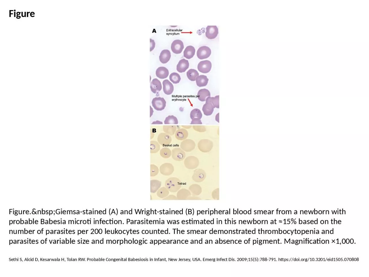
1. FigureFigure. Giemsa-stained (A) and Wright-stained (B) peripheral blood smear from a newborn with probable Babesia microti infection. Parasitemia was estimated in this newborn at ≈15% based on the number of parasites per 200 leukocytes counted. The smear demonstrated thrombocytopenia and parasites of variable size and morphologic appearance and an absence of pigment. Magnification ×1,000.Sethi S, Alcid D, Kesarwala H, Tolan RW. Probable Congenital Babesiosis in Infant, New Jersey, USA. Emerg Infect Dis. 2009;15(5):788-791. https://doi.org/10.3201/eid1505.070808