
Dr Maria A Arafah Assistant Professor Department of Pathology http facksuedusa mariaarafah courses Objectives At the end of this lecture the student should be able to Understand the pathology and predisposing factors of ectopic pregnancy and spontaneous abortion ID: 1032093
Download Presentation The PPT/PDF document "Ectopic Pregnancy, Spontaneous Abortion ..." is the property of its rightful owner. Permission is granted to download and print the materials on this web site for personal, non-commercial use only, and to display it on your personal computer provided you do not modify the materials and that you retain all copyright notices contained in the materials. By downloading content from our website, you accept the terms of this agreement.


1. Ectopic Pregnancy, Spontaneous Abortion And Gestational Trophoblastic Disease.Dr. Maria A. ArafahAssistant Professor – Department of Pathologyhttp://fac.ksu.edu.sa/mariaarafah/courses
2. Objectives At the end of this lecture, the student should be able to:Understand the pathology and predisposing factors of ectopic pregnancy and spontaneous abortion.Know the clinical presentation and pathology of hydatidiform mole and choriocarcinoma.3/31/19REPR 2242
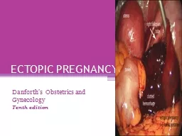
3. Ectopic PregnancyEctopic pregnancy is defined as implantation of a fertilized ovum in any site other than the endometrium of the uterine cavity.About 1% of all pregnancies are ectopic. Sites: Over 90% of ectopic pregnancies occur in the fallopian tubes (tubal pregnancy). Other sites include: ovaries, abdominal cavity and cervix.3/31/19REPR 2243
4. Clinical FeaturesA woman with an ectopic tubal pregnancy may present with pelvic pain or abnormal bleeding following a period of amenorrhea.Many present as an emergency with tubal rupture, severe acute abdominal pain and hemorrhagic shock.The diagnosis is usually confirmed by an abdominal/pelvic ultrasound gestational sac within fallopian tube or other location and a positive HCG levelsMicroscopically: placental tissue or fetal parts within the tube.3/31/19REPR 2244
5. Clinical Features3/31/19REPR 2245
6. Risk FactorsAny factor that retards passage of the ovum through the tubes predisposes to tubal ectopic pregnancy. In about half of the cases, it is due to chronic inflammation and scarring in the oviduct. 3/31/19REPR 2246
7. Risk FactorsPelvic inflammatory diseases/infections/salpingitis: is the most common cause. The inflammation can damage the ciliary activity and cause tubal obstruction, pelvic adhesions with scarring and distortion of the fallopian tubes. Women who have had pelvic infections have a five times greater risk of ectopic pregnancy (infection is usually by Neisseriae gonorrhea & chlamydia).Abdominal/pelvic surgery or tubal ligation surgery.Intrauterine tumors and endometriosis.Smoking can decreas tubal motility by damaging ciliated cells or it may predispose them to pelvic inflammatory diseases (due to the impaired immunity in smokers).3/31/19REPR 2247
8. Risk FactorsCongenital anomaly of the tubes. In-utero diethylstilbestrol (DES) exposure increases the risk of ectopic pregnancy due to abnormal tubal morphology.History of previous ectopic pregnancy.History of multiple sexual partners increases the chance of pelvic inflammatory diseases and therefore are high risk for ectopic pregnancies.Intrauterine device users are at higher risk of having an ectopic pregnancy should pregnancy occurs. 3/31/19REPR 2248
9. Risk FactorsHistory of infertility: there is higher risk of ectopic pregnancy in the infertile population. This may be due to the underlying infertility related issues or fertility drugs and treatments. In vitro fertilization has been associated with an increased risk of ectopic pregnancy including cervical pregnancies.Please note that in many tubal pregnancies, no anatomic cause is evident.Ovarian pregnancies probably result from rare instances in which the ovum is fertilized just as the follicle ruptures. Gestation within the abdominal cavity occurs when the fertilized egg drops out of the fimbriated end of the oviduct and implants on the peritoneum.3/31/19REPR 2249
10. Ectopic Pregnancy3/31/19REPR 22410
11. Spontaneous Abortion/MiscarriageIt is the spontaneous end of a pregnancy at a stage where the embryo or fetus is incapable of surviving. Miscarriages that occur:before the 6th week of gestation are called early pregnancy loss or chemical pregnancy. after the 6th week of gestation are called clinical spontaneous abortion.About 10-25% of all pregnancies end in miscarriage. Most miscarriages occur during the first 13 weeks of pregnancy. 3/31/19REPR 22411
12. Causes of Spontaneous Abortions/MiscarriagesMost miscarriages occur during the first trimester.The cause of a miscarriage cannot always be determined. Miscarriages can occur for many reasons.Chromosomal abnormalities of the fetus are the most common cause of early miscarriages.3/31/19REPR 22412
13. Causes of Spontaneous Abortions/MiscarriagesChromosomal abnormalities:Half of the 1st trimester miscarriages have abnormal chromosomes.Chromosomal abnormalities also become more common with aging, and women over the age of 35 have a higher rate of miscarriage than younger women. A pregnancy with a genetic problem has a 95% probability of ending in miscarriage. 3/31/19REPR 22413
14. Causes of Spontaneous Abortions/MiscarriagesHormonal problems: there is an increased risk of miscarriage with:Cushing’s Syndrome Thyroid disease Polycystic ovary syndrome Diabetes: good control of blood sugars during pregnancy is important. If the diabetes is not well controlled, there is an increase risk of miscarriages and also of the baby of having birth defects. Inadequate function of the corpus luteum in the ovary (which produces progesterone necessary for maintenance of the very early stages of pregnancy) leads to progeterone deficiency which may lead to miscarriage. 3/31/19REPR 22414
15. Causes of Spontaneous Abortions/MiscarriagesInfections: by Listeria monocytogenes, Toxoplasma gondii, parvovirus B19, rubella, herpes simplex, cytomegalovirus and lymphocytic choriomeningitis virus etc are associated with an increased risk of pregnancy loss.Maternal health problems can predispose to miscarriages e.g. systemic lupus erythematosus and antiphospholipid antibody syndrome 3/31/19REPR 22415
16. Causes of Spontaneous Abortions/MiscarriagesLifestyle: smoking, drug use, malnutrition and exposure to radiation or toxic substances.Maternal age: SABs increase after the age of 35 due to ovum abnormalities.Maternal trauma.3/31/19REPR 22416
17. Causes of Spontaneous Abortions/MiscarriagesAbnormal structural anatomy of the uterus can also cause miscarriages e.g. septate or bicornate uterus affect placental attachment and growth. Therefore, an embryo implanting on the septum would be at increased risk of miscarriage. Uncommonly uterine fibroids can interfere with the embryo implantation and blood supply, thereby causing miscarriage.Others: surgical procedures in the uterus during pregnancy e.g amniocentesis and chorionic villus sampling.3/31/19REPR 22417
18. Causes of Spontaneous Abortions/Miscarriages3/31/19REPR 22418
19. Diagnosis of Spontaneous Abortions/MiscarriagesA miscarriage can be confirmed:By ultrasound studyBy the examination of the passed tissue microscopically for products of conception. The products of conception include chorionic villi, trophoblasts, fetal parts and changes in the endometrium (hypersecretory).Genetic tests may also be performed to look for chromosomal anomalies.3/31/19REPR 22419
20. Products of Conception3/31/19REPR 22420
21. Products of Conception3/31/19REPR 22421
22. Normal Placenta3/31/19REPR 22422
23. Normal FertilizationNormal fertilization: a single sperm of 23 chromosomes fertilizes a normal egg of 23 chromosomes3/31/19REPR 22423
24. Types of Gestational Trophoblastic DiseasesBenign non-neoplastic trophoblastic lesions — diagnosed as an incidental finding on an endometrial curettage or hysterectomy specimen. They are:Exaggerated placental sitePlacental site noduleHydatidiform mole — result from abnormalities in fertilization. They are essentially benign, but these patients carry an increased risk of subsequently developing choriocarcinoma. They are:Complete hydatidiform molePartial hydatidiform moleInvasive mole/chorioadenoma destruens Gestational trophoblastic neoplasia (GTN) — are a group of tumors. They have potential for local invasion and metastases. They are:ChoriocarcinomaPlacental site trophoblastic tumorEpithelioid trophoblastic tumor3/31/19REPR 22424
25. Hydatidiform MoleIt is an abnormal placenta due to the excess of paternal (from father) genes. It is caused by abnormal gametogenesis and fertilization.It is the most common form of gestational trophoblastic disease; occurs in 1/1,000-2,000 pregnancies.It results in the formation of enlarged and odematous placental villi, which fill the lumen of the uterus.Passage of tissue fragments, which appear as small grapelike masses, is common. The serum HCG concentration is markedly elevated, and are rapidly increasing.3/31/19REPR 22425
26. Hydatidiform MoleRisk factors:maternal age: girls younger than 15 years of age and women over 40 are at higher risk. Ethnic background: high in Asian women. Women with a prior hydatidiform mole have a 20-fold greater risk of a subsequent molar pregnancy than the general population.There are 2 types of hydatidiform mole (HM).Complete HMPartial HM3/31/19REPR 22426
27. Complete HMIt results from the fertilization of an empty ovum that lacks maternal DNA as a result all chromosomal material is derived from the sperm. There is complete lack of maternal chromosomes. All the chromosomes come from the male/paternal side i.e. it is an androgenetic pregnancy with no maternal DNA.90% of complete moles are 46 XX, arising from duplication of the chromosomes of a haploid sperm after fertilization of an empty ovum. 10% of cases are 46 XY as a result of fertilization of an empty ovum by 2 sperm (dispermy).3/31/19REPR 22427
28. Complete HM3/31/19REPR 22428
29. Complete HMThe uterus is distended and filled with swollen/large villi with prominent trophoblastic cell proliferation. No embryo, or fetal tissue is present. Grossly it looks like a bunch of grapes.It is a genetically abnormal placenta with hyperplastic trophoblasts, without fetus or embryo. 3/31/19REPR 22429
30. Complete HM3/31/19REPR 22430
31. Complete HMSymptoms: rapid abdominal swelling (due to rapid increase in uterine size) mistaken for normal pregnancy but the uterus is disproportionately large for that stage of pregnancy. In addition patient may have vaginal bleeding, severe nausea and vomiting. HCG levels are elevated.Ultrasound: it shows a “cluster of grapes” appearance or a “snowstorm” appearance, signifying an abnormal placenta. 3/31/19REPR 22431
32. Complete HM3/31/19REPR 22432
33. Complete HMComplications: uterine hemorrhage, uterine perforation, trophoblastic embolism, and infection. Few patients develop an invasive mole. The most important complication is the development of choriocarcinoma, which occurs in about 2% of patients after the mole has been evacuated.Treatment: Evacuation of uterus by curettage and sometimes chemotherapy. With appropriate therapy cure rate is very high.3/31/19REPR 22433
34. Partial HMPartial HM results from fertilization of a normal ovum (that has not lost its maternal chromosome) by 2 normal sperms. This results in a triploid cell having 69 chromosomes (triploidy gestation), of which one haploid set (23X) is maternal and two haploid (23+23=46) sets are paternal in origin (58% are 69XXY, 40% are 69XXX, 2% are 69XYY).It almost never evolves into choriocarcinoma. 3/31/19REPR 22434
35. Partial HM3/31/19REPR 22435
36. Partial HMIt is a genetically abnormal placenta with a resultant mixture of large and small villi with slight hyperplasia of the trophoblasts, filling the uterus. In contrast to a complete mole, embryo/fetal parts may be present. But the fetus associated with a partial mole usually dies after 10 weeks' gestation and the mole (~pregnancy) is aborted shortly thereafter.Grossly the genetically abnormal placenta has a mixture of large chorionic villi and normal-appearing smaller villi. 3/31/19REPR 22436
37. Partial HMIt makes up to 15–35% of all molesThe uterine size is usually small or appropriate for gestational ageSerum HCG levels are high but not as high as complete mole.Chromosomal analysis of partial moles shows 69XXY in majority of cases (i.e. 3 haploid sets also called as triploidy). 3/31/19REPR 22437
38. Partial HMTreatment: Evacuation of uterus by curettage and sometimes chemotherapy.Prognosis: The risk for development of choriocarcinoma is very low but follow-up is mandatory.3/31/19REPR 22438
39. FEATURECMPMKaryotypeUsually diploid 46XXUsually triploidy 69XXY (most common)VilliAll villi are hydropic; no normal villi seenNormal villi may be presentFetal tissueNot presentUsually presentTrophoblastsMarked proliferationMild proliferationSerum HCGMarkedly elevatedLess elevatedInvasive moleOccurs in about 15% of CMsVery rareBehavior2% progress to choriocarcinomaVery rarely progress to choriocarcinoma3/31/19REPR 22439
40. Invasive MoleInvasive mole is when the villi of a hydatidiform mole extends/infiltrates into the myometrium of the uterus.The mole sometime enter into the veins in the myometrium, and spreads via the vascular channels to distant sites, mostly the lungs (note: death from such spread is unusual). It occurs in about 15% of complete moles and rarely in partial mole.It can cause hemorrhage and uterine perforation.3/31/19REPR 22440
41. ChoriocarcinomaDefinition: It is a malignant tumor of placental tissue, composed of a proliferation of malignant cytotrophoblast and syncytiotrophoblast, without villi formation.It is an aggressive malignant neoplasm.It is characterized by very high levels of serum HCG.Choriocarcinomas are aneuploidic.3/31/19REPR 22441
42. ChoriocarcinomaIt spreads early via blood to the lungs and other organs. It responds to chemotherapy.About half of choriocarcinoma cases are preceded by complete hydatidiform mole. Others can be preceded by a partial mole (rare), abortion, ectopic pregnancy and occasionally a normal term pregnancy.3/31/19REPR 22442
43. Choriocarcinoma3/31/19REPR 22443
44. 3/31/19REPR 22444
45. ReferenceKumar V, Abbas AK, Aster JC. Robbins Basic Pathology. 10th ed. Elsevier; 2018. Philadelphia, PA.3/31/19REPR 22445
46. Thank YouEnd of Lecture3/31/19REPR 22446