
Dr Ahmed Zeidan Prof Sunil Bhandari Iron and the Heart EudraCT Number 201400413316 Aims Background and Rationale for iron therapy in CKD Hypothesis Trial Design Objectives Primary ID: 1022834
Download Presentation The PPT/PDF document "Effects of intravenous iron in chronic k..." is the property of its rightful owner. Permission is granted to download and print the materials on this web site for personal, non-commercial use only, and to display it on your personal computer provided you do not modify the materials and that you retain all copyright notices contained in the materials. By downloading content from our website, you accept the terms of this agreement.


1. Effects of intravenous iron in chronic kidney disease and cardiac functionDr. Ahmed Zeidan Prof. Sunil BhandariIron and the Heart EudraCT Number 2014-004133-16
2. Aims Background and Rationale for iron therapy in CKDHypothesisTrial DesignObjectivesPrimary Secondary
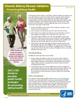
3. Chronic Kidney DiseaseProgressive loss of kidney function over a period of months or years 4-6% of the UK adult population and 8-10% in EuropeSerious implications: Complete kidney failure needing dialysis Iron deficiency Anaemia Increased risk of heart disease.
4. Cardiovascular mortality is very high in CKD patients de Jager DJ, et al. JAMA 2009;302(16):1782–1789CKD=chronic kidney disease; IV=intravenous; PY=person yearsCardiovascular Mortality in the General Population and in Dialysis Patients
5. Effect of Renal Dysfunction & Anaemia on Patient Survival No RD, no anaemiaRD, no anaemiaNo RD, anaemiaRD, anaemia + De Silva R, Bhandari S et al . European Heart Journal 2005, 27: (5), 569-581De Silva R, Bhandari S et al. American Journal of Cardiology 98: 3; 391-398. 2006
6. Uraemic Cardiomyopathy in CKDStructural Changes Fibrosis LVH reduced capillary densityCellular Changes carnitine deficiency energeticsIschaemic Vulnerability Iron deficiency EPO deficiencyMetabolic Changes calcium cycling GSK-3b/AKT/PIP3K GLUT4 and IRSemple D, Bhandari S, Seymour A. Am J Physiology 2012 Bhandari S. Frontiers in Bioscience 2011Dunja A , Bhandari S, Seymour A. Kidney International 2009 Reddy V, Bhandari S, Seymour JASN 2005Pressure and volume OverloadAnaemia
7. Why iron is important? Pivotal role in O2 uptake, transport, storage and metabolism in skeletal and cardiac muscles.Iron is important for mitochondrial function - mitochondrial ATP productionRequired for Hb production and O2 transportAlso required for:catalytic enzymesproteins for DNA synthesistransport of electronscell respirationoxidative phosphorylationTri-carboxylic acid cycleModified from Bhandari S Frontiers in Bioscience 2011Semple D, Bhandari S, Seymour AM Am J Physiol 2012 Taylor D, Seymour A, Bhandari S. Am J Physiology 2014
8. Papayannopoulou T, et al. In: Hoffman R, et al., ed. Hematology: Basic Principles and Practice. 4th ed. 2005;267-288. SCF, GM-CSF,IL-3SCF, IL-1, IL-3, IL-6, IL-11PluripotentStem CellBurst-Forming Unit-Erythroid Cells (BFU-E) Colony-FormingUnit-ErythroidCells (CFU-E)ReticulocytesRBCsErythro-blastsProerythro-blastsAbout 8 DaysIronErythropoietinErythropoiesis in CKD
9. Reduced platelets and Thrombotic PotentialHazara A & Bhandari S J Clinical Pharm & Therapeut 2014, doi: 10.1111/jcpt.12218**Post-infusion - Platelets lower at 30, 60 & 90 days versus to baseline: n=204 p = 0.01 p= 0.03 p<0.001
10. Effect of CKD on regulation of Hepcidin ProductionRed blood cellsBone marrowPlasma Fe-TfInflammation IL-6Ganz T. J Am Soc Nephrol 2007;18:394–400Katz et al Nature Genetics 2014, 46, 678-84LiverDuodenumHepcidinHepcidinSpleenHepcidinChronic oxidative StressIronBMP 6
11. Functional Vs Absolute
12. Why treat iron deficiency in CKDThe main aims are to improve symptoms and quality of life Symptomatic restless legs Cognition Physical performance Exercise tolerance Immune function reduces platelet count Beneficial effects on cardiac function via effects on mitochondrial function
13. The Heart in CKDSubject to adaptive and maladaptive changesConcentric and eccentric hypertrophy linked to FGF-23Switch from fatty acids to glucose metabolismIron and erythtropoietin deficiency Ischaemia Oxidative stress Changes in calcium cycling within the moitochondria
14. Faul C et al; J Clin Invest 2011; 121 (11) : 4393-4408FGF-23, klotho independent induced LVH
15.
16. How to treat iron deficiency of CKD
17. Macdougall I et al FIND –CKD NDT 2014, 29, 2075-2084
18. Dose dependent rise in serum ferritin and haemoglobin at 3 and 6 months post Total Dose Iron infusion in CKD patients, n=160 patients given LMW iron dextranBhandari S. (NDT Plus 2012) 3 months6 months
19. When to treat iron deficiency in CKDMost recommendations in guidelines are ‘suggestions’Limited compelling evidence for specific Hb targets KDIGO 2012: Target Hb should not exceed 115 g/L NICE: Target Hb upper limit of 120 g/LShould be considered when: SF<100 microgram/ml Or TS%< 20%Or HRC< 6% CHr<29g/L
20. Potential Risks of IV IronShort TermAnaphylaxislabile iron reactions Longer termOxidative stressRisk of infectionsRisk of iron overload
21. D Taylor, Seymour A, Bhandari S. Am J Physiology 2014Iron and Mitochondria
22. Development of HF
23. Uraemia – Iron – Oxidative Stress and Mitochondrial FunctionFaisal N PhD 2015 unpublished MUST NOT BE COPIEDFigure 19: Isolated Mitochondrial Respiratory rates in the presence of 5mM glutamate and 1mM malate (GM). Results are presented as mean +/- SEM (*p < 0.05 vs untreated).
24. Iron and the HeartFew studies examined the effects of IV iron in patients with HF, anaemia, iron deficiency, and renal dysfunctionHypothesis: iron loading leads to improvement in mitochondrial function and hence symptom improvement and cardiac function independent of haemoglobin
25. Randomised double-blind controlled study of IV iron in CHF (FAIR-HF)Cr Cl19/2019/2013/20* 19/20Diuretics6.5±3.76.6±4.42.3±0.8* 6.1± 3.8CRP0190.7±56.158±6267.5±114.980.5±8.130.8±1.72.9±0.610.2±0.5Baseline6 months6 monthsBaseline0192.3±60.960±5255.9±124.678.7±8.231.3±3.72.9±0.710.3±0.6Control (Placebo; n=20)Treatment (IV iron; n=20)184.5±58.5240.1±51.2* 6MWT 76.3± 10.4 67.5±8.2* Heart rate 450.9±248117.5±87.4* BNP 58±841±7* MLHFQ50 Hospitalisations 28.8±2.435.7±4.7* LVEF 3.3±0.62.0±0.2* NYHA 9.8±0.611.8±0.7* HbToblli et al , JACC 2007; Okonko et al , JACC 2008. 31.7±10.837.7±10.244.9±11.0* 39.8±10.1
26. Beneficial effects of long-term intravenous iron therapy with ferric carboxymaltose in patients with symptomatic heart failure and iron deficiency Ponikowski P et al CONFIRM-HF, Eur Heart J 2014
27. Ponikowski P et al , Eur Heart J 2014CONFIRM-HFTreatment of stable, symptomatic,iron-deficient HF patients with and without anaemia with i.v. ironSustainable improvement in functional capacity -6-MWT walking testconcomitant improvementImproved functional status and quality of life Reduced risk of hospital admission due to worsening HF
28. RecapIron is central in oxygen uptake, transport, storage and metabolism in both skeletal and cardiac muscle. Evidence suggest that myocardial energy production is a crucial aspect of cardiac function. The heart consumes large quantities of energy for myofibril contraction and maintenance of ionic gradients. Abnormalities in myocardial energy metabolism develop in uraemic patients. This energy production is dependent on mitochondrial function which is the main source of energy for myocyte contraction. Previous Experiments in a model of stressed hearts with CKDincrease in stage 4 respirationincrease in uncoupling proteins leading to mitochondrial dysfunctionincrease in transition pore formation leading to impaired contractile function of cardiomyocytes. Therefore a deficiency of iron may lead to impaired mitochondrial function via possible effects on transition pore opening and subsequent inhibition of the pro-survival pathway and activation of apoptotic pathways.
29. Trial Purpose - Main Research Question - HypothesisExamine whether a strategy of IV parenteral iron therapy in patients with stages 3b-5 CKD who are iron deficient (Ferritin < 100 µg/L and/or TS%<20) but NOT anaemic (haemoglobin 110-150g/L) compared to a strategy of continuing current therapy without additional iron therapy lead to:Improved functional capacity and physical capacity over a one month follow-up period, with reassessment at 3 months: 6MWTqualitative questionnaires (KDQol and MLHF) stabilisation or improvement in cardiac function hospitalization rateshaemoglobin levelsThird arm - control group of patients with CKD and no anaemia or iron deficiency will allow for baseline comparisonFourth arm - healthy patients (patients from the live donor work up program) will also serve as a baseline control (healthy volunteers with no underlying known disease or CKD as judged by their suitability for transplant donation during donor work up).
30. OTHER EFFICACY OBJECTIVESImprovement in cardiac structure and functionspeckle tracking ECHO studies (addition of physiological monitoring using pulse oximetry and cardiac harness for heart and respiratory rate)Change in New York Heart association functional classification (NYHA)Cardiac iron loading based on MRI using T2single centre in a parallel sub-study (6 patients) Improvement in renal functioneGFR
31. MECHANISTIC OBJECTIVESInflammatory mediators IL-6IL-8IL-10CRPVasoactive substances (oxidative stress) Ian Chetter - MDA, selectins and interleukins - mainly in John Greenmans lab at UoH (Lee Maddern is his post doc who was brilliant at training our research fellows in ELISA techniques etc)plasma malondialdehyde - MDAglutathione GSH/GSSH Endothelial functionPulse wave velocity (PWV) E and P selectin
32. Iron and the Heart StudyExploratory multicentre prospective double blinded randomised controlled pilot trial The effect of IV iron supplementation (Monofer®)In Iron deficient but not anaemic patientsWith CKD stages 3b or worseExamining patients’ functional status and cardiac structure and function
33. Trial DesignAn investigator led multi-centre54 participants with stage 3b or worse CKD with iron deficiency 2 further control groups: 20 with CKD, 10 healthy volunteersSufficient size to provide data on feasibility, and likely size and endpoints of a future definitive study.
34. NoExcludedNot Meeting Criteria DeclinedOther Reason 3 Month Follow-UpBaseline 1 and 3 month visits-routine tests (eGFR, FBC, BCP, urinary PCR), BP, documentation of adverse events, compliance and changes in medicationExtra tests at visits-QOL questionnaire, weight and BMI, 6-minute walk test, ECG, and bloods for C-reactive protein, cystatin-C, NT-proBNP and biomarkersYesCKD patients stage 3b-5 with iron deficiencySF<100; TS<20 and not anaemicHb 110-150g/LEligible for Study?Double Blinded Randomise1:1 ratio, N=54Experimental Arm:IV Monofer 1000mgN=27Placebo Arm:IVI Dummy DrugN=2718 month recruitment3 month follow-upGROUP 4 n=6 Monofer 1000mgUnblinded; n=6GROUP 4 Additional T2* MRIGroup 1
35. 3 Month Follow-Up for Group 2Baseline 1 and 3 month visits-routine tests (eGFR, FBC, BCP, urinary PCR), BP, documentation of adverse events, compliance and changes in medicationExtra tests at l visits-QOL questionnaire, weight and BMI, 6-minute walk test, ECG, and bloods for C-reactive protein, cystatin-C, NT-proBNP, biomarkersYesGroup 2CKD patients stage 3b-5 with NO Anaemia and NO iron deficiencySF>100; TS>20 and not anaemicHb 110-150g/LEligible for Study?N=2012 month recruitment3 month follow-upGroup 3CONTROL Live Kidney Donors patients with NO iron deficiencyand not anaemicHb 110-150g/LEligible for Study?N=10Baseline bloods onlyCONTROL ArmMECHANISTIC STUDYYes
36. Key Inclusion Criteria Key exclusion criteriaAged ≥18 years (male or female)CKD stage 3b- 5 (eGFR <30mls/minute using the MDRD equation) and not on dialysis therapyResting blood pressure (BP) ≤160/90 mmHg At least 3 months of specialist renal follow-up at the time of entry into the trialAged <18 yearsUndergoing dialysis therapyUncontrolled hypertension (>160/90mmHg) or requirement for 5 or more agents to control BPPregnancy or breastfeedingNon renal anaemiaCRP >50Eligibility
37. Primary objectiveTo compare the effect of IV iron versus placebo on changes in physical function based on: 1) 6-minute walk test 2) Qualitative questionnaires In iron deficient non-anaemic CKD patients.
38. Secondary Objectives To explore whether IV iron supplementation compared to placebo in CKD 3b+ patients leads to: Improvement in cardiac structure and function. Improvement in vascular endothelial functionCardiac iron loading Improvement in markers of oxidative stressImprovement in renal function
39. Objectives - End-pointClinicalSpeckle Echo, ECGHospitalisationsDeath and cardiac eventsThrombotic episodesinfectionSAEMagnetic Resonance Imaging - T2* MRI to measure iron loading in heart pre and after IV iron at baseline post IV iron, 1 month and 1 year follow-up (total of 4 scans per patient)54 patients with eGFR <45ml/min with Hb .110g/L and SF <100 and/or TS<20% IV IronNo ironMECHANISTICPulse wave velocity E and P selectin Inflammatory markers Cytokines - IL-6; IL-8 & IL-10, CRP Oxidative stress markers plasma malondialdehyde glutathione activity.Primary Endpoint: Difference in co-analysis of: 6MWT and Questionnaires (KD-QOL; MLHF; Restless leg)
40. Questions?
41. Trial Purpose - Main Research Question - HypothesisA reversible abnormality in muscle (cardiac & skeletal) metabolism develops in uraemic patients that is associated with:impaired cardiac functionimpaired skeletal muscle energy metabolismreported symptoms HypothesisIron loading leads to improvement in mitochondrial function and hence symptom improvement and cardiac function independent of haemoglobin.