
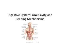
Dr Nihal Bandara BDS Hons Sri Lanka PhD Hong Kong The School of Dentistry The University of Queensland Australia Fungi A separate kingdom Neither a plant nor an animal Includes mushrooms ID: 919123
Download Presentation The PPT/PDF document "Fungi in the oral cavity: the opportunis..." is the property of its rightful owner. Permission is granted to download and print the materials on this web site for personal, non-commercial use only, and to display it on your personal computer provided you do not modify the materials and that you retain all copyright notices contained in the materials. By downloading content from our website, you accept the terms of this agreement.


Slide1
Fungi in the oral cavity: the opportunistic foes
Dr. Nihal Bandara BDS Hons (Sri Lanka), Ph.D. (Hong Kong)
The School of Dentistry
The University of Queensland
Australia
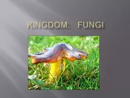
Slide2Fungi
A separate kingdom
Neither a plant nor an animal
Includes mushrooms, rusts, smuts, puffballs, truffles, morels, molds, and yeasts, A variety of sizesMicroscopic single-celled organisms e.g. yeastMulticellular macroscopic organisms. Human feet harbours over 200 species of fungi more than any other body sites . -Human Genome Research Institute in Bethesda, Maryland, USA
Slide3Opportunistic pathogens
Microorganisms
Do not cause disease in a healthy hostTake advantage of a host with a weakened immune systemE.g. some bacteria, viruses, fungi and protozoa
Slide4What are the opportunities?
Physiological factors
e.g. elderly, pregnancy and infancyLocal factors e.g. mucosal irritations, poor dental hygiene, localized radiotherapy, xerostomiaMedications e.g. broad spectrum antimicrobial therapy, cytotoxic drugs, immunosuppressive drugs, Steroid inhalers and systemic steroidsNutritional factors e.g. Iron, folate, vitamin B12 deficiencies, malnutrition
Systemic disorders e.g. Diabetes, hypothyroidism, Addison’s diseaseImmune defects e.g. HIV infection, AIDS, thymic aplasia
Malignancies e.g. acute leukaemia, agranulocytosisXerostomia due to irradiation, sjögren’s syndrome, drug therapy
Samaranayake et al 2009
Slide5Oral fungal infections
Infection
Pathogen
CandidiasisCandida albicans, C. tropicalis, C. glabrata,
C. parapsilosis, C. krusei, C. kyfer, C. dubliniensisAspergillosis
Aspergillus fumigatus
Cryptococcosis
Cryptococcus neoformans
Histoplasmosis Histoplasma capsulatum
Blastomycosis
Blastomyces dermatitidis
Zygomycosis
Orders
Mucorales
and
Entomophthorales
Coccidioidomycosis
Coccidioides
immitis
Paracoccidiomycosis
Paracoccidioides brasiliensisPenicilliosis Penicillium marneffeiSporotrichosisSporothrix schenckiiGeotrichosis Geotrichum candidum
Krishnan
PA. Indian
J Dent Res. 2012 Sep-Oct;23(5):650-9.
Slide6Candidiasis
Pseudomembranous candidiasis (Thrush)
Chronic/acute
White/Yellow plaques in mucosal surfacesConfluent or discreteReadily removable leaving raw underlying surface
http://pocketdentistry.com/
Koban et al. New
J. Phys. 12
(2010) 073039
Slide7Erythematous candidiasisAlso called atrophic candidiasisAppears as erythematous patches in the mucosaCould be chronic or acuteCommonly seen in the palate, dorsal tongue
Tongue
depapillation
Mainly associated with broad spectrum antibiotics or corticosteroidshttp://www.hivdent.org/
http://hiv.uw.edu/oral/case1/discussion.html
Slide8Chronic hyperplastic candidiasisAlso called candidal leukoplakia
White plaque present in the commissural region
Buccal commissural area, plate and tongue
Associated with dysplasia (15%) Samaranayake LP 1990 Biopsy and histopathology is necessary
http://www.tauntonmaxfax.net/html/prof_oralmed_candidalinfections_t.htm
Williams et al. Journal
of Oral Microbiology 2011,
3: 5771
Slide9Candida associated lesions
Denture associated stomatitis
A chronic inflammatory condition in denture bearing mucosa
Erythematous lesionsDenture provides ideal environment for Candida growthAttachment sitesAct as a shield for saliva and local immunity Denture hygiene is critical
http://www.studentistry.com/denture-stomatitis-classification-causes-management/
Davenport et al. British Dental Journal 189, 414 - 424 (2000)
http://
pocketdentistry.com/
Slide10Median rhomboid glossitisUncommon condition Men are affected moreRhomboid shape hypertrophic or atrophic plaque in the mid dorsal tongue
Association of
Candida
with median rhomboid glossitis is controversialhttps://en.wikipedia.org/wiki/Median_rhomboid_glossitis
http://pocketdentistry.com/3-common-oral-soft-tissue-lesions/
Slide11Angular CheilitisMixed bacteria fungal infectionsCorners of the mouth is affectedStaphylococci and streptococci are often associated withErythematous fissuring in the angle of mouth
Accompanied by a pseudomembranous covering
Can affect anterior nostril regain too
Predisposing factors: facial wrinkling, reduced occlusal vertical dimension, nutritional deficiencies ( e.g. Thiamine, Riboflavin, Iron and Folic acid)http://www.crutchfielddermatology.com/caseofthemonth/studies/l_2007_008.asp
Hunt
2013 http://www.microbiologybook.org/lecture/hiv3.htm
Slide12Diagnosis of Candida infections
Characteristic clinical appearance and symptoms e.g. burning sensation
Laboratory assays e.g.
exfoliative cytology, fungal culture, mucosal biopsy, salivary assaysDifferential diagnoses: thermal and traumatic lesions, syphilis, white keratotic lesions, erosive lichen planus, lichenoid reactions, lupus erythematosis, erythema multiforme, pernicious anaemia, and epithelial dysplasia
McIntyre 2001 Dental update;28:132-139
Slide13Treatment of oral Candida
infections
McIntyre 2001 Dental update;28:132-139
Correction of the underlying predisposing factors and habitsPharmacotherapy
Slide14Uncommon oral fungal infections
Aspergillosis
Second commonest fungal infection in human
Commonly seen with high dose of corticosteroid use, organ and marrow transplantation, increase use of immunosuppression against autoimmune diseasesLungs are commonly affectedAlso invade blood vessels causing thrombosis and infarctionsLess commonly affect maxillary sinuses Oral lesions are typically black or yellow necrotic soft tissues
Krishnan PA. Indian J Dent Res. 2012 Sep-Oct;23(5):650-9.
Aspergillus fumigatus
Slide15CryptococcosisPrimarily affects lungs and can lead to meningitisCaused by Cryptococcus neoformans, usually isolated in pigeon’s and other birds’ droppings
Cutaneous lesions : Face, neck and scalp
Oral lesions are rare; resembles superficial ulcerations, granulomas, nodules or indurated ulceration similar to carcinoma
Nonspecific chronic ulceration of the buccal mucosa due to cryptococcosisNecrosis of alveolar bone and palatal
mucosaCrispian Scully et al http://emedicine.medscape.com/
Cryptococcus
neoformans
Slide16HistoplasmosisCaused by Histoplasma capsulatum
; a dimorphic fungi
Two forms; pulmonary and mucocutaneous
Mucocutaneous form cause ulcerative/erosive lesions on tongue, plate and buccal mucosaOral lesions: single ulcers, long term and may or may not be painfulAlways misinterpreted as malignant ulcersBiopsy is mandatory
CDC/Lucille K. Georghttp://www.emedicinehealth.com/histoplasmosis/page4_em.htm
Histoplasma capsulatum
Slide17BlastomycosisCaused by Blastomyces dermatitidisWhen inhaled, spores produce disseminated or local respiratory infections
Oral lesions are rare
May produce ulcerated mucosal lesions in the oral cavity
Extensive ulceration involving the skin of the face and neck. Nonspecific papillary nodular lesion on the hard palate
Crispian Scully http://emedicine.medscape.com/article/1077685-clinical#b4
Blastomyces dermatitidis
Slide18MucormycosisCaused by a saprophytic fungi found in soil, bread mold, decaying vegetation etc.
Involvement of the oral cavity is secondary to paranasal sinuses or nasal cavity
Usually present as a palatal necrosis or ulcerations
Extends to adjacent structures causing extensive tissue necrosis and invasion of brainOrgan transplant and poorly controlled diabetic patients are susceptible Krishnan PA. Indian J Dent Res. 2012 Sep-Oct;23(5):650-9.
Rhizopus
oryzae
Slide19Diagnosis of deep seated oral fungal infectionsBiopsyPathologist should be given patients’ medical history e.g. immune suppressionPatients with deep oral fungal infections must be referred to medical specialists for further evaluationBlastomycosis
: smear/culture, Direct
immunostaining, DNA probes
Cryptococcosis: microscopy/staining, serologyHistoplasmosis: microscopy/staining, serology, skin testsMucormycosis: microscopy/Histology, smear/culture
Slide20Treatment of Oral fungal infections
Treat Guidel Med Lett. 2009 Dec;7(88):95-102
Slide21Thank you