
Brain Spinal Cord also includes 4 chambers in brain called ventricles Brain 1 Cerebral Hemispheres 2 Diencephalon 3 Brain Stem 4 Cerebellum 1 Cerebral Hemispheres Covered by ridges ID: 910410
Download Presentation The PPT/PDF document "Nervous System-Anatomy Central Nervous S..." is the property of its rightful owner. Permission is granted to download and print the materials on this web site for personal, non-commercial use only, and to display it on your personal computer provided you do not modify the materials and that you retain all copyright notices contained in the materials. By downloading content from our website, you accept the terms of this agreement.


Slide1
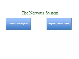
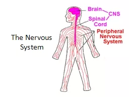
Nervous System-Anatomy
Slide2Central Nervous System
Brain
Spinal Cord
(also includes 4 chambers in brain called ventricles)
Slide3Brain
1) Cerebral Hemispheres
2) Diencephalon
3
) Brain Stem
4) Cerebellum
Slide4Slide51) Cerebral Hemispheres
Covered by ridges =
gyri
Ridges separated by grooves =
sulci
The hemispheres (right and left) are separated by a single deep
longitudinal fissure
Other shallow fissures divide each hemisphere into lobes
Lobes are named for the cranial bones over them
Slide6Slide7Ear to ear is the
central fissure
(
sulci
)
Posterior to the central fissure
in the parietal lobe is the somatic sensory area (post central
gyri
)
Impulses that travel from sensory receptors are interpreted there
Crossed pathways
Slide8Slide9Anterior to the central fissure
in the frontal lobes is the primary motor area (Pre central gyri)
Allows us to consciously move our skeletal muscles
Major voluntary motor tract that descends to the spinal cord
Crossed pathways
Slide10Slide11Areas in Cerebrum
Impulses for special senses:
Visual
= posterior occipital
Auditory
= temporal lobe (lateral fissure)
Olfactory
= deep temporal lobe
Slide12Slide13Impulses for the special senses:
Broca’s
area
= base of the pre-central
gyrus
located in left hemisphere only, gives ability to say words properly
Speech Area
= junction of temporal, parietal, and occipital lobes, allows understanding of words, spoken or read and
responses
to them
Slide14Higher Reasoning = anterior frontal lobe
Complex Memories = temporal and frontal lobes
Slide15Gray matter – of cerebral hemispheres contain the cell bodies of neurons
White matter – of cerebral hemispheres is composed of fiber tracts which carry impulses to or from the cortex
Slide16Corpus
callosum
– a very large fiber tract that connects the cerebral hemispheres and allow left and right brain to communicate
Basal nuclei or basal ganglia
are buried within the white matter and help regulate voluntary motor activities
Slide17Slide182) Diencephalon
Interbrain:
Thalamus
- encloses the 3
rd
ventricle, relay for sensory impulses
Slide19Hypothalamus
- floor of diencephalon, autonomic center.
Functions: regulates body temperature, water balance, and metabolism, contains the “
limbic system
” which is a center for many drives; thirst, appetite, sex, pleasure,
-Regulates the pituitary gland, contains
mammillary bodies
Slide20Epithalmus
- forms the roof of the 3
rd
ventricle, contains pineal body (endocrine gland), contains:
Choroid plexus
which forms CSF (cerebral spinal fluid)
Slide21Slide223) Brain Stem
Midbrain- extends from the
mammillary
bodies (in diencephalon) to the
pons
Cerebral aqueduct- connects 3
rd
ventricle to 4
th
ventricle
Corpora
Quadrigemina
- four rounded protrusions – reflex centers for vision and hearing
Slide23Slide24Slide25Pons
- just below midbrain, mostly fiber tracts, important for control of breathing
Medulla Oblongata-
most inferior part of brain stem, merges with spinal cord, mostly fiber tracts.
Functions-controls heart rate, blood pressure, breathing, swallowing, vomiting. 4
th
ventricle is posterior
Slide264) Cerebellum
Coordinates skeletal muscle activity, controls balance and equilibrium, monitors body position
Slide27Protection of the Brain
Skull
Meninges
- 3 parts:
Dura Mater
- outside tough layer
Arachnoid
Mater-
middle blood vessels
Pia
Mater-
surface of the brain layer
3) Cerebral Spinal Fluid-
CSF continuously formed by choroid plexus , cushions, protects, runs down central canal of the spinal cord
Slide28Slide29Slide30Problems of the Brain
Concussion -MS
Contusion -ALS
Aneurysm -Huntington’s
CVA (cerebrovascular
attack-stroke),
aphasia (difficulty speaking), paralysis
TIA (transient ischemic
attack-mini stroke-blockage is temporary-blood flo
w returns on its own
)
Alzheimer’s
disease -Epilepsy
Parkinson’s disease
Slide31Spinal Cord
17 inches from skull to L2
Reflex center and 2 way conduction pathway
Central canal contains CSF
Slide32Slide33Gray Matter of Spinal Cord
Dorsal Horns (posterior)-
contains interneurons and sensory neurons; enter by dorsal root (ganglion)
Ventral Horns (anterior)
- contain motor neurons, (somatic voluntary), leave by the ventral root
The dorsal and ventral roots fuse to form the spinal nerves
Slide34Slide35White Matter of Spinal Cord
Myelinated
fiber tracts, some run to other side of spinal cord, some run to higher centers
All tracts in the anterior and lateral cord are motor
All tracts in the posterior cord are sensory
Slide36Problems of the Spinal Cord
Dorsal root damage- sensory damage =
parasethesia
(numbness, tingling, pins etc.)
Ventral root damage- motor damage =
paralysis
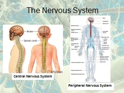
Slide37Peripheral Nervous System
Contains nerves: spinal and cranial
A nerve is a bundle of neurons found outside the CNS
Nerves are neurons bundled in connective tissue
Slide38Nerves are named like neurons:
Carry to CNS= afferent
Carry from CNS= efferent
Carrying both sensory and motor= mixed (all spinal)
Slide39Slide40Cranial Nerves- 12 pair
1.
olfactory
- sensory
2.
optic
- sensory
3.
oculomotor
-
motor
4.
trochlear
- motor
5.
trigeminal-
mixed
6.
abducens
- motor
Slide417.
facial
- mixed
8.
vestibulocochlear
- sensory
9.
glossopharyngeal
- mixed
10.
vagus
- mixed
11.
accessory
- mostly motor
12.
hypoglossal
- mixed
Slide42Slide43Spinal Nerves – 31 pairs
Formed from the fusion of the ventral and dorsal roots of the spinal cord
Divides into dorsal and ventral
rami
Both types of
rami
contain both sensory and motor nerves , just go to different places
Slide44Slide45Dorsal
rami
are smaller and serve skin and muscle of posterior body trunk
Ventral
rami
of T1- T12 form
intercostal
nerves
All other ventral
rami
form plexuses which serve limbs, neck, and diaphragm
Slide46Slide474 Plexus
Origin
Plexus
Major Nerve
Serves
C1 – C4
Cervical
Phrenic
diaphragm
C5 – C8
Brachial
Axillary
Arm
T12 , L1 – L4
Lumbar
Femoral
Lower abs, butt
L4 –L5 ,
S1 – S4
Sacral
Sciatic
Post leg
Slide48Slide49Slide50Two Divisions of the Peripheral Motor Nervous System
Somatic Nervous System
Autonomic Nervous System
Slide51Somatic Nervous System
One neuron extends to skeletal muscle
Voluntary
Slide52Autonomic Nervous System
Involuntary, Automatic
Motor control of cardiac muscle, smooth muscle, and glands
Involves a chain of two motor neurons called: pre ganglion and post ganglion
Slide53Autonomic Nervous System has two arms:
Parasympathetic
- rest and digest / homeostasis
Sympathetic-
emergency; fight or flight
Slide54Both serve the same organ
Each release different neurotransmitters
Parasympathetic= cholinergic fibers
Sympathetic= adrenergic fibers
Slide55Slide56Parasympathetic
Pre-ganglion neuron secretes acetylcholine
Post-ganglion neuron secretes acetylcholine
Slide57Sympathetic
Pre- ganglion neuron secretes acetylcholine
Post- ganglion neuron secretes
epinepherine
Slide58Parasympathetic
Rest and digest
Continued homeostasis
Slide59Sympathetic
Increased: heart rate, blood pressure and glucose
Dilation of: bronchioles and blood vessels
Close down digestive system
Activate adrenal glands
Slide60Developmental problems
Nervous system develops in the 1
st
month of pregnancy
Viruses, drugs, alcohol, smoking can affect embryo
Slide61Birth Defects
Cerebral Palsy
Hydrocephalus (fluid on brain)
Anencephaly (small brain, missing skull parts)
Spina
bifida
Microcephaly (
Zika
virus)
Slide62Continued development
Last to form = Hypothalamus
Myelination
continues through childhood
Brain reaches maximum weight in the young adult (20s)
New neural pathways can always be formed (learning)
Slide63Aging Problems
Sympathetic system becomes inefficient in the elderly
Arteriosclerosis and High Blood Pressure can cause decreased brain oxygen = senility
< 5% senility at age 65
Boxers and chronic alcoholics show shrunken brains = senility
Slide64Reversible Senility
Drug side effects, low blood pressure, depression, dehydration, and malnutrition can cause types of senility that will improve if the initial problem is corrected