
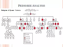
Most common Xlinked disorders Usually expressed only in males Rarely due to random Xinactivation a female will express disease called manifesting heterozygotes Pattern Of Inheritance ID: 910392
Download Presentation The PPT/PDF document "X-Linked Recessive Disorders" is the property of its rightful owner. Permission is granted to download and print the materials on this web site for personal, non-commercial use only, and to display it on your personal computer provided you do not modify the materials and that you retain all copyright notices contained in the materials. By downloading content from our website, you accept the terms of this agreement.


Slide1
X-Linked Recessive Disorders
.
Slide2Most common X-linked disorders.
Usually
expressed only in males.
Rarely, due to random X-inactivation, a female will express disease, called manifesting
heterozygotes
.
Slide3Pattern Of Inheritance:
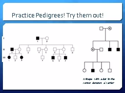
Disease usually passed on from carrier mother.
Expressed in male offspring, females are carriers. Skipped generations are commonly seen. In this case, Recurrence risk is half of sons are affected, half of the daughters are carriers.
Slide4Recurrence risk:
All the daughters are heterozygous carriers and all the sons are homozygous normal.
Slide5Disorders:
Slide6DISORDERS WITH MULTIFACTORIAL (POLYGENIC
) INHERITANCE
Involved in many physiologic characteristics of humans e.g. height, weight, hair
color.Defined as one governed by additive effect of two or more genes of small effect but conditioned by environmental, non genetic influences.
Slide7The disorder becomes manifested only when a certain number of effector genes, as well as conditioning environmental influences are involved
Rate of recurrence is 2 to 7%
Slide8COMMON DISEASES ASSOCIATED:
Diabetes mellitus
Hypertension
GoutCleft lip and palateSchizophreniaBipolar disorderCongenital heart diseaseSkeletal abnormalitiesNeural tube defectsCoronary artery disease
Slide9Cytogenetic Disorders
.
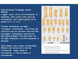
Slide10Slide11KARYOTYPING
Basic tool of
cytogeneticist
Karyotype is a photographic representation in which chromosomes are arranged in order of decreasing lengthGiemsa stain (G banding) technique—each chromosome can be seen to possess a distinctive pattern of alternating light and dark bands of variable widths
Slide12Shorthand of
Cytogenetics
:
Short arm denoted as p, long arm denoted q.
Each arm divided into numbered
regions
from the
centromere
onwards.
Each region numerically arranged into
bands.
For e.g., 5p24 would denote chromosome 5, short arm, region 2 and band 4.
Slide13Cytogenetic disorders may result from structural or numeric abnormalities of chromosomes
It may affect autosomes or sex chromosomes
Slide14Numeric Abnormalities:
Normal Chromosomal number is 46. (2
n
=46). This is called euploid state. (Exact multiple of haploid number).Polyploidy: posession of more than two sets of homologous chromosomes. Chromosomal numbers like 3n or 4n. (Incompatible with life); generally results in spontaneous abortionAneuploidy: Any Chromosomal number that is not an exact multiple of haploid number . E.g 47 or 45.
Slide15Aneuploidy:
Most common cause is
nondisjunction
of either a pair of homologous chromosomes during meiosis I or failure of sister chromatids to separate during meiosis II.The resultant gamete will have either one less chromosome or one extra chromosome.
Slide16Fertilization of such gamete will result in zygote being either
trisomic
( 2
n+1 ) or monosomic ( 2n-1 ).Monosomy in autosomes is incompatible with life. Trisomy of certain autosomes and monosomy of sex chromosomes is compatible with life.
Slide17Mosaicism
The presence of two or more types of cell populations in the same individual.
Postzygotic
mitotic nondisjunction will result in one trisomic and one monosomic daughter cell.
The descendants of these cells will produce a
mosaic.
Slide18Structural Abnormalities:
Usually result from chromosomal breakage, resulting in loss or rearrangement of genetic material.
Patterns of breakage:
Translocation.Isochromosomes.Deletion.Inversions.Ring Chromosomes.
Slide19TRANSLOCATION
Transfer of a part of one chromosome to another chromosome
Translocations are indicated by t
E.g. 46,XX,t(2;5)(q31;p14)Balanced reciprocal translocation is not harmful to the carrier, however during gametogenesis, abnormal gametes are formed, resulting in abnormal zygotes
Slide20Centric fusion type or
Robertsonian
translocation:The breaks occur close to the centromere, affecting the short arms of both choromosomesTransfer of the chromosome leads to one very large and one extremely small chromosomeThe short fragments are lost, and the carrier has 45 chromosomesSuch loss is compatible with survivalHowever, during gametogenesis difficulties arise
Slide21Slide22ISOCHROMOSOMES
Result when one arm of a chromosome is lost and the remaining arm is duplicated, resulting in a chromosome consisting of two short arms only or of two long arms.
DELETION
Loss of a portion of chromosomeThis can be terminal (close to the end of the chromosome on the long arm or the short arm), or it can be interstitial (within the long arm or the short arm). A ring chromosome is a variant of deletion. It occurs when break occurs at both the ends of chromosome with fusion of the damaged ends.
Slide23INVERSIONS
Occur when there are two breaks within a single chromosome with inverted reincorporation of the segment.
Since there is no loss or gain of chromosomal material, inversion carriers are normal.
An inversion is paracentric if the inverted segment is on the long arm or the short arm . The inversion is pericentric if breaks occur on both the short arm and the long arm .
Slide24General Features of Cytogenetic Disorders:
Associated with absence, excess, or abnormal rearrangements of chromosomes.
Loss of genetic material produces more severe defects than does gain.
Abormalities of sex chromosomes generally tolerated better than those of autosomes.
Slide25Sex chromosomal abnormalities are usually
subtle(faint & mysterious)
and are not detected at birth.
Most cases are due to de novo changes (i.e. parents are normal and recurrence in siblings is low).
Slide26Cytogenetic Disorders involving Autosomes
.
Slide27Trisomy 21/Down’s Syndrome
:
Most common chromosomal disorder.
Down syndrome is a chromosomal abnormality characterized by the presence of an extra copy of genetic material on the 21st chromosomeTrisomy 21 is caused by a meiotic non disjunction event.
Slide28With
non disjunction
, a gamete (
i.e., a sperm or egg cell) is produced with an extra copy of chromosome 21; the gamete thus has 24 chromosomesWhen combined with a normal gamete from the other parent, the embyo now has 47 chromosomes, with three copies of chromosome 21.About 4% of cases are due to Robertsonian translocations.Maternal age has a strong influence.
Slide29Karyotype for trisomy Down syndrome. Notice the three copies of chromosome 21
Slide30Other Trisomy Syndromes:
Trisomy
18 :Edwards Syndrome.Around half will die within two weeks &only around one in every five will live at Least 3 months. Around one in every 12 babies born
with
Edwards
' syndrome survive beyond one year, and they
will
live with severe physical and mental disabilities.
.
Slide31Trisomy
13 :
Patau Syndrome
Slide32Marfan
Syndrome
Slide33It is a common heritable disorder ( 1 : 3000-5000 people ) affects
chromosome15 q21.1
, which affect s fibrillin-1 production which is necessary for the production of
c.t. It affects adults & children alike irrespective of gender, race or ethnic back ground.It affectsc various body systems including skeleton, NS, eyes, skin, lung, heart & blood vessels. 50000 in USA have this disease.
Slide34Cytogenetic Disorders involving Sex Chromosomes
.
Slide35Extreme karyotypic variations seen frequently with Sex Chromosomes, with females having 4-5 extra X Chromosomes.
Males with two to three Y chromosomes have also been identified.
Slide36Klinefelter’s
Syndrome
:
Defined as Male Hypogonadism, develops when there are at least two X chromosomes and one or more Y chromosomes.Usual karyotype is 47,XXY. The extra X may be maternal or paternal.
Slide37Results from
non disjunction
of sex chromosome during meiosis.
Risk factors include advanced maternal age and a history of exposure to radiation in either parent.
Slide38Clinical Manifestations:
Increase in body length between soles and pubis.
Reduced facial, body and pubic hair. Gynecomastia.
Testicular atrophy.Infertility.Mild mental retardation.
Slide39Turner Syndrome:
Primary
hypogonadism
in phenotypic females.Results from partial or complete monosomy of the X chromosome.
Slide40Most common cause is absence of one X chromosome.
Less commonly, mosaicism, or deletions on the short arm of the X chromosome.
Slide41Slide42Slide43DIAGNOSIS OF GENETIC DISEASE
Conventional Cytogenetic Analysis
FISH
Molecular Analysis