
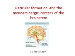
RF is formed of large number of neurons present through the entire brainstem it extends upward to the level of the thalamus and downward to be continuous with the interneurons of the spinal cord ID: 914836
Download Presentation The PPT/PDF document "RETICULAR FORMATION THE RETICULAR FORMAT..." is the property of its rightful owner. Permission is granted to download and print the materials on this web site for personal, non-commercial use only, and to display it on your personal computer provided you do not modify the materials and that you retain all copyright notices contained in the materials. By downloading content from our website, you accept the terms of this agreement.


Slide1
RETICULAR FORMATION
Slide2THE RETICULAR FORMATION
RF is formed of large number of neurons
present through the entire brainstem, it
extends upward to the level of the thalamus
and downward to be continuous with the
interneurons
of the spinal cord.
Slide3RETICULAR FORMATION
Slide4Slide5NUCLEI OF RETICULAR FORMATION
Slide6Numerous connections.
It is connected to almost all parts of the nervous system directly or indirectly.
The pathways involved are both:---
Ascending and descending
Crossed and uncrossed
Somatic and visceral.
It is NOT merely a relay station of these pathways.
It has an important REGULATORY role, both
facilitatory
and inhibitory.
CONNECTIONS OF RETICULAR FORMATION
Slide7AFFERENT CONNECTIONS
EFFERENT CONNECTIONS
DESCENDING PROJECTIONS
ASCENDING PROJECTIONS
RETICULAR PATHWAYS
Slide81.SPINAL CORD
Via the spino reticular tract and via collaterals from all ascending tracts.
2.BRAIN STEM
Afferents from the cranial nerves including vestibular.
3.TECTORETICULAR
(SUPERIOR AND INFERIOR COLLICULI)
CONVEYING VISUAL AND AUDITORY IMPULSES
AFFERENTS TO RETICULAR FORMATION
Slide94. CEREBELLUM
cerebelloreticular
5.BASAL ganglia
directly and indirectly
6.NEOCORTEX
corticoreticular
fibres
from the motor, sensory cortex, orbital, parietal and temporal lobes,
cingulate
gyrus and collaterals from the
corticofugal
fibres
.
7.LIMBIC SYSTEM
Including the
amygdaloid
, hippocampus
Slide10Efferent connections are:
1.To the spinal cord
The descending
reticulospinal
tracts (medial inhibitory and lateral
facilitatory
) connect with the anterior horn cells either directly or through
internuncial
neurons.
They also connect to the lateral horn cells which are the cells of origin of sympathetic nervous system.
EFFERENTS FROM RETICULAR FORMATION
Slide112.To brain stem
The reticulobulbar
fibres
connect to the cranial nerve motor nuclei.
3.To the cerebellum.
4.To the red
nucleus,substantia
nigra
and
tectum
in the midbrain.5.To the thalamus, subthalamus
and hypothalamus.
6.To the corpus striatum,
neocortex
and limbic system indirectly through the thalamus and hypothalamus
Slide12CORTICO-RETICULAR-SPINAL PATHWAYS
CEREBELLORETICULAR CONNECTIONS
ASCENDING RETICULAR ACTIVATING SYSTEM
CONNECTIONS OF RETICULAR FORMATION
Slide13The Reticular formation receives impulses from the motor and other areas of the cerebral cortex and relays them to the spinal cord through the MEDIAL and LATERAL RETICULOSPINAL TRACTS.
The
cortico
-reticular
fibres
descend along with
cortico
-spinal
fibres
.
They terminate mainly in relation to the oral and caudal reticular nuclei of the
pons
and the giganto-cellular nucleus of the medulla.
Cortico
-
Reticulo
-Spinal pathways
Slide14The Medial
Reticulo-spinal Tract originates from the oral and caudal pontine
reticular nuclei and the
gigantocellular
reticular nucleus of medulla.
Pontine
fibres
descend mainly
ipsilaterally
in the ventral
funiculus
of the cord.
Medullary fibres descend both ipsilaterally
and
contralaterally
in the ventral
funiculus
and the ventral part of the lateral
funiculus
.
These
fibres have many collaterals.
Slide15Two-thirds of these
reticulospinal neurons that reach the cervical cord also descend to lumbosacral
levels.
These
fibres
terminate widely in spinal grey
mater,but
the exact lamina of termination is controversial.
Majority of the terminals of medial
reticulospinal
fibres
are distributed to laminae six yo eight.
Slide16Alpha and Gamma motor neurons are influenced by
reticulospinal fibres through polysynaptic and monosynaptic connections.
Reticulo
-spinal
fibres
from
pontine
sources excite motor neurons of axial and limb muscles.
Medullary
fibres
excite , or inhibit motor neurons of cervical muscles and excite motor neurons of axial muscles.
Slide17Functionally Medial
Reticulospinal tract is concerned with posture, the steering of head and trunk movements in response to external stimuli, and crude, stereotyped movements of the limbs.
Slide18The Lateral
Reticulo-spinal Tract arises from the neurons of the ventrolateral
part of reticular formation of the
pons
(CAUDAL and ORAL
pontine
reticular nuclei).
The
fibres
cross to the opposite side of medulla oblongata and run in the lateral
funiculus
of the spinal cord.
Axons of this tract terminate in laminae one, five and six .
This pathway is involved in the control of pain perception and in motor functions.
Slide19RETICULO-CEREBELLAR FIBRES
The reticular formation receives
fibres
from and sends
fibres
to cerebellum.
Impulses passing from the cerebellum to the reticular formation are relayed to the spinal cord and to cranial nerve nuclei through
reticulospinal
and
reticulonuclear
pathways; and to the thalamus through
reticulothalamic
fibres.
Connections between cerebellum and reticular formation
Slide20The cerebellum receives
fibres mainly from three nuclei in the reticular formation.
1. Lateral reticular nucleus in the medulla
2.
Paramedian
reticular nucleus (lying in lower part of medulla in medial longitudinal fasciculus).
3.Nucleus
reticularis
tegmenti
pontis
.(NRTP)Paramedian reticular nucleus sends
fibres
to the entire
cerebellar
cortex.
The lateral and NRTP give collaterals to
cerebellar
nuclei(
fastigial
nucleus mainly).
Slide21Cerebellar
nuclei project to the lateral reticular nucleus and the NRTP.Fibres
to the lateral reticular nucleus are mainly from
fastigial
nucleus.
Some of these
fibrtes
reach the reticular formation through the descending branch of the superior
cerebellar
peduncle.
Fibres
from the
fastigial nucleus also reach the tegmentum of middle brain (including the dorsal
tegmental
nucleus,the
central grey)the
raphe
nucleus and the locus
coeruleus
.
Fibres
from the
dentate,emboliform
and
globose
nuclei end in the medial reticular formation of the
pons
and medulla and in the NRTP (
mainly from emboliform
nucleus)
CEREBELLO-RETICULAR CONNECTIONS
Slide22Various ascending tracts passing through the brainstem are intimately related to the reticular formation.
Many of the fibres
in these tracts give off collaterals to it.
These come from the
spinothalamic
tracts, from secondary trigeminal pathways and from auditory pathways.
These collaterals terminate predominantly in lateral reticular formation.
ASCENDING RETICULAR ACTIVATING SYSTEM (ARAS)
Slide23Fibres
arising here project to the intralaminar and reticular nucleiof
the thalamus.
These nuclei in turn project to widespread areas of the cerebral cortex.
These pathways form part of the ascending reticular activating system which is believed to be responsible for maintaining a state of alertness.
Slide24ASCENDING RETICULAR ACTIVATION SYSTEM - ARAS
Receives fibers from the sensory pathways via long ascending spinal tracts.
Alertness, maintenance of attention and wakefulness.
Emotional reactions, important in learning processes.
Tumor or lession – sleeping sickness or coma.
Slide25Major afferents of reticular formation
Slide26Major
efferents
of reticular formation
Slide27Control of somatic and visceral sensations
Control of ANS
Influence the biologic clock
The reticular activating system
Control of endocrine nervous system
Control of skeletal muscles
FUNCTIONS OF RETICULAR FORMATION
Slide28SOMATO MOTOR CONTROL
Reticular formation has an influence on fine control of movements including those involved in postural adjustments, skilled use of the
hands,speech
etc. through its direct connections with the spinal cord and indirectly through the corpus striatum, the cerebral cortex and the cerebellum
FUNCTIONS OF RETICULAR FORMATION
Slide29The reticular formation influences conduction through
somatosensory pathways.Similar effects may also be exerted on visual and auditory pathways.
SOMATOSENSORY CONTROL
Slide30Stimulation of certain areas in the reticular formation of the medulla has great influence on respiratory and cardiovascular function.
The region influencing respiratory activity corresponds approximately to the
gigantocellular
nucleus and
parvocellular
nucleus.
Stimulation of the
gigantocellular
nucleus and the upper part of the ventral reticular nucleus causes depression of vasomotor activity while stimulation of other areas has a
pressor
effect. These effects are mediated through connections between the reticular formation and autonomic
centres
in the brainstem and spinal cord,but
the pathways concerned are not well defined.
VISCERAL CONTROL
Slide31Reticular formation influences activity of the
adenohypophysis and of the neurohypophysis
through its connections with the hypothalamus.
It also influences the pineal body. pineal gland secretes the hormone melatonin which shows a marked circadian rhythm which appears to be strongly influenced by exposure of animal to light.
Activity is greater in darkness.
NEUROENDOCRINE CONTROL
Slide32Reticular formation controls arousal and the state of consciousness through the ARAS.
ARAS is also known as extrathalamic
control
modulatory
system or simply reticular activating system (RAS).
RAS is a collection of different nuclei- more than 20 on each side in the upper
brainstem,the
pons
, medulla and posterior hypothalamus.
The most significant components of the ARAS include;-
Slide33Serotonergic
nuclei-dorsal raphe nucleus and median
raphe
nucleus (RAPHE NUCLEI).
Dopaminergic
nuclei-ventral
tegmental
area
Noradrenergic nuclei-Locus
coruleus
Histaminergic
nuclei-
tuberomammillary nucleus
Cholinergic nuclei-
pontine
tegmental
nuclei
Slide34DESCENDING RETICULAR ACTIVATION SYSTEM - DRAS
INHIBITORY
Smoothness and accuracy of voluntary movements;
Reflex movements;
Regulates muscle tone;
Maintenance of posture;
Control
of
vegetative functions.
FACILITATORY
Ma
i
ntains
the muscle tone;
Facilitates autonomic functions;
Activates ARAS.
Slide35REGULATION OF SLEEP
, thus, the maintenance of the
SLEEPING cycle or CIRCADIAN
rhythm
;
Filtering of incoming stimuli to discriminate irrelevant background stimuli;
It’s crucial to maintain the state of
CONSCIOUSNESS
related to the circadian rhythm –
MELATONIN effects on RAS
;
ANS control
– respiratory rate, heart rate, GIT activity.
Slide36DISORDERS ASSOCIATED WITH RETICULAR FORMATION
NARCOLEPSY
Associated with excessive sleepiness, sleep paralysis, hallucinations and in some cases episodes of cataplexy (loss of muscle control often triggered by strong emotion such as laughter.
SCHIZOPHRENIA
Mental disorder
charecterised
by abnormal social
behaviour
and failure to understand what is real.
There is
overactivity
of reticular formation.
Slide37PARKINSONS DISEASE
It is degenerative disorder characterized by shaking rigidity, slowness of movement and difficulty with walking.
This is because of decrease in dopamine in the area of
substantia
nigra
of midbrain.