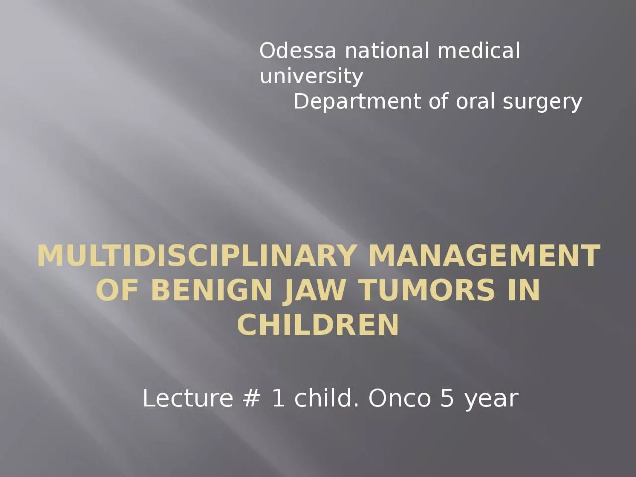
Children Lecture 1 child Onco 5 year Odessa national medical university Department of oral surgery A tumor is defined in brief as abnormal growth of tissue tumoral formations are classified ID: 1038842
Download Presentation The PPT/PDF document "Multidisciplinary Management of Benign J..." is the property of its rightful owner. Permission is granted to download and print the materials on this web site for personal, non-commercial use only, and to display it on your personal computer provided you do not modify the materials and that you retain all copyright notices contained in the materials. By downloading content from our website, you accept the terms of this agreement.
1. Multidisciplinary Management of Benign Jaw Tumors inChildrenLecture # 1 child. Onco 5 yearOdessa national medical university Department of oral surgery
2. A tumor is defined, in brief, as abnormal growth of tissue; tumoral formations are classifiedunder two main headings, benign and malignant. The oro-facial region including the jawbones, maxilla and mandible, is a site for a multitude of neoplastic conditions. Odontogenictumors (OTs) constitute a wide range and diverse kind of lesions derived from tooth formingapparatus and its remnants. OTs originate from epithelium or ectomesenchyme or from both,showing varying degrees of inductive interaction between these embryonic components of thedeveloping tooth germ.
3. Etiology of odontogenic tumors
4. According to current literatures, it is known that the potential sources for development of an odontogenic tumor are varied, and these include:
5. 1. The pre-functional dental lamina (odontogenic epithelium with ability to produce atooth), which is more abundant distal to the lower third molars.2. The post-functional dental lamina, a concept that covers those epithelial remnants suchas Serre´s epithelial rests, located within the fibrous gingival tissue; the epithelial cell restsof Malassez in the periodontal ligament and the reduced enamel organ epithelium, whichcovers the enamel surface until tooth eruption.
6. 3. The basal cell layer of the gingival epithelium, which originally gave rise to the dentallamina.4. The dental papilla, origin of the dental pulp, which has the potential to be induced toproduce odontoblasts and synthesize dentin and/or dentinoid material.
7. 5. The dental follicle.6. The periodontal ligament, which has the potential to induce the production of fibrousand cemento-osseous mineralized material
8.
9. From a biological point of view, some of these lesions represent hamartomas with varying degrees of differentiation, while the rest are benign or malignant neoplasms with variable aggressiveness and potential to develop metastases. These tumors constitute a heterogeneous group of diseases with diverse clinical and histopathological features
10. Primary jaw tumors are broadly classified into odontogenic and nonodontogenic groups. The World Health Organization (WHO) classified this group of lesions in 1971 and 1992. In 2005, the WHO published the latest updated edition of the classification of OTs. There were 6 major changes in this schema from the previous versions namely:
11. 1. parakeratinized variant of odontogenic keratocyst is now classified as a benign tumor and termed KCOT2. adenomatoid odontogenic tumor (AOT) originates from the odontogenic epithelium with mature fibrous stroma and without ectomesenchyme
12. 3. calcifying odontogenic cyst (COC) is divided into 2 benign and 1 malignant groups4. clear cell odontogenic tumor is a malignant lesion and termed clear cell odontogenic carcinoma (CCOC)
13. 5. odontogenic carcinosarcoma is not included due to the lack of evidence for the existence of this type and6. some changes were made regarding terminology and subtypings
14. Classification of pediatric jaw tumorsThe various classifications systems proposed by authors are enumerated as below:1. Jaw Tumors in Children1.Classification of non-odontogenic jaw tumors in children
15. I. Benign mesenchymal tumorsa) Giant cell lesionsb) Fibro-osseous lesionsc) Myxoma
16. II. Hematopoietic and reticuloendothelial tumorsa) Langerhans cell histiocytosisb) Burkitt’s lymphomac)Lymphoma
17. III. Neurogenic tumorsa) Neurofibromab) Neurilemmomac) Neuromad)Ganglioneuromae) Neuroblastomaf)Melanotic neuroectodermal tumor
18. IV. Vascular lesionsa)Vascular malformation (capillary, lymphatic, venous, arterial, combined)b)Hemangiomac)Aneurysmal bone cyst
19. V. Malignant mesenchymal tumorsa) Osteogenic sarcomab) Chondrosarcomac) Fibrosarcomad) Ewing’s sarcoma
20. VI. Malignant epithelial tumorsa)Squamous cell carcinomab)Mucoepidermoid carcinomac)Adenoid cystic carcinomad)Adenocarcinoma
21. 2. Classification of odontogenic jaw tumors in children
22. I. Epithelial tumorsa) Ameloblastoma (Peripheral, Unicystic, Solid, Multicystic)b) Adenomatoid odontogenic tumorc) Calcifying epithelial odontogenic tumor
23. II. Mesodermal tumorsa)Cementomab)Periapical cemental dysplasiac)Cementifying fibromad)Cementoblastomae)Odontogenic fibroma
24. III. Mixed tumorsa)Ameloblastic fibromab) Odontoma
25. 3. Classification of Small Round Cell Tumors in children
26. 1. Soft tissue RhabdomyosarcomaSoft tissue (extraosseous) Ewing’s sarcomaHemangiopericytoma
27. 2. Osseous Ewing’s sarcomaSmall cell osteosarcomaMesenchymal chondrosarcomaHemangiopericytoma of bone
28. 3. NeuralNeuroblastomaPeripheral neuroectodermal tumors- Askin’s tumor- NeuroepitheliomaPheochromocytoma
29. 4. International classification of childhood cancer
30. Malignant Bone tumorsa) Osteosarcomasb) Chondrosarcomasc) Ewing tumor and related sarcomas of boned) Other specified malignant bone tumorse) Unspecified malignant bone tumors
31. Soft tissue and other extraosseous sarcomasa) Rhabdomyosarcomasb) Fibrosarcomas, peripheral nerve sheath tumors and other fibrous neoplasmsc) Kaposi’s sarcomad) Other specified soft tissue sarcomase) Unspecified soft tissue sarcomas
32. Clinical sign and symptoms of pediatric jaw tumors
33. Pediatric patients encompass a very interesting study group, as several long term physiological changes take place in the maxillofacial area. During the mixed dentition period, children can refer with a complaint of swelling in the maxillofacial area, which may or may not be associated with pain. These mostly include both hard and soft tissue pathologies.
34. When involving bone, only odontogenic cysts or odontogenic tumors as a category have been considered. Intraosseouspediatric jaw lesions can present in diverse clinical patterns and their diagnoses can vary from odontogenic to non-odontogenic pathogeneses, which can rarely include connective tissue pathology. The great majority of pediatric jaw tumors are non-odontogenic
35. During the mixed dentition period, children can report with complaint of swelling in the maxillofacial area, which may or may not be associated with pain. A history of trauma also needs to be elicited, because they are prone to falling down during playing, which can affect the jaws. Because of the complex anatomy and development of the head and neck, neoplasms during infancy and childhood arising at this site represent the most difficult challenges in clinical practice.
36. Diagnosis of pediatric jaw tumors
37. The odontogenic cysts and tumors are a diverse group of lesions that represent deviation from normal odontogenesis. The physical signs and symptoms of odontogenic cysts and tumors will depend to a certain extent on the dimensions of the lesion. A small lesion is unlikely to be diagnosed on a routine examination of the mouth because signs will not be demonstrable. Such lesions are only likely to be detected at an early stage as the result of routine radiographic examination.
38. Clinically evident expansion is often a late finding, especially in lesions developing within the ramus or angle of the mandible or within the maxillary sinus. Lesions in these areas may become extremely large before expansion is observed clinically. Masses in the neck confront the pediatrician with greater opportunities for evaluation before a decision regarding biopsy or excision is reached. Signs of systemic involvement must also be determined
39. Radiographic and imaging studies
40. The primary goals of radiographic assessment are to more precisely define the primarylesion and to detect metastatic disease for clinical staging• Chest radiographs are useful screens for mediastinal lymphadenopathy• Ultrasound is able to differentiate a solid from a cystic mass, and give general relationshipsof the mass to adjacent structures
41. • Axial and coronal computerized tomography (CT) allows documentation of bone erosion and invasion of adjacent structures• Magnetic resonance imaging (MRI) offers improved tissue contrast and definition
42. • Angiography delineates the blood supply to a lesion, and offers the ability to embolize specific factors to decrease blood loss associated with excision of vascular lesions• Bone scans and liver spleen scans offer modalities to detect systemic disease. A tissue diagnosis becomes necessary in order to diagnose and initiate proper therapy.
43. Biopsy
44. • Biopsy of OTs allows histologic evaluation of the mass• Excisional biopsy is often therapeutic as well as diagnostic• Incisional biopsy is required in cases where the lesion is large, or the lesion is relativelyinaccessible
45. • Fine needle aspiration for cytologic study is useful in salivary gland and thyroid gland lesions. However, its generalized use for all pediatric head and neck masses is limited due to the rarity of squamous or glandular neoplasms developing in children• Large bore needle biopsy has no established role in the evaluation of head and neck malignancies in children and has been reported to cause seeding along the needle tract in children.
46. Odontogenic tissue is programmed to produce dentin and enamel due to active interactions between odontogenic mesenchyme and epithelium. Tooth formation is achieved viaodontogenic mesenchyme and epithelium stage- and spatial-specific differentiation from early tooth development to late maturation
47. Treatment of pediatric jaw tumors
48. Treatment consists of a range of surgical methods, from surgical curettage to hemimandibulectomy and reconstruction with bone graft. Generally, surgical excision, curettage, cryosurgery or en bloc resection are adequate for treatment of these tumors. However, some patientsneed multiple treatment because of its specific criterias such as the clinical behavior and extentof the lesion.
49. Odontogenic lesions encompasses a wide spectrum of lesions and their variants, which either can be a cyst or a tumor. Odontogenic cysts are derived from the epithelium associated with the development of the dental apparatus while a tumor forms through someaberration from the normal pattern of odontogenesis. But the fact, that these lesions can mimiceach other can complicate the diagnosis.
50.
51.
52.
53.
54.
55.
56.
57.
58.
59.
60.