
Limb Mgr Veronika Mrkvicová physiotherapist Examination Methods in Rehabilitation 26102020 Nerves of the Lower Limb The Lumbar Plexus Iliohypogastricus ID: 914970
Download Presentation The PPT/PDF document "Nerves of the Lower" is the property of its rightful owner. Permission is granted to download and print the materials on this web site for personal, non-commercial use only, and to display it on your personal computer provided you do not modify the materials and that you retain all copyright notices contained in the materials. By downloading content from our website, you accept the terms of this agreement.


Slide1
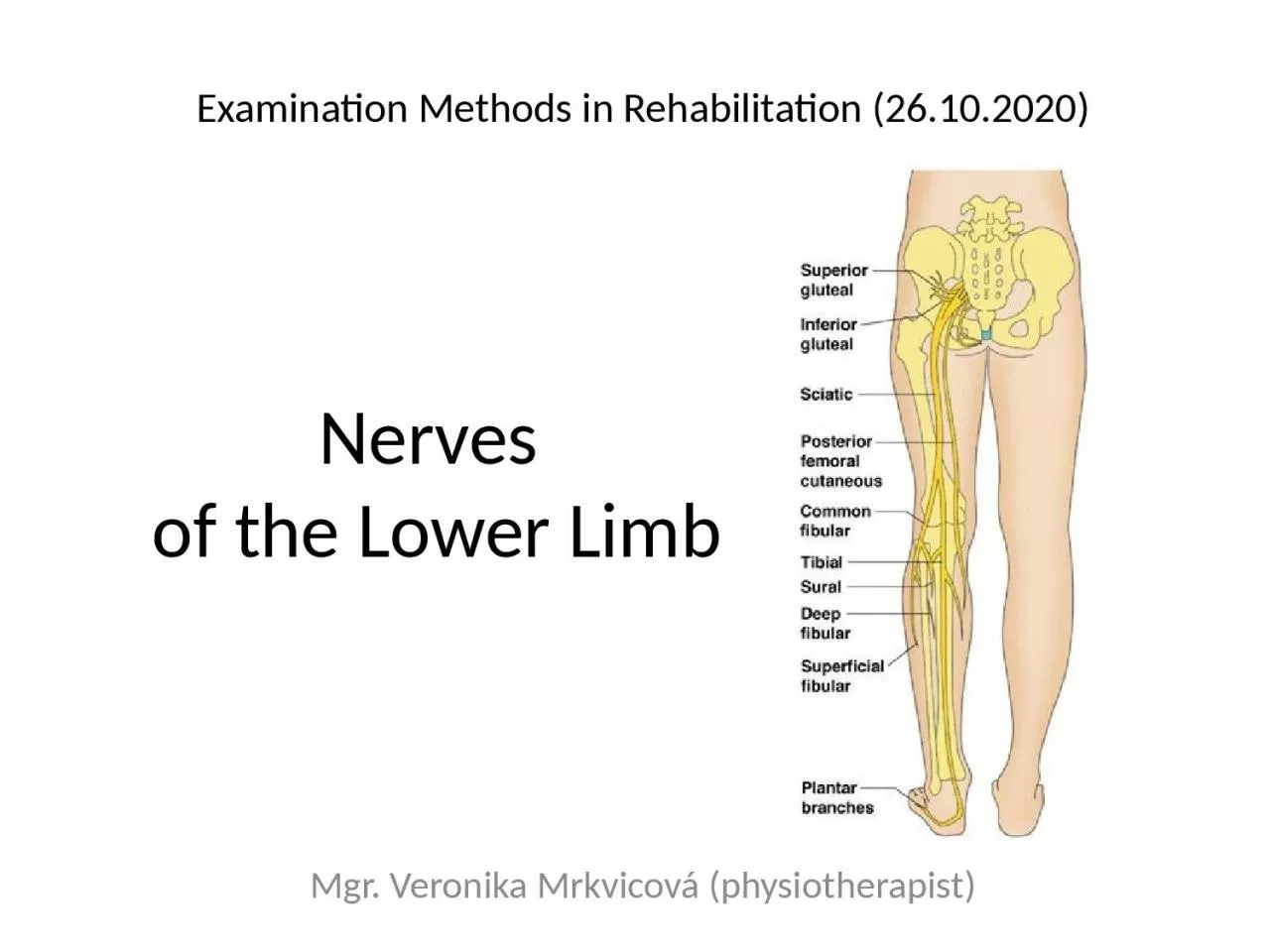
Nerves of the Lower Limb
Mgr. Veronika Mrkvicová (physiotherapist)
Examination Methods in Rehabilitation (26.10.2020)
Slide2Nerves of the Lower
LimbThe Lumbar Plexus
Iliohypogastricus nerveIlioinguinalis nerveLateral Cutaneous Femoral
nerve
Obturator
nerve
Femoral
nerve
The
Sacral
Plexus
Sciatic
nerve
Tibial
nerve
Common
Peroneal
nerve
Slide3Spinal Nerves
Slide4Slide5The Lumbar Plexus
Slide6Slide7Slide8The Lumbar Plexusa nervous plexus in the lumbar region of the body
which forms part of the lumbosacral plexusit is formed by the divisions of the four lumba
r nerves (L1-L4) and from contributions of the subcostal nerve (T12)additionally, the ventral rami of the fourth lumbar nerve pass communicating branches, the
lumbosacral
trunk, to the sacral plexus
t
he nerves of the lumbar plexus pass in front of the hip joint and mainly support
the anterior part of the thigh
Slide9The Lumbar Plexus
it is formed lateral to the intervertebral foramina and passes through psoas majorits smaller motor branches
are distributed directly to psoas majorwhile the larger branches leave the muscle at various sites to run obliquely downward through the pelvic area to leave the pelvis under the inguinal ligamentwith the exception of the
obturator
nerve which exits the pelvis through the
obturator
foramen
Slide10The Iliohypogastric Nerve
it runs anterior to the psoas major on its proximal lateral border to run laterally and obliquely on the anterior side of quadratus lumborumlateral
to this muscle, it pierces the transversus abdominis to run above the iliac crest between that muscle and abdominal internal obliqueit gives off several motor branches to these muscles and a
sensory branch
to the skin of the lateral hip
i
ts
terminal branch
then runs parallel to the inguinal ligament to exit the
aponeurosis
of the abdominal external oblique above the external inguinal ring where it supplies the skin above the inguinal ligament (i.e. the
hypogastric
region) with the
anterior
cutaneous
branch
Slide11The Ilioinguinal Nerveit
closely follows the iliohypogastric nerve on the quadratus lumborum, but then passes below it to run at the level of the iliac crestit pierces the lateral abdominal wall and runs medially at the level of the inguinal ligament where it supplies
motor branches to both transversus abdominis and sensory branches through the external inguinal ring to the skin over the pubic
symphysis
and the lateral aspect of the labia
majora
or scrotum
Slide12The Genitofemoral Nerve
it pierces psoas major anteriorly below the former two nerves to immediately split into two branches that run downward on the anterior side of the musclethe lateral femoral branch
is purely sensory. It pierces the vascular lacuna near the saphenous hiatus and supplies the skin below the inguinal ligament (i.e. proximal, lateral aspect of femoral triangle)the genital branch differs in males and femalesi
n males it runs in the spermatic cord and in females in the inguinal canal together with the
teres
uteri ligament
i
t then sends
sensory branches
to the scrotal skin in males and the labia
majora
in females. In males it supplies motor
innervation
to the
cremaster
Slide13Genitofemoral nerve paralysismost commonly caused by surgical trauma
other causes reported include direct trauma to the inguinal region and tight clothingSymptomsa pain and a burning sensation in the groin, which radiates to the inner thigh (aggravating factors including walking, stooping and hyperextension of the hip)
tenderness and possible hyperaesthesia along the inguinal canalprovocative testing involves internal or external rotation of the hip joint
Slide14The lateral cutaneous femoral nerve it
pierces psoas major on its lateral side and runs obliquely downward below the iliac fasciamedial to the anterior superior iliac spine it leaves the pelvic area through the lateral muscular lacuna it enters the thigh by passing behind the lateral end of the inguinal ligament
in the thigh it briefly passes under the fascia lata before it breaches the fascia and supplies the skin of the anterior thigh
Slide15The lateral cutaneous femoral nerve
Involvement of the lateral cutaneous branch of the nerve may produce:
- painful paraesthesiae of the thigh (meralgia paraesthetica)
- m
ild
pain near the inguinal ligament may be experienced
Slide16The obturator nerveit
leaves the lumbar plexus and descends behind psoas major on it medial side, then follows the linea terminalis into the lesser pelvis, and finally leaves the pelvic area through the obturator canal
in the thigh, it sends motor branches to obturator externus before dividing into an anterior and a posterior branch, both of which continues distally
t
hese
branches are separated by adductor
brevis
and supply
all thigh adductors with motor
innervation
:
pectineus
, adductor
longus
, adductor
brevis
, adductor
magnus
, adductor
minimus
, and
gracilis
t
he anterior branch contributes a terminal,
sensory branch
which passes along the anterior border of
gracilis
and supplies the skin on the medial, distal part of the thigh
Slide17The femoral nerveit is the largest and longest of the plexus' nerves
it gives motor innervation to iliopsoas, pectineus,
sartorius, and quadriceps femorisand sensory innervation to the anterior thigh, posterior lower leg, and hindfoot
i
n the pelvic area, it runs in a groove between
psoas
major and
iliacus
giving off branches to both muscles, and exits the pelvis through the medial aspect of muscular lacuna
i
n the thigh it divides into
numerous sensory and muscular branches
and the
saphenous
nerve
, its long sensory terminal branch which continues down to the foot
Slide18Slide19Slide20Femoral nerve paralysisSymptoms:
Slide21Obturator nerve paralysisSymptoms:
hypoaesthesia, paraesthesia or pain in the medial thigh, groin or pubic boneweakness and a feeling of leg instabilityweakness or wasting of the adductor muscles and a decrease in hip adduction and internal rotation of the hipa circumducting gait secondary to an externally rotated hip
Slide22The Sacral Plexus
Slide23Slide24Slide25The Sacral Plexusit
is a nerve plexus which provides motor and sensory nerves for the posterior thigh, most of the lower leg and foot, and part of the pelvisit is part of the lumbosacral plexus and emerges from the lumbar vertebrae and sacral vertebrae (L4-S4)
a sacral plexopathy is a disorder affecting the nerves of the sacral plexus, usually caused by trauma, nerve compression, vascular disease, or infection. Symptoms may include pain, loss of motor control, and sensory deficits
Slide26The Sacral Plexus
The sacral plexus is formed by:the lumbosacral trunkthe anterior division of the first sacral nerveportions of the anterior divisions of the second and third sacral nervesThe nerves forming the sacral plexus converge toward the lower part of the greater sciatic foramen, and unite to form a flattened band, from the anterior and posterior surfaces of which several branches arise
The band itself is continued as the sciatic nerve, which splits on the back of the thigh into the tibial nerve and common fibular nerve; these two nerves sometimes arise separately from the plexus, and in all cases their independence can be shown by dissection
Slide27Slide28Slide29Slide30Slide31Tibial nerve paralysisSymptoms
:Sensation changes on the bottom of the footNumbness, tingling, or other abnormal sensationsBurning sensationPainWeakness of the knee or foot, difficulty with walking
Slide32Slide33Slide34Peroneal nerve paralysis
SymptomsNumbness or tingling on the anterior side or on top of the footReduced sensation to touchWeakness with lifting the foot in an upward direction and turning it outwardsLoss of function of the footSevere cases of
peroneal nerve injury results in footdrop meaning the inability of a person to lift the foot up when ambulatingPresence of a slapping gait where the foot slaps on the ground during ambulation due to inadequate control over muscles
Slide35Peroneal nerve paralysisExamination of the legs may show:
Loss of muscle control in the lower legs and feetAtrophy of the foot or foreleg musclesDifficulty lifting up the foot and toes and making toe-out movements
Slide36Slide37Slide38E-sources, literature:
https://en.wikipedia.org/wiki/Lumbar_plexushttps://en.wikipedia.org/wiki/Sacral_plexus http://accessphysiotherapy.mhmedical.com/data/Multimedia/grandRounds/lumbar/media/lumbar_print.html http://antranik.org/peripheral-nervous-system-spinal-nerves-and-plexuses/
http://www.gpnotebook.co.uk
Slide39Thank you for your attention