
By Rasha Mohamed Elshinety Ass Prof of Anatomy amp Embryology Prostate It is a firm partly glandular and partly muscular organ Site In the pelvic cavity between lower border of symphysis pubis anteriorly and ampulla of rectum posteriorly ID: 1046197
Download Presentation The PPT/PDF document "Prostate, Seminal Vesicles & Ejacula..." is the property of its rightful owner. Permission is granted to download and print the materials on this web site for personal, non-commercial use only, and to display it on your personal computer provided you do not modify the materials and that you retain all copyright notices contained in the materials. By downloading content from our website, you accept the terms of this agreement.


1. Prostate, Seminal Vesicles & Ejaculatory DuctsBy,Rasha Mohamed ElshinetyAss. Prof. of Anatomy & Embryology
2. Prostate- It is a firm partly glandular and partly muscular organ.Site: In the pelvic cavity, between lower border of symphysis pubis anteriorly and ampulla of rectum posteriorly
3. • Shape: Conical in shape and presents a base, an apex, and an anterior, posterior, and 2lateral surfaces.• The base: directed upwards, surrounds the neck of urinary bladder, and penetrated by the urethra.• Apex: directed down, and in direct contact with the superior fascia of the urogenital diaphragm.• Posterior surface:lies in direct contact with the ampulla of the rectum and can be palpated by per-rectal (P/R) examination in the living • Anterior surface: connected to the pubic bone by the puboprostatic ligament. The urethra emerges from this surface above and in front of the apex of the gland.• Inferolateralsurfaces (one on each side):related to the 2 free borders of the levator ani muscle. The most anterior part of this muscle (Levatore prostatæ) passes backwards from pubis and embrace the sides of the prostate to reach the perineal body.
4.
5. Structures inside the prostate:1. Prostatic urethra.2. Ejaculatory ducts; run down and forwards one on each side to open into the prostatic urethra.3. Prostatic utricle: extends up and backwards from the prostatic urethra into the median lobe.
6. Anatomical lobes of the prostate:1.Median lobe: lies behind the prostatic urethra, and bounded by an ejaculatory duct on each side.- It projects up behind the internal urethral orifice to form the uvula of the bladder.2- Right and Left lateral lobes: lie on each side of the prostatic urethra.3. Anterior lobe (isthmus): connects the lateral lobes in front of the urethra and contains few glandular tissue.4. Posterior lobe.
7. Prostatic capsule1. Inner true capsule: a firm fibromuscular adherent to the prostate substance.2. Outer false capsule (prostatic sheath): formed by pelvic fascia.Prostatic venous plexus is situated between the 2 capsules.
8. • Arterial supply: inferior vesical and middle rectal arteries.• Venous drainage: prostatic venous plexus Drains into the internal iliac vein. This venous plexus is also connected with the vertebral venous plexus by valveless veins (this is how prostate cancer spreads to the vertebrae).• Lymph drainage: sacral and internal iliac lymph nodes.
9.
10. Benign enlargement of the prostate is common in men older than 50 years (senile enlargement). The median lobe of the gland enlarges upward and encroaches within the sphincter vesicae, located at the neck of the bladder.The leakage of urine into the prostatic urethra causes an intense reflex desire to micturate.The enlargement produces distortion of the urethra so that the patient experiences difficulty in passing urine and the stream is weak.
11. The enlargement of the uvula vesicae (owing to the enlarged median lobe) results in the formation of a pouch of stagnant urine behind the urethral orifice within the bladder. The stagnant urine frequently becomes infected, and the inflamed bladder (cystitis) adds to the patient's symptoms
12. Vas DeferensIs a thick walled cord-like duct, it has a very narrow lumen.Beginning:at the lower end of the tail of the epididymis.Course: - It ascends along the posterior border of the testis, medial to epididymis.- It runs up in the posterior part of the spermatic cord, where it is surrounded by the pampiniform venous plexus.- It enters the inguinal canal, within the spermatic cord.- Enters the pelvis by passing through the deep inguinal ring, here it curves lateral to the inferior epigastric artery
13. - It descends along the lateral pelvic wall crossing obturator vessels and nerves, obliterated umbilical artery, and external iliac vessels.- Close to the base of urinary bladder, it crosses the front of the terminal part of the ureter.- It presents a dilated ampulla which lies medial to the seminal vesicle behind the base of the urinary bladder.- At the base of prostate it narrows and joins the duct of the seminal vesicle to form the ejaculatory duct.
14. Arterial supply: Artery of vas, which arises from the superior or inferior vesical artery.
15.
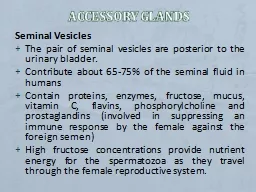
16. Seminal vesicles There are 2 large, sacculated pouches, each is about 5cm long- Site: on the base of the urinary bladder, anterior to the rectum, and lateral to the vas deferens.- Its lower end joins the vas deferens to form the ejaculatory duct.- Its lower end lies anterior to the terminal part of the ureter.- Arterial supply: inferior vesical and middle rectal arteries.
17.