
239 Corresponding Department of Speech Therapy School of Paramedical Sciences Mashhad University of Medical Sciences Mashhad IranTel 98 513 8846711The basal ganglia BG controls different ID: 955792
Download Pdf The PPT/PDF document "Role of Basal Ganglia in Swallowing Proc..." is the property of its rightful owner. Permission is granted to download and print the materials on this web site for personal, non-commercial use only, and to display it on your personal computer provided you do not modify the materials and that you retain all copyright notices contained in the materials. By downloading content from our website, you accept the terms of this agreement.


239 Role of Basal Ganglia in Swallowing Process, Zahra Ghayoumi AnarakiTherapy, School of Rehabilitation, Tehran University of Medical Sciences, Tehran, Iran. 2. Department of Speech Therapy, School of Department of Speech Therapy, School of Rehabilitation Sciences, Hamadan University of Medical Sciences, Hamadan, Iran. * Corresponding Department of Speech Therapy, School of Paramedical Sciences, Mashhad University of Medical Sciences, Mashhad, Iran.Tel: +98 (513) 8846711The basal ganglia (BG) controls different patterns of behavior by receiving Basal ganglia, Swallowing, Ghaemi H, Sobhani-Rad D, Arabi A, Saifpanahi S, Ghayoumi AnarakiZ. Role of Basal Ganglia in Swallowing Process. Iranian Journal of Rehabilitation. 2016; 14(4):239-246. https://doi.org/10.18869/nrip.irj.14.4.239 : : https://doi.org/10.18869/nrip.irj.14.4.239 CrossMark I ranian Rehabilitation Journal Use your device to scan and read the ar�cle online1. Introduction wallowing is de�ned as a complex sensonated function of muscles located around the mouth, tongue, larynx, pharynx, and esophagus in order to transport food from . The volitional and auThe volitional and au, could be divided into three inter-related physiological stages: the oral stage that is voluntary and highly variable in the pharyngeal stage during which trans-location of bolus occurs from the oropharynx (throat) esopha-geal stage in which the bolus moves through the lower December 2016, Volume 14, Number 4 Sobhani-Rad D, et al. Role of Basal Ganglia in Swallowing Process: A Systematic Review. IRJ. 2016; 14(4):239-246.esophageal sphincter into the stomach under the control of autonomic nervous system [5-10]. Accurate control of the swallowing function relies on the exact innervations of several areas in the brain, including the neo-cortex, sub-cortical regions, brainstem, and peripheral nervous [11]. The brainstem swallowing center is known as the �rst level of swallowing control while the subcormus, amygdala, and tegmental area of the midbrain, are in the next levels, and �nally, there are supra-bulbar cor .BG is a group of interconnected nuclei including striatum (further subdivided into putamen and caudate nuclei), Sub-Thalamic Nucleus (STN), globus pallidus external and internal segments (GPe and GPi, respecticulate (SNc and SNr, respectively). BG plays a vital role in a variety of motor, cognitive, and limbic functions by integrating the information derived from multiple cortical regions and conveying it back to frontal cortical . Although varied investigations have clari�ed the various functions of the BG, its exact role in the swallowing stance, a number of reports have indicated that damages and lesions of BG might lead to some degree of swallowing disorders; but, little has been discussed about the probable mechanisms and pathways in this aspect. Hence, we decided to conduct a systematic review based on a number of c
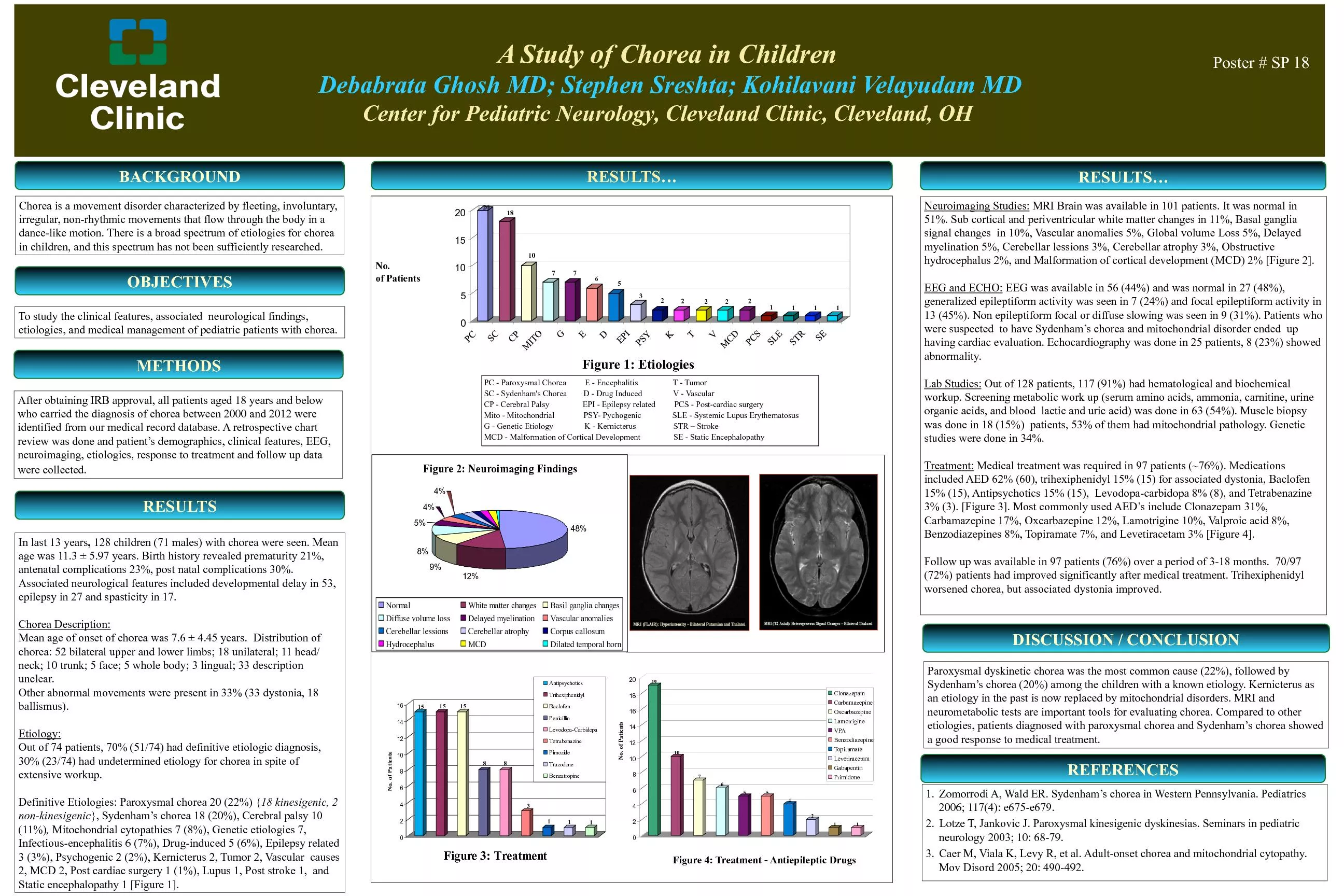
linical studies for a better understanding of BG’s function in swallowing.lished up to December 2015 and included keywords like swallowing, basal ganglia, swallowing neurology, neuro-imaging, dysphasia, and neurogenic dysphasia in their title or abstract were extracted from databases such as PubMed, Willy, Springer, and Elsevier (Medline, EBMR, Google Scholar, Science Direct, and ProQuest). cortical or BG relevant to swallowing disorders” were �ltered out in accordance with our pre-decided inclusion criteria. Conversely, papers with no focus on BG and tensive data published on swallowing neurology, only a few studies have focused on the topic considered here. For instance, the role of BG in the process of swallowing , stroke [16-18], dementia, and traumatic brain injury [19, . Some studies have also focused on the role of the extra-pyramidal syndrome, such as Parkinson’s, Huntington’s, and Wilson’s diseases in the swallowing pros diseases in the swallowing pro. In addition, there are a few reports on the . These limited publications indicate that swallowing is a sophisticated process with several unknown aspects. ments in neuroimaging techniques have improved our lowing. As summarized in Table 1, neuroimaging studies have con�rmed the fact that swallowing involves the activation of multiple areas in the human brain, including rimotor, supplementary motor, pre-motor, associative and limbic cortices through functionally related loops , and such somatotopic organization supports . BG receives input from sensorimotor areas of the cerebral cortex, wesultsImaging TechniqueReferencesRecognized swallow-associated augmented blood in the putamenHartnick et al. t al. wise in swallow-induced local ac�vity in putamen, globus pallidus, and substan�a nigraf-MRISuzuki et al. t al. The right putamen was introduced as a focal point for ac�va�onf-MRIaar�n et al. -MRIDiscovered .G ac�va�on through water oral combina�onf-MRI PETIamdy et al. 240 241 Sobhani-Rad D, et al. Role of Basal Ganglia in Swallowing Process: A Systematic Review. IRJ. 2016; 14(4):239-246. Figure 1. Cortical and subcortical functional loops. Striatum including GABAergic neurons (red), as globus pallidus externa (GPe) too, globus pallidus interna (GPi) and tions of neurons to tectum and brainstem motor centers. The indirect loop is represented by the GPe, the subthalamic nucleus (STN), and the output level (SNr/GPi). The striatal neurons of the direct pathway to SNr/GPi express D1R and substance P (D1/SP) while the indirect pathway neurons in striatum express D2R and enkephalin (D2/Enk). which are primary and secondary somato-sensory, primary motor cortex, and premotor areas premotor areas 38]. In ad-dition, BG has extensive connections with the thalamus 40]. As presented in Figure 1, BG (striatum) receives input from all cortical
areas and projects through the thalamus to prefrontal, premotor, and supplementary motor areas that are involved in motor planning. In the BG-thalamo-cortical circuits, the thalamus acts as a sensory-relay station and conveys information about the sensation of eating and swallowing to other cortical and sub-cortical structures . Both voluntary and involuntary movements happen-ing during ingestion are further modi�ed by the feedbacks received from kinesthetic images and other afferents converging onto the BG, which further monitor and re�ne the movement progression in order to ensure the temporal and spatial accuracy accuracy . Finally, BG noti�es about the movement-related cortices involved in the preparation for next .4. DiscussionSwallowing is a multi-faceted performance with active els. Studies on functional magnetic resonance imaging (fMRI) have recognized the anatomic sections, which gulate cortex, insula, operculum, prefrontal and inferior . As mentioned earlier, automatic movements of swallowing are controlled by BG that establishes accurate timing 38]. Although the rhythmic pattern of mastication is con-trolled by the central pattern generator in the brainstem, programmed movement patterns based on expectations and sensory feedbacks in conjunction with BG. The production level of BG includes active GABAergic neurons arising from two nuclei, GPi and SNr. The sub-populations of GABAergic neurons from the structure have outgic neurons from the structure have out. As these neurons are tonically energetic at rest, . Therefore, starting a motor program similar to swal-lowing depends on the elimination of such tonic reti-cence; hence, the pallidal output neurons must be inhib-ited from the input layer of BG [51, 53, 57-59]. Beyond jected by pallidum returns to the groups of cells inside the thalamus and are further projected back to the cortex. The pallido-thalamo-cortical loop controls emotions, . It is kinson’s, the cortex is out of the loop, and hence, all the actions of BG are done straightforwardly over brainstem targetsgets.It is also essential to review the BG-nuclei that man-age the output level as discussed in previous studies. , the projection neurons in the striatum are divided into two groups. Firstly are the tion level (SNr/GPi) and get involved in the beginning of motor programs of swallowing, and secondly are the dopamine D2 receptors (D2R) that project to GPe - a neural structure that contributes with STN and restrain . The subpopulation of D1R projected neurons control mus and cortex/pallium, which further veri�es whether rons will participate in the instigation of a given motor 65]. The tonic level of dopamine discharge resolves the responsiveness of the striatal neurons, and thus, the negligible dopamine makes it served in Parkinson’s disease s disease . Dopamine neurons of BG have another characteristic importance in the motor tant in promo
ting the knowledge of swallowing behavior 68]. Gespite their signi�cance, our understanding of neural circuits, which are responsible for the value-based changes in dopamine discharge, is not complete ge, is not complete . It is, therefore, important to mention the dissimilar grams. The swallowing process depends on the input from pallium/cortex, thalamus and the dopamine system rect pathways (D2). The production cells from GPi and 71]. The selection of speci�c units depends on the excitatory input from thalamus and pallium/cortex along with the degree of tonic dopamine activity, which collectively de�ne the prototype of BG’s behavior. In addition varied other motor patterns can be shared, for instance, one can swallow and chew simultaneously while one can only turn left or Sobhani-Rad D, et al. Role of Basal Ganglia in Swallowing Process: A Systematic Review. IRJ. 2016; 14(4):239-246. 242 243 Therefore, mechanisms must be discovered for governing different behaviors of BG. Nevertheless, it is obvious that the BG plays a key role in making a �at series of move . Therefore, movement skills are compromised in patients with Parkinson’s disease, and there is a propens disease, and there is a propen.In this review study, we aimed at clarifying the role of the BG in the swallowing process for the �rst time. Reviewing of the published data on neurological control in swallowing process revealed that BG is one of the most complicated neurological structures, partially due to its location in the brain with indistinct performances. On the other hand, few published studies have focused on neurological aspects of swallowing, indicating that it is a sophisticated process with several unknown aspects. However, with the help of neuroimaging techniques, it has been con�rmed that BG is linked to neural structures that support motor and cognitive functions such as the one involved in swallowing. BG receives input from all the cortical areas and projects to prefrontal, pre-motor, and supplementary motor areas through the thalamus. In BG-thalamo-cortical circuits, the thalamus conveys the ing to other structures while BG monitors the movement of progression to ensure the accuracy of swallowing from its different aspects. This research did not receive any speci�c grant from for-pro�t sectors. The authors would like to thank Mr. Con�ict of InterestThe authors declare that there is no con�ict of interest ReferencesencesErtekin C, Aydogdu I. Neurophysiology of swallow-ing. Clinical Neurophysiology. 2003; 114(12):2226-44. doi: Ertekin C. Physiological and pathological aspects of oropharyngeal swallowing. Movement Disorders. 2002; Matsuo K, Palmer JB. Anatomy and physiology of feeding and swallowing: Normal and abnormal. Physical Medicine Jones B. Normal and abnormal swallowing: Imaging in di-agnosis and therapy. Philadelphi
a: Springer; 2003.[5] Bakheit AMO. Management of neurogenic dysphagia. Post-graduate Medical Journal. 2001; 7(913):694–9. doi: 10.1136/pmj.77.913.694 Ertekin C, Palmer JB. Chapter 19 Physiology and electro-myography of swallowing and its disorders. Supplements González-Fernández M, Ottenstein L, Atanelov L, Christian AB. Dysphagia after stroke: An overview. Current Physical Medicine and Rehabilitation Reports. 2013; 1(3):187–96. doi: Logemann JA. Swallowing disorders. Best practice & research Clinical gastroenterology. 2007; 21(4):563-73. doi: O’Rourke F, Vickers K, Upton C, Chan D. Swallowing and Malandraki G, Robbins J. Dysphagia. In: Michael PB, DaDaMistry S, Hamdy S. Neural control of feeding and swallow-ing. Physical Medicine and Rehabilitation Clinics of North Bronfeld M, Bar-Gad I. Tic disorders what happens in the basal ganglia? The Neuroscientist. 2013; 19(1):101-8. doi: Mink JW. The basal ganglia and involuntary move-ments: impaired inhibition of competing motor patterns. Snell RS. Clinical neuroanatomy. Philadelphia: Lippincott Herrero MT, Barcia C, Navarro J. Functional anatomy of thalamus and basal ganglia. Child’s Nervous System. 2002; Schepp SK, Tirschwell DL, Miller RM, Longstreth W. Swal-lowing screens after acute stroke a systematic review. Stroke. Kim IS, Han TR. In�uence of mastication and salivation Martinez-Biarge M, Diez-Sebastian J, Wusthoff CJ, Law-rence S, Aloysius A, Rutherford MA, et al. Feeding and com-munication impairments in infants with central grey matter lesions following perinatal hypoxic–ischaemic injury. European Journal of Paediatric Neurology. 2012; 16(6):688-96. doi: Potulska A, Friedman A, Królicki L, Spychala A. Swal-lowing disorders in Parkinson’s disease. Parkinsonism & Sobhani-Rad D, et al. Role of Basal Ganglia in Swallowing Process: A Systematic Review. IRJ. 2016; 14(4):239-246. Hamdy S, Mikulis DJ, Crawley A, Xue S, Lau H, Henry S, Diamant NE. Cortical activation during human volitional can Journal of Physiology. 1999; 277(1):219–225. PubMed: Sutton JP. Dysphagia in Parkinson’s disease is respon-sive to levodopa. Parkinsonism & Related Disorders. 2013; Michou E, Baijens L, Rofes L, Cartgena PS, Clavé P. Oro-pharyngeal swallowing disorders in Parkinson’s disease: Argolo N, Sampaio M, Pinho P, Melo A, Nóbrega AC. Crary M, Sura L, Madhavan A, Carnaby-Mann G. Dys-phagia in the elderly: Management and nutritional consid-erations. Clinical Interventions in Aging. 2012; 7(287):98. doi: Forster A, Samaras N, Gold G, Samaras D. Oropharyn-geal dysphagia in older adults: A review. European Geriatric Omari T, Kritas S, Cock C, Besanko L, Burgstad C, Thompson A, et al. Swallowing dysfunction in healthy older people using pharyngeal pressure�ow analysis. NeurogastroenterpressureKocdor P, Siegel ER, Giese R, Tulunay‐‐‐-Ugur OE. Charac-teristics of dysphagia in older patients evaluated
at a tertiary Daniels SK. Neurological disorders affecting oral, phar-yngeal swallowing. GI Motility Online. 2006. doi: 10.1038/gimo34[29] Hartnick CJ, Rudolph C, Willging JP, Holland SK. Func-tional magnetic resonance imaging of the pediatric swallow: Imaging the cortex and the brainstem. Laryngoscope. 2001; Martin RE, MacIntosh BJ, Smith RC, Barr AM, Stevens TK, Gati JS, et al. Cerebral areas processing swallowing and tongue movement are overlapping but distinct: A functional Troche MS, Brandimore AE, Foote KD, Morishita T, Chen Nakano K, Kayahara T, Tsutsumi T, Ushiro H. Neural cir-cuits and functional organization of the striatum. Journal of Kelly RM, Strick PL. Macro-architecture of basal ganglia loops with the cerebral cortex: Use of rabies virus to reveal multisynaptic circuits. Brain Mechanisms for the Integration Haber SN. The primate basal ganglia: Parallel and inte-grative networks. Journal of Chemical Neuroanatomy. 2003; Alberts MJ, Horner J, Gray L, Brazer SR. Aspiration af-ter stroke: lesion analysis by brain MRI. Dysphagia. 1992; Humbert IA, Robbins J. Normal swallowing and functional Chakravarthy VS, Joseph D, Bapi RS. What do the basal ganglia do? A modeling perspective. Biological Cybernetics. Cunnington R, Iansek R, Bradshaw JL, Phillips JG. Move-ment-related potentials in Parkinson’s disease Presence and predictability of temporal and spatial cues. Brain. 1995; Suntrup S, Warnecke T, Kemmling A, Teismann IK, Ha-macher C, Oelenberg S, et al. Dysphagia in patients with acute striatocapsular hemorrhage. Journal of Neurology. 2011; Grasso M, Mazzini L, Schieppati M. Muscle relaxation in Leopold NA, Kagel MC. Dysphagia—ingestion or deglu-tition?: A proposed paradigm. Dysphagia. 1997; 12(4):202-6. Prosiegel M. Neurology of swallowing and dysphagia. medical radiology. Dysphagia: Springer. 2012; 83-106. doi: Kern MK, Birn R, Jaradeh S, Jesmanowicz A, Cox R, Hyde J, Shaker R. Swallow-related cerebral cortical activity maps are not speci�c to deglutition. American Journal of Physiol Martin RE, Goodyear BG, Gati JS, Menon RS. Cerebral cor-tical representation of automatic and volitional swallowing in Lowell SY, Poletto CJ, Knorr-Chung BR, Reynolds RC, Si-monyan K, Ludlow CL. Sensory stimulation activates both motor and sensory components of the swallowing system. Malandraki GA, Sutton BP, Perlman AL, Karampinos DC, related tasks in healthy young adults: An attempt to separate the components of deglutition. Human Brain Mapping. 2009; Soros P, Lalone E, Smith R, Stevens T, Theurer J, Menon RS, Sobhani-Rad D, et al. Role of Basal Ganglia in Swallowing Process: A Systematic Review. IRJ. 2016; 14(4):239-246. 244 245 Swanson LW. Cerebral hemisphere regulation of moti-vated behaviour. Brain Research. 2000; 886(1-2):113–164. doi: Grillner S, Georgopoulos P, Jordan LM (1997). Selection and initiation of motor behavior. In: Stein PSG, Grillner S, Hikosaka O, Takikawa Y & Kawago
e R. Role of the basal ganglia in the control of purposive saccadic eye movements. Takakusaki K, Saitoh K, Harada H & Kashiwayanagi M. Role of basal ganglia–brainstem pathways in the control of motor behaviors. Neuroscience Research. 2004; 50(2):137–151. M´enard A, Auclair F, Bourcier-Lucas C, Grillner S & phalic locomotor region in the lamprey Petromyzon marinus. The Journal of Comparative Neurology. 2007; 501(2):260–273. M´enard A & Grillner S. Diencephalic locomotor region in the lamprey–afferents and efferent control. Journal of Takakusaki K. Forebrain control of locomotor behaviors. Stephenson-Jones M, Samuelsson E, Ericsson J, Robertson Brudzyn´ski SM, Mogenson GJ. Association of the mesen-cephalic locomotor region with locomotor activity induced by injections of amphetamine into the nucleus accumbens. Brain Grillner S, Hellgren J, M´enard A, Saitoh K, Wikstrom M. Mechanisms for selection of basic motor programs – roles for the striatum and pallidum. Trends in Neuroscience. 2005; Kozlov A, Huss M, Lansner A, Kotaleski JH, Grillner S. plex patterns of motor behavior. Proceedings of the National Alexander GE, DeLong MR, Strick PL. Parallel organiza-tion of functionally segregated circuits linking basal ganglia Bjursten LM, Norrsell K, Norrsell U. Behavioural repertory of cats without cerebral cortex from infancy. Experimental DeLong MR. Primate models of movement disorders of ba-sal ganglia origin. Trends in Neurosciences. 1990; 13(7):281–5. Kravitz AV, Freeze BS, Parker PRL, Kay K, Thwin MT, iours by optogenetic control of basal ganglia circuitry. Nature. Lacey CJ, Bolam JP, Magill PJ. Novel and Distinct Op-erational Principles of Intralaminar Thalamic Neurons and Their Striatal Projections. Journal of Neuroscience. 2007; Doig NM, Moss J, Bolam JP. Cortical and Thalamic Inner-vation of Direct and Indirect Pathway Medium-Sized Spiny Neurons in Mouse Striatum. Journal of Neuroscience. 2010; Gerfen CR, Surmeier DJ. Modulation of Striatal Projection Systems by Dopamine. Annual Review of Neuroscience. 2011; Redgrave P, Gurney K. The short-latency dopamine signal: Schultz W. Multiple dopamine functions at different time courses. Annual Review of Neuroscience. 2007; 30(1):259–88. Hikosaka O. The habenula: From stress evasion to value-based decision-making. Nature Reviews Neuroscience. 2010; Habel SJ, Proulx CD, Trias A, Murphy RT, Malinow R. tory, aversive, and suppressed by serotonin. Neuron. 2012; Stephenson-Jones M, Ericsson J, Robertson B, Grillner S. served throughout vertebrate phylogeny. The Journal of Ericsson J. Cellular and synaptic properties in the lamprey [72] Dziewas R, Sörös P, Ishii R, Chau W, Henningsen H, RinImage. 2003; 20(1): 135–44. doi: 10.1016/s1053-8119(03)00285-4 Sobhani-Rad D, et al. Role of Basal Ganglia in Swallowing Process: A Systematic Review. IRJ. 2016; 14(4):239-246 I ranian Rehabilitation Journal December 2016, Volume 14, Nu