
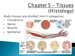
DrDuran Kala 4 2 Tissues and Histology What is a Tissue A collection of similar cells and noncellular substances extracellular matrix secreted by the cells Tissue Level of Organization Epithelial ID: 917348
Download Presentation The PPT/PDF document "Chapter 3 Histology: The Study of Tiss..." is the property of its rightful owner. Permission is granted to download and print the materials on this web site for personal, non-commercial use only, and to display it on your personal computer provided you do not modify the materials and that you retain all copyright notices contained in the materials. By downloading content from our website, you accept the terms of this agreement.


Slide1
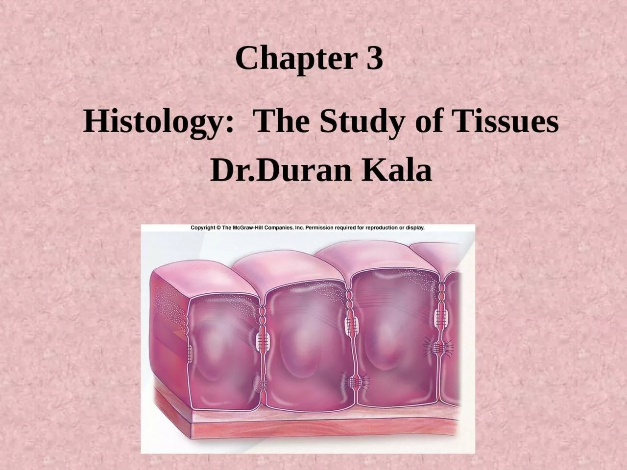
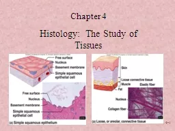
Chapter 3
Histology: The Study of Tissues
Dr.Duran Kala
Slide24-
2Tissues and Histology
What is a Tissue?
A collection of similar cells and noncellular substances (extracellular matrix) secreted by the cells
Tissue Level of Organization
Epithelial
ConnectiveMuscleNervousHistology: Microscopic Study of Tissues
Slide34-
3Main characteristics of the four basic types of tissues.
Tissues
Cells
Extra cellular matrix
Main functions
Epithelial
A
ccumulate
d
polyhedral cells
Small amount
Lining of surface or body cavities, glandular secretion
Connective
Several types of fixed and wandering cells
Abundant amount
Support and protection
Muscle
Elongated contractile cells
Moderate amount
Movement
Nervous
elongated
None
Transmission of nervous impulses
Slide44-
41. EPITHELIAL TISSUE
Epithelial tissues form boundaries between environments.Epithelia are thus made of sheets of tightly connected cells.Types:
Covering & lining epithelium.
Outer layers of skin.
Linings of cavities.
Glandular epithelium.Glands
Slide54-
5Epithelium Characteristics
Consists almost entirely of cells
Covers
body
surfaces and forms glands
Has free and basal surfaceAvascularUndergoes mitosis6. Regenerative.
7. Limited – layer or layers
Fig. 4.1
Slide64-
6ROLE OF EPITHELIA
Protection.SkinAbsorption.
GI
Filtration.
Kidney
Secretion.GlandsSensory Perception.Skin, GI
Slide74-
7Classification of Epithelium:
How to Make Sense of It All?
There are 2 basic features (or criteria) for classification of epithelium:
Number of cell layers
Simple
Single layerStratifiedMore than 1 layerExceptions?
Pseudostratified
Single layer; only some cells reach free surface
Transitional
Number of cell layers decreases as it is stretched
Fig. 4.2
Slide84-
8Classification of Epithelium
Shape of cells
Squamous (=scaly)
Cells flattened
Cuboidal
Cells cube-shapedColumnarCells are taller than wide
Fig. 4.2
Slide94-
9Types of Epithelial Tissues
Simple squamous: Lining of vessels (endothelium). Serous lining of cavities; pericardium, pleura, peritoneum (mesothelium).
Functions:
Facilitates the movement of the viscera (mesothelium), active transport by pinocytosis (mesothelium and endothelium), secretion of biologically active molecules (mesothelium)
Cell shapes are polygonal.
Slide104-
10Types of Epithelial Tissues
Simple cuboidal: Covering the ovary, thyroid.Functions:
Covering, secretion.
Slide114-
11Types of Epithelial Tissues
Simple columnar: Lining of intestine, gallbladder
Functions:
Protection, lubrication, absorption, secretion.
In the small intestine the apical surface of these cells have microvilli present on their surface and specialized gland cells called goblet cells which produce and secrete mucus.
microvilli on apical surface
Slide124-
12Types of Epithelial Tissues
Pseudostratified columnar ciliated:layers of cells with nuclei at different levels; not all cells reach surface but all adhere to basal lamina
.
Lining of trachea, bronchi, nasal cavity
, and lining of the Fallopian tubes (oviducts) of female.Functions:Protection, secretion; cilia-mediated transport of particles trapped in mucus out of the air passages.
Slide134-
13Types of Epithelial Tissues
Stratified squamous epithelial non-keratinized (moist)
lining of
Mouth, esophagus, larynx, vagina, anal canal.
Funtions:
Protection, secretion; prevents water loss.Cells can be cuboidal or columnar over the basal lamina but they are flattened near the surfaces.
Slide144-
14Types of Epithelial Tissues
Stratified squamous epthithelium keratinized(dry)
:
Location:
Epidermis.Function:Protection; prevents water loss.Keratin is a layer a waterproof protein on the apical surface of stratified squamous keratinized epithelium (skin).
Slide154-
15
Slide164-
16Stratified Cuboidal Epithelium
Sweat glands, developing ovarian follicles.Protection,
Secretes sweat; ovarian hormones & produces sperm
Slide174-
17Stratified columnar epithelium
Multiple layers of cells in which the surface layer is cuboidal or columnar.
Function:
Protection or secretion.
Location
: Conjunctiva (mucus membrane on the eyeball and eyelids) ,Male Urethra, Sweat Glands
Slide184-
18Types of Epithelial Tissues
Stratified Transitional: This epithelium is unique to the urinary bladder, ureters
and
renal calces.
Functions:
Protection, expandability. It has the unique property of expansion and contraction. This allows the tissue to adjust to the urinary bladder’s expansion and contraction when it is full or empty.
Slide194-
19Glands
Secretory organs made mostly of epithelium
Form as invaginations (ingrowths) of outer layer of epithelium in embryo
Two basic types:
Exocrine
:Have ducts lined with epithelium
Salivary glands, sweat glands, mucus gland
Endocrine:
Have no ducts
Examples include pituitary gland, pancreas, thyroid gland
Slide204-
20
Epithelial Cell Junctions
Slide214-
21
Junctional Complexes
Slide224-
22
Tight Junctions by TEM
Slide234-
23
Tight Junctions by Freeze Fracture
Slide244-
24
Tight Junction diagram
Slide254-
25
Gap Junction by TEM
Slide264-
26
Gap Junction
Connexin molecules
Open pores for ions, molecules, etc
2 nM gap between membranes
Slide274-
27
TEM of Zonula Adherens (red arrows)
TEM of a Desmosome (blue arrow)
Slide284-
28
TEM (colorized) of Adherens Junctions
Slide294-
29
TEM of a Desmosome with tonofibrils
Slide304-
30
Desmosome Structure
Slide314-
31The macromolecular components of basal laminae
Laminin ( large glycoprotein molecules),
Type IV collagen
(
m
onomers of type IV collagen contain three polypeptide chains ) Entactin, (a glycoprotein) Perlecan, a proteoglycan (mucopolysaccharide that containing polysaccharide and amino acid) with heparan sulfate side chains.These glycosylated proteins and others serve to link together the laminin and type IV collagen sheets.
Slide324-
32Specializations of Cell Surface
MicrovilliFound mainly on absorptive cellsBrush border, 1
m
high
Cilia / flagella
Cylindrical, motile structures, 5-10m highContain microtubulesBasal bodiesSterocilia:arelong apical processes of cells in other absorptive epithelia such as that lining the epididymis and ductus deferens. These structures are much longer and less motile than microvilli
Slide334-
33Microvilli
Apical region of an intestinal epithelial cell seen with TEM. Filaments that constitute the core of the microvilli are clearly seen. An extracellular cell coat (glycocalyx) is bound to the plasmalemma of the microvilli. x45,000.
Slide344-
34
Slide354-
35Stereocilia
Slide364-
36Cilia
Slide374-
37MEDICAL APPLICATION
Both benign and malignant tumors can arise from most types of epithelial cells.
C
arcinoma
(Gr.
karkinos, cancer, + oma, tumor) is a malignant tumor of epithelial cell origin Malignant tumors derived from glandular epithelial tissue are usually called adenocarcinomas (Gr. adenos, gland, + karkinos); these are by far the most common tumors in adults. Under certain abnormal conditions, one type of epithelial tissue may undergo transformation into another type in another reversible process called metaplasia.Examples are
1-
In heavy cigarette smokers, the ciliated pseudo-stratified epithelium lining the bronchi can be transformed into stratified squamous epithelium.
2-
In individuals with chronic vitamin A deficiency, epithelial tissues of the type found in the bronchi and urinary bladder are gradually replaced by stratified squamous epithelium
Slide384-
38