
167 DOI 104274tpa4668 cranial magnetic resonance imaging revealed occipitalmeningocele thinning of corpus callosum Dandy Walkervariant chest tomography revealed high left diaphragmaand esophag ID: 952722
Download Pdf The PPT/PDF document "AA case of Edwards syndrome with e" is the property of its rightful owner. Permission is granted to download and print the materials on this web site for personal, non-commercial use only, and to display it on your personal computer provided you do not modify the materials and that you retain all copyright notices contained in the materials. By downloading content from our website, you accept the terms of this agreement.


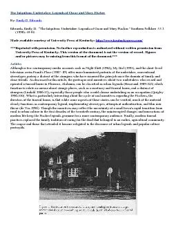

AA case of Edwards syndrome with e 167 DOI: 10.4274/tpa.46.68 cranial magnetic resonance imaging revealed occipitalmeningocele, thinning of corpus callosum, Dandy Walkervariant; chest tomography revealed high left diaphragmaand esophageal graphy with barium revealed esophagealatresia. Other laboratory tests were found to be normal. The infant was internalized in the neonatal intensivecare unit and intubated. Ventilatory support was provided. Esophageal atresia and accompanying tracheoesophageal fistula were corrected adequately. Noproblem related to the operation was experienced. Patentductus arteriosus was closed by giving oral ibuprofen.Lung infection and cardiac failure developed during thefollow-up. Although appropriate antibiotics and inotropictreatment were administered, full recovery could not beachieved and oxygen requirement persisted. The infantwas referred to a secondary care n
eonatal unit for maintenance of care and treatment on the 102nd day oflife by receiving informed consent from the family.Genetic consultancy was provided for the family. 16688Gülaet al.Edwards syndrome Picture 2: Meningocele in the occipital region Picture3: Flexion anomaly of the fingers Picture 4: External prominence of the heels Picture 1: General appearance of the infant diagnosed as Edwards syndrome Picture 5: Cariotype of our patient demostrating 47,XX+18 anomaly Edwards syndrome was defined by Edward and histhought that advanced maternal age, environmental 1.Smith DM. Trisomy 18 Syndrome. In: Jones KL ed. Smiths2.Descartes M, Caroll AJ. Cytogenetics. In: Behrman RE,of Pediatrics 18th ed. Philadelphia: Saunders, 2007: 510-1. 3.Tekin N, Akit A, Gürpnar M. Trizomi 18 sendromlu bir olguda4.Baty BJ, Blackburn BL, Carey JC. Natural history of trisomy 185.Pant SJ, Robbins JM, B
ird TM, et al. Congenital malformations6.Edwards Jh, Harnden Dg, Cameron Ah, Crosse Vm, Wolff Oh.7.Weber WW. Survival and sex ratio in trisomy 17-18. Am J Hum8.Bhanumathi B, Neelam AG, Mishro ZA. Trisomy 18 in a 50 yearold female. Indian J Hum Genet 2006; 12: 146-7.9.Parker MJ, Budd JL, Draper ES, Young ID. Trisomy 18 in a10.Naguib KK, Al-Awadi SA, Bastaki L, et al. Clustering of trisomy11.Gonzales-Zamora JF, Villegas-Alvares F. Esophagial atresiaand chromosomal abnormalities in a Mexican children 12.Suranyi A, Bito T, Vojda G, et al. Unusual clinical history of a13.Steffensen TS, Opitz JM, Gilbert-Barness E. Congenital 14.Bick D, Markowitz RI, Horwich A. Trisomy 18 associated with15.Scorta A, Franceschini P, Pilotti G. [On a case of trisomy 18with spina bifida and meningocele]. Minerva Ginecol 1966; Tuurrkk AArrcchh PPeedd 22001111;; 4466:: 116677--99Gülaet al.Edwards syndrome