
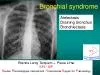
Atelectasis Draining b ronchus Bronchiectasis Etienne Leroy Terquem Pierre LHer SPI ISP S outien P neumologique I nternational I nternational S upport for P ulmonology Atelectasis Cons ID: 936798
Download Pdf The PPT/PDF document "Bronchial syndrome" is the property of its rightful owner. Permission is granted to download and print the materials on this web site for personal, non-commercial use only, and to display it on your personal computer provided you do not modify the materials and that you retain all copyright notices contained in the materials. By downloading content from our website, you accept the terms of this agreement.


Bronchial syndrome Atelectasis Draining b ronchus Bronchiectasis Etienne Leroy Terquem â Pierre LâHer SPI / ISP S outien P neumologique I nternatio
nal / I nternational S upport for P ulmonology Atelectasis Consequence of the obstruction of a bronchus intrinsic ( tumor , foreign body, inflammato
ry stenosis ) or extrinsic (compression by adenopathy or tumor ) The alveolar air gradually disappears and the lung tissue is retracted . The
retraction can involve one segment, one lobe or the whole lung Main etiologies of atelectasis ⢠Bronchial cancer ⢠Tuberculosis ⢠Extrinsic comp
ression by adenopathy TB (++ children ) or malignant ⢠Foreign body ( + + + young children , not always Radio opaque) Less common causes: Asthma
Chronic bronchitis Viral or bacterial pneumonia Atelectasis after thoracic or abdominal surgery , after trauma Many other rare etiologies : benign
tumor , endobronchial metastasis , lymphoma , granulomatous inflammation regardless of etiology , bronchiolitis , cystic fibrosis , ... The radiographic a
ppearance is an opacity , like a consolidation but: Systematized (in contact with a fissure) Retractile ( decrease volume) Homogeneous Without ai
r bronchogram With a variable size : a segment, a lobe, the lung ATELECTASIS M 59 y. Hemoptysis Smoking = 40 pack - years AFB sputum negative Atelectasis
of the right upper lobe M 59 y. Hemoptysis Smoking = 40 pack - years AFB sputum negative Endoscopy : neoplastic bud in the RUL bronchus Small fis
sure in the normal position, horizontal, in pneumonia Ascension of the small fissure = Retraction of RUL Cancer Male ,75 years old , smoker , chronic cou
gh and one episode of hemoptisis Right lower lobe atelectasis by bronchial cancer Male ,75 years old , smoker , chronic cough and one episode of
hemoptisis Retractile opacity who does not erase the edge of the heart Middle lobe atelectasis Partial atelectasis of the right superior lobe by
cancer Man, 56 years old . High fever , right abdominal and thoracic pain, Muscular defense of the right hypochondrium , X - ray : Middle lobe atelectasis
Liver abcess: the reduction of right hemidiaphragm mobility leads to atelectasis above the diaphragm «passive atelectasis» Smoker 60 pack - years . Hemoptysis , thor
acic pain and dyspnea AFB sputum negative Left upper lobe atelectasis by cancer RUL c onsolidation by cancer Atelectasis in the process of co
nstitution A.Khallil and coll. EMC 386 C10 2005 Notice the association with a big hilar round mass Smoker 40 pack - years Hemoptisis Left anterior
thoracic pain and cough . Recent weight loss & asthenia - AFB sputum negative Left upper lobe atelectasis by cancer Smoker 40 pack - years H
emoptisis Left anterior thoracic pain and cough . Recent weight loss & asthenia - AFB sputum negative Fissure Retraction of LUL Notice the r
ound mass on the left hilus Atelectasis + round hilar mass: ï In adults = most often cancer ï In children = most often TB 1 year
old child TB primary 1 year old child Bilateral adenopathies , Left lower lobe atelectasis : compression of the left inferior bronchus by
mediastinal adenopathies TB primary But not always ⦠Look for AFB in sputum systematiquely Cancer W 37 years old , cough and dyspnea . AFB
+ Classic retractile evolution with TB treatment Opacity of the whole left lung field Do you think this is an atelectasis of the left l
ung ? Opacity of the whole left lung field Do you think this is an atelectasis of the left lung ? No The mediastinum is pushed by the o
pacity = Great abundance pleurisy Whole left lung atelectasis Retraction Atelectasis Retraction Pushing back Compressive pleural effusion
Draining bronchus - TB cavern +++ - bacterial nonTB abcess +/ - Small TB cavern with a draining bronchus associated with a lower lobe
TB pneumonia Silhouette s ign Opacity does not erase the left side of the heart TB cavity Notice the draining bronchus and right axillar infil
trate Bronchiectasis Bronchial disease characterized by a permanent increase in bronchial caliber The cartilaginous framework of the bronchial wall
is destroyed or broken up Barker A. N Engl J Med 2002;346:1383 - 1393 ï Located ⢠Pulmonary TB ⢠Bacterial / viral infection in children ( mea
sles , whooping cough ..) ⢠Foreign body ⢠Bronchial stenosis , extrinsic compression( adenopathy ) ⢠D iffuses ⢠Bacterial or viral infection in
children ( measles , whooping cough ..) ⢠TB ⢠Cystic fibrosis ⢠Other congenital diseases : Situs inversus , Imotile cilia Syndrome ⢠Dy
sglobulinemia , chronic immune deficiency , autoimmune diseases ... Bronchiectasis : etiologies ⢠Bronchopulmonary infections repeated ⢠Hemoptysis
⢠Significant , Chronic and often purulent sputum , with AFB negative ⢠Frequent history of TB or Severe respiratory infection in early child
hood Bronchiectasis : clinical features Bronchiectasis is a frequent and underestimated pathology , especially in countries with high incidence of TB and
childhood lung diseases Bronchiectasis radiological features ⢠Round or cylindric images ( clarity with thick or thin wall ) ⢠Sometimes with
fluid level if active infection ⢠Localized in a lobe or a segment, or diffuse The lipiodol bronchography is replaced by the scanner Bronchectasis (opa
cification with iodin hydrosoluble solution) â Ampullary Saccular â Bronchiectasis (opacification with iodin hydrosoluble solution) â Cylindrical â
Typical railway picture in middle lobe & RLL with associated round cavities : Bronchiectasis W. chronic cough with morning abundant sput
um . Repeted bronchial infections and frequent antibiotic treatments . Railway picture : Middle lobe bronchiectasis Another case Railway p
icture : Middle lobe bronchiectasis With CT scan the diagnosis is obvious . Bronchography is not available now , you donât have scanne
r . You must be able to identify Bronchiectasis on CXR Unilateral bronchiectasis of the left lower lobe Chronic purulent sputum Bilateral
bronchiectasis Digital hippocratism is often associated with bronchiectasis Female , 25 y, chronic cough and purulent sputum * Measles at the a
ge of 6 years * Bronchorrhea Young woman , 20 y, repeated bronchus infections from a very early age , and gradual respiratory insufficiency
MUCOVISIDOSIS (1 case/ 2000 births in Europa) Right severe bronchiectasis , Retractile sequela of extensive TB of the right lung CXR f
rom Lao TB Prevalence survey Bronchiectasis is very frequent and underestimated , in countries with high TB incidence Dilation of bronchus , Bronc
hiectasis is a common disease with many etiologies : ï¼ Sequelae of TB ï¼ Effects of early childhood infections ( Measles + + +, pertussis ),
ï¼ Congenital malformations (rare) Not to be confused with TB cavitations Bronchial syndrome You must be able to identify ï¼ Atelectasis ï¼ Drai