
ZOO 103 Structure They are c losely packed cells Specialized to cover external surfaces or line internal cavities Epithelial cells are packed tightly together with almost no intercellular ID: 917351
Download Presentation The PPT/PDF document "THE EPITHELIAL TISSUE Lab 2" is the property of its rightful owner. Permission is granted to download and print the materials on this web site for personal, non-commercial use only, and to display it on your personal computer provided you do not modify the materials and that you retain all copyright notices contained in the materials. By downloading content from our website, you accept the terms of this agreement.


Slide1
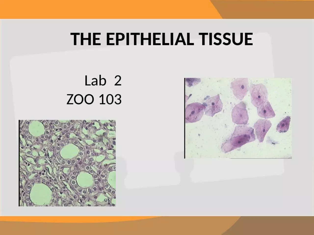
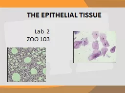
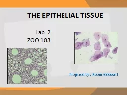
THE EPITHELIAL TISSUE
Lab 2
ZOO 103
Slide2Structure
They are c
losely packed cells.
Specialized
to cover external surfaces or line internal cavities.Epithelial cells are packed tightly together, with almost no intercellular spaces.
Slide3Rest on
Basement Membrane
thin sheet of connective tissue
provides structural support to the epithelium binds it to neighboring structures There are lots of tight junctions and It provides tissues with strength and stability.
T
hey also have
the ability of Renewal
(
Stem and
germinative
cells)
Slide4Slide5Types of Epithelial Tissue
Slide6Number of layers
simple epithelium
(one layer)
squamous epitheliumcuboidal epitheliumcolumnar epitheliumStomach
Intestine
Collecting tubule
Thyroid gland
Lining of
Mouth
Top view
Bowman’s capsule
Side view
stratified epithelium
(more than one layer)
Squamous
,
cuboidal
, columnar
Skin of Toad
( Frog
)
Slide7Covering “surface” epithelium
.
Glandular epithelium
Neuro
-epithelium
Types of Epithelial Tissue Epithelial tissue can be divided into two groups depending on their function of which it is composed.
Simple Stratified
epithelium of cells specialized to produce
secretion
(Goblet cell)
Slide8Simple epithelium
Its
subdivided according to the
shape and function
of its cells. Squamous epitheliumThey have the appearance of thin, flat plates.
They
tends to have
horizontal flattened, elliptical nuclei.
They form the
lining of cavities such as the
mouth, blood vessels, heart and lungs
and
outer layers of the skin.
Wall of blood vessels =
Endothelium
Wall of peritoneum and pleura
=
Mesothelium
Slide9S
quamous epithelium
Slide10Simple Cuboidal Epithelium
C
uboidal
cells are roughly
square
or cuboidal in shape. Each cell has a spherical central nucleus.
It is found in glands and ducts.
They are found lining of kidney tubules.
They
constitute the
germinal epithelium
which produces the egg cells in the ovary and the sperm cells in the testes.
Slide11Simple Cuboidal Epithelium
Slide12Simple Columnar Epithelium
The cells are
elongated
and
column-shaped
. The nuclei are elongated and are usually oval and basal. Its form the lining of the stomach
and
intestines
.
Some columnar cells are
specialized
for
sensory reception
such as
“sensory epithelium”
in the
nose, ears and the taste buds
.
Goblet cells (unicellular glands) are found between the columnar epithelial cells of the duodenum.
They secrete mucus or slime, a lubricating substance which keeps the surface smooth
.
They may be
ciliated
or
non-ciliated
Slide13Simple Columnar Epithelium
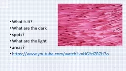
Slide14Stratified squamous epithelium
Slide15E
pithelium of cells
specialized to produce secretion
. All glands are composed of epithelium.
Secretion –
Exocytotic release of products, not metabolic wastes.Columnar and cuboidal epithelial cells often become specialized as gland cells which are capable of synthesizing and secreting certain substances such as enzymes, hormones, milk, mucus, sweat, wax and saliva.
Glandular Epithelium
Slide161
2
Glandular Epithelium
Slide17Under The Microscope
Slide18Simple Squamous
Epithelium ( Lining of mouth )1- Cell membrane
2- Cytoplasm
3- Nucleus123
Slide19Squamous
Epithelium
Side view
glomerulus
Bowman’s capsule
19Simple Squamous Epithelium(Bowman’s capsule )
Slide20Simple
Cuboidal
Epithelium 1-Nucleus
2-Basement
membrane
3-Cuboidal cell 4-Collecting Tubules2431
T.S of (collecting tubule
(
in the kidney
T.S of Thyroid gland
Slide21Simple
Columnar Epithelium 1-Nucleus
2-Plasma membrane3-Basement membrane
1
3
2T.S of intestine
Slide22Stratified
Squamous Epithelium 1-Basement membrane
2-Malpighian Layer3-Spongy Layer
Cells
4-Squamous1324
Slide23Epithelium tissues (Goblet cells )
1-Goblet cells
2-Villi
1
2
Slide24Thank you
for your attention