
wwwwfsahqorgresourcesanaesthesia tutorial of the week ATOTW 361 Coronary Artery Angiography 5 th Sept 2017 Page 1 of 6 B A S I C S C I E N C E Tutorial 361 Coronary A rtery ID: 941443
Download Pdf The PPT/PDF document "Subscribe to ATOTW tutorials by visiting" is the property of its rightful owner. Permission is granted to download and print the materials on this web site for personal, non-commercial use only, and to display it on your personal computer provided you do not modify the materials and that you retain all copyright notices contained in the materials. By downloading content from our website, you accept the terms of this agreement.


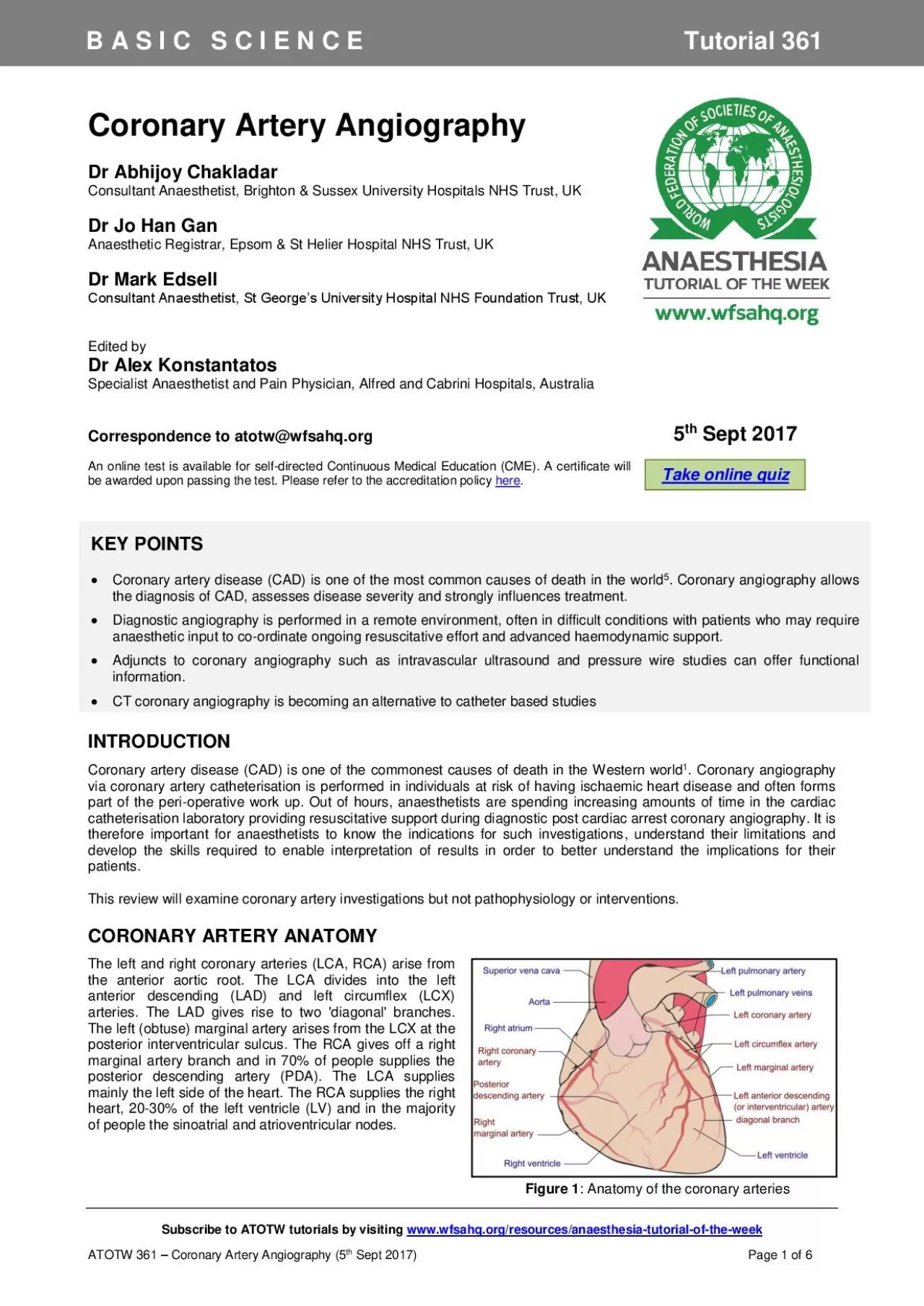
Subscribe to ATOTW tutorials by visiting www.wfsahq.org/resources/anaesthesia - tutorial - of - the - week ATOTW 361 â Coronary Artery Angiography ( 5 th Sept 2017 ) Page 1 of 6 B A S I C S C I E N C E Tutorial 361 Coronary A rtery A ngiograph y Dr Abhijoy Chakladar Consultant Anaesthetist , Brighton & Sussex University Hospitals NHS Trust , UK Dr J o Han Gan Anaesthetic Registrar, Epsom & St Helier Hospital NHS Trust, UK Dr Mark Edsell Consultant Anaesthetist, St Georgeâs University Hospital NHS Foundation Trust, UK Edited by Dr Alex Konstantatos Specialist Anaesthetist and Pain Physician, Alfred and Cabrini Hospitals, Australia Correspondence to atotw@wfsahq.org An online test is available for self - directed Cont inuous Medical Education (CME). A certificate will be awarded upon passing the test . Please refer to the accreditation policy here . INTRODUCTION Coron ary artery disease (CAD) is one of the common est causes of death in the Western world 1 . Coronary angiography via coronary artery catheterisation is performed in individuals at risk of having ischaemic heart disease and often forms part of the peri - operative work up. Out of hours, anaesthetists are spend ing increasing amounts of time in the cardiac cathe te risation laboratory providing resuscitative support during diagnostic po st cardiac arrest coronary angiography. It is therefore important for anaesthetists to know the indications for such investigations , understand their limitations and develop the skills required to enable interpretation of results in order to better underst and the implications for their patients. This review will examine coronary artery investigations but not pathophysiology or interventions. CORONARY ARTERY ANAT OMY The left and right coronary arteries (LCA, RCA) arise from the anterior aortic root. The L CA divides into the left anterior descending (LAD) and left circumflex (LCX) arteries. The LAD gives rise to two 'diagonal' branches . The left (obtuse) marginal artery arises from the LCX at the posterior interventricular sulcus . The RCA gives off a right marginal artery branch and in 70% of people supplies the posterior descending artery (PDA). The LCA supplies mainly the left side of the heart . The RCA supplies the right heart, 20 - 30% of the left ventricle (LV) and in the majority of people the sinoatrial and atrioventricular nodes. Figure 1 : Anatomy of the coronary arteries 5 th Sept 2017 KEY POINTS ï· Coronary artery disease (CAD) is one of the most common cause s of death in the world 5 . Coronary angiography allows the diagnosis of CAD, assess es disease severity and strongly influences treatment. ï· Diagnostic angiography is performed in a remote environment, often in difficult conditions with patient s who may require anaesthetic input to co - ordinate ongoing resuscitative effort and advance d haemodynamic support. ï· Adjuncts to coronary angiography such as intravascular ultrasound and pressure wire stud ies c an offer functional information. ï· CT coronary angiography is becoming a n alternative to catheter based studies Take online quiz Subscribe to ATOTW tutorials by visiting www.wfsahq.org/resources/anaesthesia - tutorial - of - the - week ATOTW 361 â Coronary Artery Angiography
( 5 th Sept 2017 ) Page 2 of 6 The artery responsible for supplying the PDA determines the 'dominance' of the coronary circulation. The PDA supplies the atrioventricular node ( AVN ) and as such interruptio n of its blood supply can lead to heart block and atrio - ventricular dissociation. Patient presenting with chest pain and having an elevated cardiovascular risk profile are at a higher risk of non - fatal myocardial infarction ( MI ) and all - cause mortality if they have a left dominant circulation 2 . CORONARY ARTERY CATH ETERISATION AND ANGI OGRAPHY 1. P racticalities and risks Coronary angiography is performed in a catheter laboratory, typically in patients who are not sedated . The radial or femoral artery is cannulated with a sheath using the Seldinger technique. U niquely shaped catheter s are then used to engage the LCA or RCA ostia. Radiocontrast agents are injected through catheter s over 3 to 5 seconds under continuous fluoroscopy to delineate the coronary a rterial anatomy. Rotating the X - ray source during radiocontrast injection allows multiple views to be achieved to give optimal visualisation of arteries in different planes. The main views obtained during coronary angiography can be seen in Figure 2 . Pe rcutaneous coronary interventions (PCI) are sometimes performed during coronary angiography. This is where angioplasty balloons or coronary stents are used to dilate stenotic segment in the coronary artery identified during angiography . R isks of cardiac ca theter angiography are summarised in Figure 3 . In addition to patient risk s , the anaesthetist should be vigilant to the challenges of working in the angiography suite . With mounting evidence for primary PCI, anaesthetists are increasingly involved in lea ding resuscitation efforts and providing physiological support during emergency coronary angiography. These patients are frequently sedated, ventilated and require complex critical care in remote location s . The environment is often challenging, owing to di fferent and complex monitoring applications, staff unfamiliar with assistance and teamwork required for the provision of safe anaesthesia, physical restrictions imposed by x - ray machines, radiation exposure, difficult patient access, and the need for ongoi ng manual or mechanical CPR. Anaesthetists working in these environments should be familiar with their local resuscitation guidelines and resuscitation equipment available to them. (Appendix 1) Figure 2 : Standard Angiography Views Image Courtesy of Dr Gerald Yong, Fiona Stanley Hospital, Australia Figure 3 : Complications of coronary angiography Complications of Coronary Angiography Access Bleeding Infection Haematoma formation Arterial damage or occlusion Distal l imb i schaemia Emboli Cardiac Myocardial ischaemia Myocardial infarction Coronary artery dissection Systemic Allergic response to radiocontrast agent Stroke Radiation e xposure â 4.6 - 15.8 mSv (~230 - 790 PA chest radiographs) Subscribe to ATOTW tutorials by visiting www.wfsahq.org/resources/anaesthesia - tutorial - of - the - week ATOTW 361 â Coronary Artery Angiography ( 5 th Sept 2017 ) Page 3 of 6 2. Access site Vascular access for diagnostic angiography and primary PCI has traditionally been via the
femoral arter ies . This access site has significant risk of bleeding. Pooled data from several trials de scribes bleeding complications in patients undergoing PCI are a ssociated wit h increased long - term mortality 3 . While multiple strategies have been employed to reduce bleeding i.e. mechanical compression, drugs, access sheaths, bleeding still remains a si gnificant problem for patients. This has led to the introduction of the trans - radial approa ch for coronary catheterisation. I ncidence of bleeding has fallen significantly since the adoption of trans - radial technique compared to the trans - fem oral approach (0.05% vs 2.3%) 4 . However, this technique requires the acquisiti on of additional skills over time and has greater risk of radial artery spasm and/or occlusion. 3. Post - procedure haemostasis Post - procedure haemostasis is minimized via manual or mechanical compression or vascular closure devices which deploy collagen plugs, sutures or staples to the arterial puncture site. The anaesthetist should monitor these sites for bleeding while transferring patient post procedure to and from critical care areas. One should also be vigilant for distal limb ischaemia due to vasos pasm or vessel injury. 4. Describing lesions A r theromatous lesions are described by their location, degree of stenosis , length and the relative spread of the narrowing , for example focal versus diffuse disease. Lesions are described as involving proxima l, mid - or distal parts of the coronary arteries and are visually inspected during angiography to give a percentage that grad es the degree of stenosis. Less than 50% stenosis is considered mild disease, 50 - 70% stenosis moderate disease and greater than 70% as severe disease. Severe disease should be considered for PCI angioplasty, clot removal and/or stent placement. Coronary angiograp hy is not a dynamic or functional study. It cannot predict the stability of any plaques that may be identified. It provides a 2D image of a 3D structure and it may occasionally be difficult to correlate anatomical disease with patient symptom atology . C l inical implications of a stenotic lesion depend on the territory of the heart that may be render ed ischaemic. For example, RCA ischaemia is more likely to lead to bradycardia and heart block, whereas left sided lesions are more likely to lead acute left ve ntricular failure and hypotension. 5. Thrombosis in Myocardial Infarction ( TIMI ) Scoring 'TIMI Grade Flow' is a scoring system from 0 - 3 referring to the quality of coronary blood flow assessed during percutaneous coronary angioplasty 5 . It was developed by the TIMI study group to semi - quantitatively assess coronary artery perfusion beyond point s of occlusion on coronary angiography. Determination of TIMI flow grade after coronary reperfusion yields important prognostic information in patients with acute myocardial infarction. Higher TIMI grades are associated with improved mortality and morbidity figures. Figure 4 : TIMI Flow Grade A DJUNCTS TO CORONARY ANGIOGRAPHY 1. Pressure wire studies and their interpretation As an adjunct to visual assessment of the degree of coronary artery stenosis, pressure wire studies and measurement of fractional flow reserve (FFR) provide a functional assessment of stenosis detected durin g angiography 7 . A wire with an in built pressure t
ransducer at its tip is passed through a stenotic area and the pressure measured distal to it. The wire is then pulled back and pressure measured proximal to the narrowing. FFR = D istal coronary pressu re - right atrial (RA) pressure P roximal corona ry pressure - RA pressure It is quantification of how much flow would occur through the blood vessel if the stenosis did not exist . A FFR score of 1.0 indicates no obstruction to flow. It is important to acknowledge the transient iatrogenically induced effects to coronary vascular tone during these studies , and as such maximal hyperaemia is induced with intravenous adenosine 6 . A FFR of less than 0.75 - 0.8 suggests that stent placement may be of prognostic benefit and is especially useful for guiding treatment of visually assessed intermediate and severe coronary lesions. FFR measurement can also be used to identify 'hot' lesions responsible for myocard ial ischaemia in those patients with widespread multi - vessel disease. TIMI Grade Angiographic Appearance 0 Absence of any antegrade flow beyond a coronary occlusion 1 Faint antegrade coronary flow beyond the occlusion 2 Delayed or sluggish antegrade flow with complete filling of the distal territory 3 Normal flow which fills the distal coronary bed completely Subscribe to ATOTW tutorials by visiting www.wfsahq.org/resources/anaesthesia - tutorial - of - the - week ATOTW 361 â Coronary Artery Angiography ( 5 th Sept 2017 ) Page 4 of 6 2. Intravascular ultrasound Standard X - ray angiography provides a qualitative 2 dimensional assessment of the calibre of the interior surface of coronary arteries in the long axis. In travascular ultrasound (IVUS) can provide additional information regarding vessel geometry (diameter, cross - sectional area) and plaque dimensions 8 ( Figure 5 ) . IVUS is performed by passing a coronary artery catheter with an inbuilt ultrasound array over a g uide wire. A cross - sectional image of the coronary artery wall is displayed showing the relationship of the intima, media and adventitia, which in health are closely associated. Measurements obtained by IVUS are more accurate than angiography par ticularly in the left main stem and ostial lesions where vessels overlap. Due to its higher resolution, IVUS may reveal presence of plaques that have not yet expanded into the lumen or ruptured. This occult disease is clinically significant as the minimally occlu sive plaque has been shown to be more likely to rupture and cause acute cardiac ischaemia 8 . The main disadvantage of IVUS is the cost and the length of time it adds to any diagnostic procedure. There is also increased risk of intimal damage to the coronary artery which must be balanced against the additional data gained from an IVUS study. Figure 5 : IVUS to assess stent apposition. Image c ourtesy of Dr Viknesh Jayapalen, Kingâs College Hospital, UK 3. Ventriculography and valvular assessment Ventriculography can be performed before or after angiography. A catheter is advanced through the aortic valve into the LV and radiocontrast injected. The contraction of the LV is then assessed to detect any wall motion abnormalities and provides data on s troke volume, ejection fraction and cardiac output. Gross diagnoses of aortic and mitral valve regurgitation can be made during left ventriculography.
OTHER IMAGING MODALI TIES FOR CORONARY AN GIOGRAPHY 1. Coronary c omputed tomography angiography (CCTA) Coronary CT angiography is a non - invasive method of examining the coronary arteries. No arterial catheterisation is required and instead intravenous radiocontrast is injected and timed scans of the heart performed. In the past, CCTA was limited by the numb er of (0.5 mm) slices a scanner could image per rotation of its emitter/detector tube, leading to movement artefact from the heart and respiratory cycle. The advent of sub - second tube rotations and 64, 128, 256 and 320 slice scanners enables the heart to be imaged quickly and have a high enough resolution to allow accurate assessment of CAD 9 . Radiation doses are comparable to invasive, catheter angiography but can be significantly reduced with technologies such as prospective ECG gating where CCTA images a re obtained only in diastole, when the heart is motionless 1 0 . Images can be reconstructed to give a 3D representation of the heart. A disadvantage of CCTA is that images can become difficult to interpret in the presence of significant vessel wall calcific ation and , in the event of discovering an amenable lesion, no intervention is possible 11 . Figure 6 : CT Coronary angiography demonstrating a distal LAD stenosis. Coronary angiography confirms findings. Image c ourtesy of Dr Viknesh Jayapalen, Kingâs College Hospital, UK Subscribe to ATOTW tutorials by visiting www.wfsahq.org/resources/anaesthesia - tutorial - of - the - week ATOTW 361 â Coronary Artery Angiography ( 5 th Sept 2017 ) Page 5 of 6 2. Magnetic resonance (MR) coronary angiography At present, the indications for cardiac magnetic resonance angiography (MRA) are limited. This is mainly d ue to relatively long scan times of five to 15 mi nut es versus fraction of a second with CCTA. Image resolution is also considered inferior; 1 to 1.5 mm versus a s lice thickness of 0.5 mm with CCTA 12 . However, cardiac MRA emits no ionising radiation making it suitable for children requiring multiple scans . Furthermore, it is not affected by vessel wall calcification . Advances in MR scanner field strength, use of MR contrast and the number of coils means that more of the heart can be image d simultaneously thereby improving resolution and decreasing scan ti mes. Currently cardiac MRA is mainly used for diagnosing ano malous coronary artery origins ( in children) and coronary artery aneurysms in Kawasaki Disease . This tutorial is estimated to take 1 hour to complete. Please record time spent and report th is to your accrediting body if you wish to claim CME poin ts. ACKNOWLEDGEMENT We would like to thank Dr Viknesh Jayapalen (Kingâs College Hospital, UK) and Dr Gerald Yong (Fiona Stanley Hospital, Australia) for sourcing the images . Image Copyright Figure 1: Creative Commons Attribution - Share Alike 3.0 Unported license. Created by Patrick Lynch and adapted by Mikael Häggström. Downloaded from https://commons.wikimedia.org/wiki/File:Coronary_arteries.svg To take the onlin e test accompanying this tutorial , please click on this link. This tutorial is estimated to take 1 hour to complete. Please record time spent and report this to your accrediting body if you wish to claim CME points REFERENCES AND FURTH ER R
EADING 1. World Health Organisation. Cardiovascular diseases. http://www.who.int/mediacentre/factsheets/fs317/en/ (Accessed 26 Feb 2017) 2. Veltman CE, van der Hoeven BL, Hoogslag GE et al. Influence of coronary vessel dominance on short - and long - term outcome in patients after ST - segment elevation myocardial infarction. Eur Heart J 2015; 36(17): 1023 - 1030 3. Eikelboom JW, Mehta SR, Anand SS, Xie C, Fox KAA, Yusuf S. Adverse impact of bleeding on prognosis in patients with acute coronary syndromes. Circulation 2006; 114: 774 â 782 4. Jolly SS, Amlani S, Hamon M, Yusuf S, Mehta SR. Radial versus femoral access for coronary angiography or intervention and the impact on major bleeding and ischemic events: a systematic review and meta - analysis of randomized trials. Am Heart J. 2009; 157(1): 132 - 40 5. The Thrombolysis in Myoca rdial Infarction (TIMI) trial. Phase I findings. TIMI Study Group N Engl J Med. 1985; 312(14): 932 - 6 6. Blows LJ, Redwood SR. The Pressure wire in practice. Heart . 2007; 93(4): 419 â 422. 7. Pijls NH, De Bruyne B, Peels K, Van Der Voort PH, Bonnier HJ, Bartunek J Koolen JJ, Koolen JJ Measurement of fractional flow reserve to assess the functional severity of coronary - artery stenoses. N Engl J Med. 1996; 334(26):1703 - 8. 8. Nissen SE, Yock P. Intravascular Ultrasound: Novel Pathophysiological Insights and Current Clinical Applications, Circulation. 2001;103:604 - 616 9. Sabarudin A, Sun Z. Coronary CT angiography: Diagnostic value and clinical challenges. World J Cardiol. 2013; 5(12): 473 â 483 10. S human WP, Branch KR, May JM, Mitsumori LM, Lockhart DW, Dubinsky TJ, Warren BH, Caldwell JH. Prospective versus Retrospective ECG Gating for 64 - Detector CT of the Coronary Arteries: Comparison of Image Quality and Patient Radiation Dose. Radiology. 2008;24 8(2):431 - 7. SUMMARY Diagnostic coronary angiography is becoming increasingly common as the incidence of coronary artery disease rises. Anaesthetists are often called upon to provide anaesthesia and resuscitative support in the catheter lab during routine and emergency procedures. A functional knowledge of the indication s , risk s and common terminology used in diagnostic angiography will help in interpreting the main findings , improve team communication and tailor the anaesthetic technique to the individual patient. To take the online test accompanying this tutorial, please click here Subscribe to ATOTW tutorials by visiting www.wfsahq.org/resources/anaesthesia - tutorial - of - the - week ATOTW 361 â Coronary Artery Angiography ( 5 th Sept 2017 ) Page 6 of 6 11. Brodoefel H, Burgstahler C, Tsiflikas I, Reimann A, Schroeder S, Claussen CD, Heuschmid M, Kopp AF. Dual - source CT: effect of heart rate, heart rate variability, and calcification on image quality and diagnostic accuracy. Radiology. 2008; 247:3 46 - 55. 12. Sakuma H. Coronary CT versus MR Angiography: The Role of MR Angiography. Radiology 2011; 258:340 â 349 13. Resuscitation Council (UK). Adult Advanced Life Support Guidelines. Accessed on 15 th March 2017. Available from: https://www.resus.org.uk/EasySiteWeb/GatewayLink.aspx?alId=6442 Appendix 1 13 This work by WFSA is licensed under a Creative Commons Attribution - NonCommercial - NoDerivitives 4.0 International License. To view this license, visit https://creativecommons.org/licenses/by - nc - nd/4.0/