
Back 43 Mahlon R DeLong THE BASAL GANGLIA CONSIST of four nuclei portions of which play a major role in normal voluntary movement Unlike most other comof the motor system however they do not have ID: 937357
Download Pdf The PPT/PDF document "The Basal Ganglia" is the property of its rightful owner. Permission is granted to download and print the materials on this web site for personal, non-commercial use only, and to display it on your personal computer provided you do not modify the materials and that you retain all copyright notices contained in the materials. By downloading content from our website, you accept the terms of this agreement.


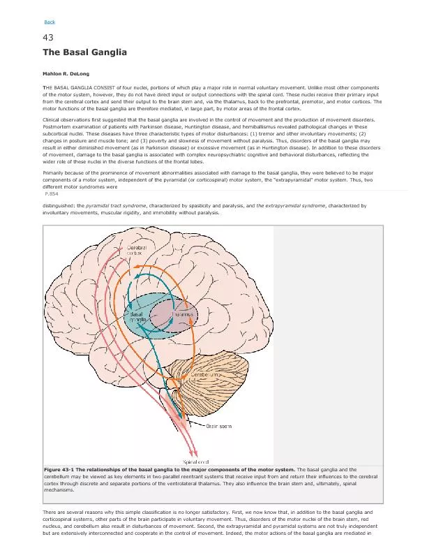


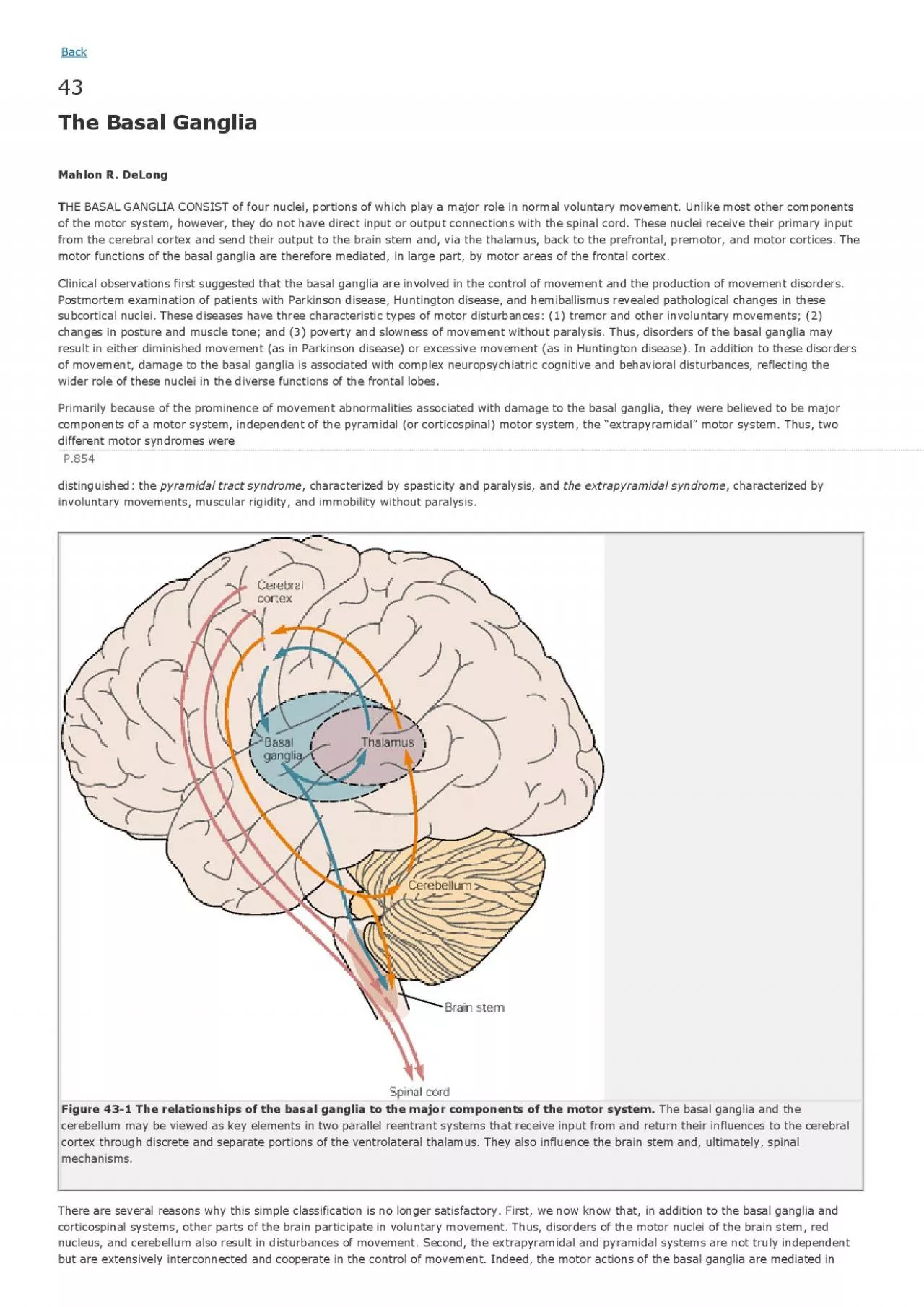
Back 43 The Basal Ganglia Mahlon R. DeLong THE BASAL GANGLIA CONSIST of four nuclei, portions of which play a major role in normal voluntary movement. Unlike most other comof the motor system, however, they do not have direct input or output connections with the spinal cord. These nuclei receive thefrom the cerebral cortex and send their output to the brain stem and, via the thalamus, back to the prefrontal, premotor, and momotor functions of the basal ganglia are therefore mediated, in large part, by motor areas of the frontal cortex. Clinical observations first suggeste
d that the basal ganglia are involved in the control of movement and the production of movemof movement, damage to the basal ganglia is associated with complex neuropsychiatric cognitive and behavioral disturbances, reflwider role of these nuclei in the diverse functions of the frontal lobes. Primarily because of the prominence of movement abnormalities associated with damage to the basal ganglia, they were believed to P.854 distinguished: the pyramidal tract syndrome, characterized by spasticity and paralysis, and the extrapyramidal syndrome, characterized by Figure 43-
1 The relationships of the basal ganglia to the major components of the motor system. The basal ganglia and the cerebellum may be viewed as key elements in two parallel reentrant systems that receive input from and return their influences tcortex through discrete and separate portions of the ventrolateral thalamus. They also influence the brain stem and, ultimately,mechanisms. There are several reasons why this simple classification is no longer satisfactory. First, we now know that, in addition to the large part through the supplementary, premotor, and motor cortices vi
a the pyramidal system. Because they are so common, disorders of the basal ganglia have always been important in clinical neurology. Parkinson disease wdisease of the nervous system to be identified as a molecular disease caused by a specific defect in transmitter metabolism. Therelationship of transmitters to disorders of mood, cognition, and nonmotor behavior, topics that will be considered in detail in Chapters 60 and 61 . The use of a variety of anatomical, molecular, and neural imaging techniques as well as animal models of basal ganglia disordemajor advances in un
derstanding the organization and function of the basal ganglia. These insights have, in turn, led to new phaneurosurgical approaches to treatment of diseases of the basal ganglia. The Basal Ganglia Consist of Four Nuclei The basal ganglia consist of several interconnected subcortical nuclei with major projections to the cerebral cortex, thalamus, Figure 43-1 ). Thus, the basal ganglia are major components of large cortical- P.855 subcortical reentrant circuits linking cortex and thalamus. Figure 43-2 This coronal section shows the basal ganglia in relation to surroundin
g structures. (Adapted from Nieuwenhuys et al. 1981 .) The four principal nuclei of the basal ganglia are (1) the striatum, (2) the globus pallidus (or pallidum), (3) the substantia n Figure 43-2 ). The striatum consists of three important subdivisions: the caudate nucleus, the putamen, and the ventral striatum (which includes the nucleus accumbens). Except at its most anterior pole,internal capsule, a major collection of fibers that run between the neocortex and thalamus The striatum is the major recipient of inputs to the basal ganglia from the cerebral cortex, thala
mus, and brain stem. Its neuroand internal segments. The internal pallidal segment is related functionally to the pars reticulata of the substantia nigra, whimidbrain on the medial side of the internal capsule. The cells of the internal pallidal segment and pars reticulata use g-aminobutyric acid (GABA) as a neurotransmitter. Just as the caudate nucleus is separated from the putamen by the internal capsule, the internal pallidal In addition to its reticular portion, the substantia nigra also has a compact zone (pars compacta). This zone is a distinct nucl The subthalamic
nucleus is closely connected anatomically with both segments of the globus pallidus and the substantia nigra. It Figure 43-3 The anatomic connections of the basal ganglia-thalamocortical circuitry, indicating the parallel direct and indirectpathways from the striatum to the basal ganglia output nuclei. Two types of dopamine receptors (D1 and D2) are located on different sets of output neurons in the striatum that give rise to the direct and indirect pathways. Inhibitory pathways are shown as gray arrows; pink arrows. GPe = external segment of the globus pallidus; GPi = i
nternal segment of the globus pallidus; SNc = substantia nigra pars compacta; STN = subthalamic nucleus. P.856 The Striatum, the Input Nucleus to the Basal Ganglia, Is Heterogeneous in Both Its Anatomy and Function All areas of cortex send excitatory, glutaminergic pro-jections to specific portions of the striatum. The striatum also receives Although the striatum appears homogeneous on routine staining, it is anatomically and functionally highly heterogeneous. It conssubstantia nigra pars compacta. Although the striatum contains several distinct cell types, 90-95% of them
are GABA-ergic medium-spiny projection neurons. Theseboth major targets of cortical input and the sole source of output. They are largely quiescent except during movement or in resp The striatum also contains two types of local inhibitory interneurons: large cholinergic neurons and smaller cells that contain neuropeptide Y, or nitric oxide synthetase. Both classes of inhibitory interneurons have extensive axon collaterals that reduce The Striatum Projects to the Output Nuclei via Direct and Indirect Pathways The two output nuclei of the basal ganglia, the internal palli
dal segment and the substantia nigra pars reticulata, tonically in P.857 in an excitatory glutaminergic projection ( Figure 43-3 ). The projection from the subthalamic nucleus is the only excitatory intrinsic connection of the basal ganglia; all others are GABA-ergic and inhibitory. The neurons in the two output nuclei discharge tonically at high frequency. When phasic excitatory inputs transiently activate tdirect pathway indirect pathway transiently increases inhibition of the thalamus, as can Figure 43-3 ). Thus, the direct pathway can provide positive feedback and th
e indirect pathway negative feedback in the circuit between the basal ganglia and The two striatal output pathways are affected differently by the dopaminergic projection from the substantia nigra pars compacta Although their synaptic actions are different, the dopaminergic inputs to the two pathways lead to the same effect—reducing inhiand in the cortex, could further modulate the actions of the direct and indirect pathways from the striatum. Figure 43-4 The frontal lobe targets of the basal ganglia-thalamocortical circuits. ACA = anterior cingulate area; DLPC = do
rsolateral prefrontal cortex; FEF = frontal eye field; LOFC = lateral orbitofrontal cortex; MC = primary motor cortex; MOFC = medial orbitofrontal cortex; PMC = premotor cortex; SEF = supplementary eye field; SMA = supplementary motor area. The Basal Ganglia Are the Principal Subcortical Components of a Family of Parallel Circuits Linking the Thalamus and Cerebral Cortex The basal ganglia were traditionally thought to function only in voluntary movement. Indeed, for some time it was believed that areas. It is now widely accepted, however, that through their interaction w
ith the cerebral cortex the basal ganglia also contribehaviors other than voluntary movement, including skeletomotor, oculomotor, cognitive, and even emotional functions. Several observations point to diversity of function. First, certain experimental and disease-related lesions of the basal gangli P.858 highly organized connections with virtually the entire cerebral cortex, as well as the hippocampus and amygdala. Finally, a wide The basal ganglia may be viewed as the principal subcortical components of a family of circuits linking the thalamus and cerebraskeletomotor ci
rcuit begins and ends in the precentral motor fields (the premotor cortex, the supplementary motor area, and the motor cortex); the oculomotor circuit, in the frontal and supplementary eye fields; the prefrontal circuits, in the dorsolateral prefrontal and lateral orbitofrontal cortices; and the limbic circuit, in the anterior cingulate area and medial orbitofrontal cortex Figure 43-4 ). Each area of the neocortex projects to a discrete region of the striatum and does so in a highly topographic manner. Associatareas project to the caudate and rostral putamen; sensorimoto
r areas project to most of the central and caudal putamen; and limb The concept of segregated basal ganglia-thalamocortical circuits is a valuable anatomic and physiologic framework for understandwithin, rather than between, the five identified Within each of these subunits there may even be discrete pathways responsible for different aspects of motor processing. Injecti Figure 5-9 for technique). Virus transported in the anterograde direction was labeled in distinctly separate regions of the putamen. Given the substantia nigra pars reticulata. The Skeletomotor Circuit
Engages Specific Portions of the Cerebral Cortex, Basal Ganglia, and Thalamus Since movement disorders are prominent in diseases of the basal ganglia, it is appropriate here to focus on the skeletomotor cirTopographically organized projections from each cortical area result in a somatotopic organization of movement-related neurons iThe leg is represented in a dorsolateral zone, the orofacial region in a ventromedial zone, and the arm in a zone between the tw Figure 43-5 ). Each of these representations extends along virtually the entire rostrocaudal axis of the putamen. R
ecent anatomical and physiol Output neurons in the putamen project topographically to the caudoventral portions of both segments of the pallidum and to the c Figure 18-4 for the organization of the thalamic nuclei). The skeletomotor circuit is then closed by projections from the ventral lateral and ventral anterior (pars mag P.859 the ventral lateral and centromedian nuclei to the precentral motor fields. Single Cell Recording Studies Provide Direct Insight into the Role of the Motor Circuits The contribution of the basal ganglia to movement can be assessed most directly
by studying the activity of neurons within the sserial processing occurs within the basal ganglia-thalamocortical circuits and that much of the activity within During the execution of a specific motor act, such as wrist flexion or extension, the normally high rate of spontaneous discharg Little is known about how movement-related signals from the direct and indirect pathways are integrated in the internal pallidal possibly smoothing the movement, while those in the direct pathway simultaneously facilitate the movement. This reciprocal regulconsistent with the basal gang
lia's apparent role in scaling the amplitude or velocity of movement. Alternatively, the direct and indirect inputs configuration, the skeletomotor circuit might play a dual role in modulating voluntary movements by both reinforcing the selectethe direct pathway) and suppressing potentially conflicting patterns (via the indirect pathway). This dual role could result in focusing the neural Figure 43-5 The somatotopic organization of the basal ganglia-thalamocortical motor circuit is illustrated in these mesial and lateral views of a monkey brain, as well as the basal gang
lia and thalamus. The motor circuit is divided into a “face” representation (blue), “arm” representation (dark green), and “leg” representation (light green). Arrows show subcircuits within the portion of the motor circuit concerned with the arm. CM = centromedian nucleus of the thalamus; GPe = external segment of the globus pallidus; GPi = internal segment of the globus pallidus; MC = primary motor cortex; PMC = prefrontal motor cortex; SMA = supplementary motor area; STN = subthalamic nucleus; VApc = parvocellular portion of the ventral ant
erior nucleus of the thalamus; VLo = pars oralis of the ventrolateral nucleus of the thalamus. Neuronal activity within the skeletemotor circuit has been examined in monkeys performing a variety of motor tasks. At all stage(cortical, striatal, and pallidal) the activity of substantial proportions of movement-related neurons depends upon the directio P.860 P.861 activity. These directional cells comprise 30-50% of the movement-related neurons in the supplementary motor area, motor cortex, Studies combining behavioral training and single-cell recording indicate that the ske
letomotor circuit is involved not only in t Chapter 38 ). Directionally selective activity before movement also occurs within the putamen and the internal segment of the pallidum. Indivieither preparatory (set-related) or movement-related responses, suggesting that the preparation and support the idea that the skeletomotor circuit is composed of distinct subcircuits that connect to different precentral motor fisupplementary motor area, and arcuate premotor area). These subcircuits may have distinctive roles in motor control and in the p specific motor signs and symptoms t
hat occur in Parkinson disease and other diseases of the basal ganglia. Figure 43-6 (Opposite) The basal ganglia-thalamocortical circuitry under normal conditions and in Parkinson disease, hemiballism, and chorea. Inhibitory connections are shown as gray and black arrows; excitatory connections, as pink and red. Degeneration of the nigrostriatal dopamine pathway in Parkinson disease leads to differential changes in activity in the two strprojections, indicated by changes in the darkness of the connecting arrows (darker arrows indicate increased neuronal activity and light
er arrows, decreased activity). Basal ganglia output to the thalamus is increased in Parkinson disease and decreases in ballism and choreGPe = external segment of the globus pallidus; Gpi = internal segment of the globus pallidus; SNc = substantia nigra pars compacta; STN = subthalamic nucleus. Studies of the Oculomotor Circuit Provided Important Insight Into How the Skeletomotor Circuit Operates The oculomotor circuit is involved in the control of saccadic eye movements. It originates in the frontal and supplementary motothe substantia nigra pars reticulata disinhibits o
utput neurons in the deep layers of the superior colliculus whose activity is saccades. Inactivation of neurons in the pars reticulata results in involuntary saccades to the contralateral side. These observ Some Movement Disorders Result From Imbalances in the Direct and Indirect Considerable progress has been made in understanding the mechanisms underlying the major movement disorders of the basal ganglia (of which Parkinson disease is the best-known example) are characterized by impaired initiation of movement (akinesia) bradykinesia). They are usually accompanied by m
uscular rigidity (increased Hyperkinetic disorders (exemplified by Huntington disease and hemiballismus) are characterized by excessive motor activity, the dyskinesias) and decreased muscle tone (hypotonia). The involuntary movements may take several violent, large-amplitude, proximal limb movements (ballism), and more sustained abnormal postures and slower movements with undecocontraction of agonist and P.862 antagonist muscles (dystonia). Various types of involuntary movements often occur in combination and some appear to have a commo In recent years the development o
f primate models of both hypo- and hyperkinetic disorders, induced by systemic or local adminiscircuit. Normal motor behaviors depend on a critical balance between the direct and indirect pathways from the striatum to the psimplest of terms, overactivity in the indirect pathway relative to the direct pathway results in hypokinetic disorders, such as Figure 43-6 ). Overactivity in the Indirect Pathway Is a Major Factor in Parkinsonian Signs Parkinson disease, first described by James Parkinson in 1817, is one of the most common movement disorders, affecting up to one …
;involuntary tremulous motion, with lessened muscular power, in parts not in action and even when supported, with a propensity t The cardinal symptoms of the disease include a paucity of spontaneous movement, akinesia, bradykinesia, increased muscle tone (r Parkinson disease is the first example of a brain disorder resulting from a deficiency of a single neurotransmitter. In the mid symptoms. The subsequent demonstration by George Cotzias that gradual increases in oral administration of L-DOPA could provide sand continuous benefit began the modern era of pharmacologic the
rapy. Even with the development of newer and more effective Research in Parkinson disease was recently revitalized by William Langston's discovery that drug addicts exposed to the meperidi activity in the indirect pathway and decreased activity in the direct pathway (see Figure 43-6 ) because of the different actions of dopamine on the two pathways (via D1 and D2 receptors, respectively). Both of these changes lead to increased activity in the internal palliresults in increased inhibition of thalamocortical and midbrain tegmental neurons and thus the hypokinetic feature
s of the disea Experiments with MPTP-treated monkeys have shown significant changes in neuronal activity along the indirect pathway. For exampladministration of the dopamine receptor agonist apomorphine. The excessive activity in the indirect pathway at the subthalamic nto be an important factor in the production of parkinsonian signs, since lesioning of the subthalamic nucleus, which reduces the P.863 excitatory drive on the internal pallidal segment, markedly ameliorates parkinsonian signs in MPTP-treated monkeys. Selective in portion of either the subthalamic nucleus o
r the internal pallidal segment is sufficient to ameliorate the cardinal parkinsonia Figure 43-7 ). Surgical lesions of the posterior (sensorimotor) portion of the internal pallidal segment (pallidotomy) in patients with advanced, medically intractable cases of Parkinson disease is also highlater). Figure 43-7 Sites of surgical intervention in Parkinson disease. Lesions of the subthalamic nucleus (left) or internal segment othe globus pallidus (right) effectively reduce parkinsonian signs and dyskinesias by respectively normalizating or eliminating segment of the globus p
allidus; STN = subthalmic nucleus; SNc = substantia nigra pars compacta. Thus the hypokinetic features of Parkinson disease appear to result from increased (inhibitory) output from the internal pallidaresult of increased (excitatory) drive from the subthalamic nucleus. Accordingly akinesia and bradykinesia are no longer viewed In addition to the increase in tonic output of the internal pallidal segment in MPTP-treated monkeys, phasic activity also changpattern of discharge in basal ganglia output are likely to be equally as important as the changes in the rate of dischar
ge. Indeed, rec P.864 increased synchronization of oscillatory discharge within the basal ganglia nuclei. Differences in spatial temporal patterns and The Level of Dopamine in the Basal Ganglia Is Decreased in Parkinson Disease Measurements of dopamine in the striatum and the metabolic activity of individual basal ganglia nuclei in patients with Parkinso earlier by direct biochemical assays and more recently by uptake of the precursor 18F-DOPA measured by positron emission tomography (PET) (see Chapter 19 ). Imaging of patients with Parkinson disease has shown less synap
tic activity (as measured by activated blood flow in the contralateral putamen, the anterior cingulate, the supplementary motor area, and the dorsolateral prefrontal cortex) both when tmoving a joystick and when they were resting. Administration of dopamine agonists increased the blood flow to the supplementary restore activity in the supplementary motor and premotor areas during this same movement task. These neuroimaging studies lend sadditional support to the importance of the pallidothalamocortical portion of the motor circuit in normal movement and the produ Underact
ivity in the Indirect Pathway Is a Major Factor in Hyperkinetic Disorders Involuntary movements in patients with basal ganglia disorders may result either from clear-cut lesions of these nuclei or from form. Experimental lesions of the subthalamic nucleus in monkeys show that dyskinesias result only when lesions are made selectively ileaving intact the adjacent projections from the internal pallidal segment to the thalamus. More recent studies combining selectreduced in hemiballism, as expected if the projection from the subthalamic nucleus is segment and decrease the pha
sic responses of these neurons to limb displacement. Thus hemiballism may result from disinhibition thalamus due to reduction in the tonic (and perhaps phasic) output from the internal pallidal segment. Reduced inhibitory input Huntington Disease Is a Heritable Hyperkinetic Disorder The other hyperkinetic disorder most often associated with dysfunction of the basal ganglia is Huntington disease. This disease The Gene for Huntington Disease Has Been Identified Huntington disease is one of the first complex human disorders to be traced to a single gene, which was identifi
ed by mapping ge Box 3-3 ). The disease is a highly penetrant, autosomal dominant disorder with a gene defect on chromosome 4. This gene encodes a large protein, huntingtin, the function of which has yet to be determined ( Chapter 3 ). The protein normally is located in the cytoplasm. As we have seen P.865 in Chapter 3 , the first exon of the gene contained repeats of the trinucleotide sequence CAG, which encodes the amino acid glutamine. Whereanormal subjects have less than 40 CAG repeats in the first exon, patients with Huntington disease have more than 40 repeats. Th
ooffspring than in the parent. To determine why the CAG repeats in the first exon caused disease, the first exon from the mutant human huntingtin protein has bin mice where it was found to be sufficient to cause a progressive neurological phenotype. In these mice, exon formed multiple i A Drosophila model of Huntington disease has been developed by expressing an amino terminal fragment of the human huntingtin protein Finally, a cellular model of Huntington disease has been created by transfecting the mutant Huntington's gene into cultured striapoptosis. However, this apo
ptotic death did not correlate with the formation of intranuclear inclusions. Full length huntingtinvery rarely, raising the possibility that intranuclear inclusions may not play a causal role in mutant huntingtin's induced deat Although Huntington disease is characterized by widespread loss of neurons in the brain, the pathology is seen earliest in the sfunctional inactivation of the rigidity and akinesia in advanced Huntington disease are associated with the loss of the striatal neurons that project to the insegment. This loss would reduce inhibition in the internal pa
llidal segment and thus increase firing in these neurons. Drug-induced dyskinesias, which closely resemble chorea, are a side effect of dopamine replacement therapy for Parkinson diseaseneurons and excessive inhibition of the subthalamic nucleus by overactive neurons in the external pallidal segment. The decreasethe subthalamic nucleus would lower the output from the internal pallidal segment in a manner similar to that seen after direct Glutamate-Induced Neuronal Cell Death Contributes to Huntington Disease Glutamate is the principal excitatory transmitter in the centra
l nervous system. It excites virtually all central neurons and isterminals at high concentration (10-3 M). In normal synaptic transmission the extracellular glutamate rises transiently, and this P.866 rise is restricted to the synaptic cleft. In contrast, sustained and diffuse increases in extracellular glutamate kill neurons. death occurs primarily by the persistent action of glutamate on the N-methyl-D-aspartate (NMDA) type of glutamate receptors and the resulting excessive influx of Ca2+ ( Chapter 12 ). Excess Ca2+ has several damaging consequences that lead to cytoto
xicity and death. First, it can activate calcium-dependent proteases (calpains). Second, Ca2+ activates phospholipase A2, which liberates arachidonic acid, leading to the production of eicosanoids, substances that produce inflammation and free radicals that cause tissue damage. Toxic changes produced by glutamate, called glutamate excitotoxicity, are thought to cause cell damage and death after acute brain injury such as stroke or excessive convulsions. In addition, excitotoxicity may contribute to chronic degenerative diseases of the brain, su The Basal Ganglia Also Have
a Role in Cognition, Mood, and Nonmotor Behavior Some circuits in the basal ganglia are involved in nonmotor aspects of behavior. These circuits originate in the prefrontal and The dorsolateral prefrontal circuit originates in Brodmann's areas 9 and 10 and projects to the head of the caudate nucleus, which then projects Chapter 19 ). These include cognitive tasks such as organizing behavioral responses and using verbal skills in problem solving. Damage to the dorsolateral prefrontal cortex The lateral orbitofrontal circuit arises in the lateral prefrontal cortex and p
rojects to the ventromedial caudate nucleus. The pathway from the socially appropriate responses. Damage to this area is associated with irritability, emotional lability, failure to respond to sempathy. A neuro-psychiatric disorder thought to be associated with disturbances in the lateral orbitofrontal cortex and circuit Chapter 61 ). The anterior cingulate circuit arises in the anterior cingulate gyrus and projects to the ventral striatum. The ventral striatum also receives inputs through the ventral tegmental areas and the substantia nigra pars compacta. These inputs ma
y play a major role in procedu(see Chapter 62 ). Damage to the anterior cingulate region bilaterally can cause akinetic mutism, a condition characterized by profound impairment of movement initiation. In general, the disorders associated with dysfunction of the prefrontal cortex and corticobasal ganglia-thalamocortical circuits Chapter 61 ). These observations suggest that the neural mechanisms underlying complex behavioral disorders might be analogous to the dysfunct P.867 An Overall View In 1949 Linus Pauling revolutionized medical thinking by coining the term “mo
lecular disease.” He and his collaborators observedelectrophoretic mobility of hemoglobin S and reasoned that sickle cell anemia, a disease known to be genetic, could be explainedhemoglobin S, where a glutamic acid residue is replaced by a valine. This change from a single negatively charged residue in norto a neutral one explains the altered molecular properties of hemoglobin S, and these in turn account for the intermolecular dif While the explanation for other diseases may not be as simple, it is a fundamental principle of modern medicine that every disormutant ge
ne for Huntington disease, as yet we have no idea about the function of the protein that the wild-type gene encodes. Itrational treatment for diseases of transmitter metabolism requires a good understanding of synaptic transmission in the affected Selected Readings Albin RL. 1995. The pathophysiology of chorea/ballism and parkinsonism. Parkinsonism and Related Disorders 1:3–11. Brooks DJ. 1995. The role of the basal ganglia in motor control: contributions from PET. J Neurol Sci 128:1–13. Chesselet MF, Delfs JM. 1996. Basal ganglia and movement disorders: an upda
te. Trends Neurosci 19:417–422. Graybiel AM. 1995. Building action repertoires: memory and learning functions of the basal ganglia. Curr Opin Neurobiol 5:733–74 Wichmann T, DeLong MR. 1996. Functional and pathological models of the basal ganglia. Curr Opin Neurobiol 6:751–758. References Albin RL, Young AB, Penney JB. 1989. The functional anatomy of basal ganglia disorders. Trends Neurosci 12:366–375. Alexander GE, Crutcher MD, DeLong MR. 1990. Basal ganglia-thalamocortical circuits: parallel substrates for motor, oculomotor, ‘ Baron MS, Vitek JL,
Bakay RAE, Green J, Kaneoke Y, Hashimoto T, Turner RS, Woodard JL, Cole SA, McDonald WM, DeLong MR. 1996. Bergman H, Wichmann T, DeLong MR. 1990. Reversal of experimental parkinsonism by lesions of the subthalamic nucleus. Science Gash DM, Zhang Z, Ovadia A, Cass WA, Yi A, Simmerman L, Russell D, Martin D, Lapchak PA, Collins F, Hoffer BJ, Gerhardt GA. 1996 Gerfen CR. 1995. Dopamine receptor function in the basal ganglia. Clin Neuropharmacol 18:S162-S177. Hikosaka O, Matsumara M, Kojima J, Gardiner TW. 1993. Role of basal ganglia in initiation and suppression of saccad
ic eye movemeRole of the Cerebellum and Basal Ganglia in Voluntary Movement. Amsterdam: Elsevier. Hoover JE, Strick PL. 1993. Multiple output channels in the basal ganglia. Science 259:819–821. Kordower JH, et al. 1995. Neuropathological evidence of graft survival and striatal reinnervation after the transplantation of f Marsden CD, Obeso JA. 1994. The functions of the basal ganglia and the paradox of sterotaxic surgery in Parkinson's disease. Bra Nieuwenhuys R, Voogd J, van Huijzen C. 1981. The Human Central Nervous System: A Synopsis and Atlas. 2nd ed. Berlin: Spri
nger. UROPHYSI OF REWAR gure 13D ination ard magn by stria urons. (a) asingres in a cau euron to uction c edicting asing ma es of re 12,0.18, l). (b) Dec g respon a ventra atum neu rewards ncreasin mes. Dat Cromwel hultz 20 nu. Rev. Psychol. 20 6SCHULTZFigure informat ontained ediction ng on do neu-ron the part behavio sk,the p lity of ,and thu eward pr on,incre ith incr numbers rials af e lastre eaching 0 after rewarded s. Accor dopamine respons rewardi nt decre ver cons e trials( curve),an negative nse to a warding becomes ominent ( curve). D e averag m 32
dop neurons dby Naka t al. (2 ll. Pre gure 12A hetical e utilit tion. EV ted valu both g with out of 1 an d 4 and U,expect lity. Se t for de ion.Annu. Rev. Psychol. 20 EORY AND PHYSIOLO REWARDC gure 8Co f predic rrors by ine neur specifi digms.(a) ing test of resp o absenc eward fo g the bl stimulus positive al to de of rewa eft),in st to co trials w learneds s (right) from Wa t al. 20 ) Conditi nhibitio . Lack o nse to a of rewa lowing t mulus pr ng no re op),even stimulus ired wit therwise d-predic timulus ( le,summa st),but s activati reward f ng a sti pred
icti reward (b These re s contra h those wing the al contr mulus (r Datafr ler et a 3).A . Psychol. 200 4SCHULTZFigur uisition pamine r e to rew edicting lus is ned by p ion erro ral lear s blocke the rew predict nother s s (left) intact same ne hen rewa unpre-di n contro ls with ent stim ght). The n has th city tor to rewa dicting i (top l d discri s agains ward-ed i (top The add of a sec imulus r in main e andacq on of re ,respect (middle). ng the a timulus sabsence arning w e reward ready pr d by a p sly cond ed stim bottom l Data fro ti et al 01).An
nu. Rev. Psychol. 20 EORY AND PHYSIOLO REWARDC e legend xt pageAnnu. Rev. Psychol. 20 2SCHULTZFigure of resp n dopami ron to t ditioned lus fol- withhol f reward manipul violates ontiguit ire-ment currence ward and lus) and es a neg predicti r that b down the iative s h of the lus. The ngencymo ward the al situa Data fro er et al 3).Annu. Rev. Psychol. 20 EORY AND PHYSIOLO REWARDC gure 2Te the cont require or assoc learnin isition r-al res in a sin pamine n during a learning de. Each of dotsr nts a tr ch dot r nts the f the di e of the ine neur vertical indicat t
ime of imulus a ce rewa the pic bove the shows t ual cond d stimul sented t monkey o mputer s Chronolo trials i top to . The to l shows tivity o neuronwh e animal he stimu r the fi me in it ,whereas d previo e-rience the liqu ard. Dat Waelti ( . Psychol. 200 nal us nal us nal us nal us nal us nal us nal us nal us nal us u(xi));iD1,n;nDnumberofrewards.Assessingtheexpectedutilityallowscomparisonsbetweengamblesthathaveseveraloutcomeswithdifferentvaluesoccurringatdifferentprobabilities.Notethatagamblewithasinglerewardoccurringatap1actuallyhastwoout-comes,therewardoccur
ringwithpandthenonrewardwith(1Ðp).AgamblewithonlyonerewardatpD1.0iscalledasafeoption.Riskreferssimplytoknownprobabilitiesof1.0anddoesnotnecessarilyinvolveloss.Riskygambleshaveknownprobabilities;ambiguousgambleshaveprobabilitiesunknowntotheagent.Theshapeoftheutilityfunctionallowsustodealwiththeinßuenceofun-certaintyondecision-making.Letusassumeanagentwhosedecisionmakingischaracterizedbyaconcaveutilityfunction,asshowninFigure12,whoperformsinagamblewithtwooutcomesofvalues1and9atpD0.5each(eitherthelowerorthehigheroutcomewilloccur,withequalprobability).TheEVofthegambleis5(vert
icaldottedline),andtheutilityu(EV)(horizontaldottedline)liesbetweenu(1)andu(9)(horizontallines).Interestingly,u(EV)liesclosertou(9)thantou(1),suggestingthattheagentforegoesmoreutilitywhenthegambleproducesu(1)thanshewinswithu(9)overu(EV).Giventhatoutcomes1and9occurwiththesamefrequency,thisagentwouldproÞtmorefromasaferewardatEV,withu(EV),overthegamble.Sheshouldberiskaverse.Thus,aconcaveutilityfunctionsuggestsriskaversion,whereasaconvexfunction,inwhichanAnnu. Rev. P nal us nal us nal us nal us nal us nal us nal us ev. P nal us ev. P nal us nal us nal us nal us nal us nal us