
4288 European Review for Medical and Pharmacological Sciences2018 22 42884298 UOC Nutrizione Clinica Dipartimento di Scienze Gastroenterologiche EndocrinoMetaboliche e NefroUrologiche Fondazio ID: 945678
Download Pdf The PPT/PDF document "AbstractMitochondrial diseases are a gro..." is the property of its rightful owner. Permission is granted to download and print the materials on this web site for personal, non-commercial use only, and to display it on your personal computer provided you do not modify the materials and that you retain all copyright notices contained in the materials. By downloading content from our website, you accept the terms of this agreement.


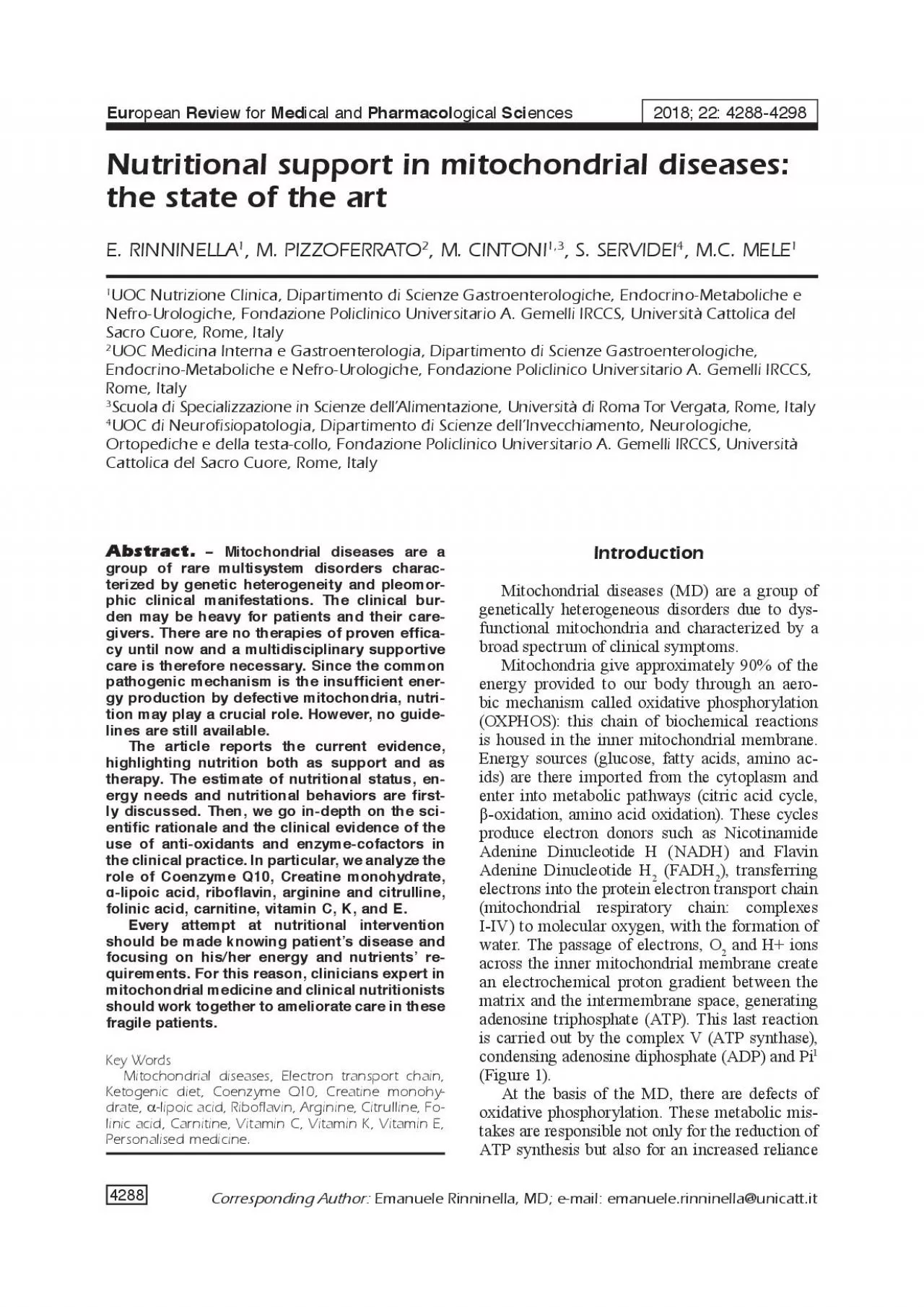
4288 Abstract.Mitochondrial diseases are a group of rare multisystem disorders characterized by genetic heterogeneity and pleomorphic clinical manifestations. The clinical burden may be heavy for patients and their caregivers. There are no therapies of proven ef�cacy until now and a multidisciplinary supportive care is therefore necessary. Since the common pathogenic mechanism is the insuf�cient ener European Review for Medical and Pharmacological Sciences2018; 22: 4288-4298 UOC Nutrizione Clinica, Dipartimento di Scienze Gastroenterologiche, Endocrino-Metaboliche e Nefro-Urologiche, Fondazione Policlinico Universitario A. Gemelli IRCCS, Università Cattolica del Sacro Cuore, Rome, ItalyUOC Medicina Interna e Gastroenterologia, Dipartimento di Scienze Gastroenterologiche, 4289 on non-aerobic metabolic pathways –such as glycolysis and glycogenosis – to provide ATP, which in turns may result in elevated serum lactate. On the other hand, uncoupled oxygen may generate reactive oxygen species (ROS), contributing to oxidative cell damage. Mitochondria are the only organelles that host their own DNA. Thus, the pathogenic mechanisms that produce mitochondrial dysfunction rely on mutations that can affect either the mitochondrial DNA (mtDNA) or the nuclear DNA (nDNA). nDNA mutations are inherited in a Mendelian way with autosomal recessive, dominant or more rarely X-linked transmission. Conversely, mtDNA mutations derive almost exclusively from the mother given the lack of mtDNA contribution of the sperm to the zygote cell (matrilineal, non-mendelian transmission). As far as the mtDNA changes, the entity of disease depends on the rate of mutant and wild-type mtDNA genomes housed in the maternal egg cell and with the mitotic segregation randomly distributed in each cell and in the different tissues (mitochondrial heteroplasmy. For the same reason, there is an extreme clinical variability of the disease expression among different individuals carrying the same mutation even in the same family.MD are overall the most common inherited metabolic disorders and are among the most common inherited neurological disorders. Their incidence is around 11.5/100,000 worldwide. Instead, the prevalence of childhood-onset (16 years of age) disorders varies from 5 to 15 cases per 100,000 individuals in different countries, while in the adult population from the North East of England is about 9.6 cases per 100,000 individuals for MD due to mutations in mtDNA and 2.9 cases per 100,000 for mutations in nDNA. Furthermore, it was calculated that 10.8 per 100,000 individuals were potentially at risk of developing mtDNA-linked disorders having an affected �rst-degree relativeMD may be syndromic or non-syndromic but, given the ubiquitous distribution of mitochondria (except in red blood cells), are mostly multisystem. However, because muscle and nervous cells have elevated energy needs, they are the main targets of these diseases. Clinical presentation may vary including muscle weakness, lactic acidosis, intolerance to exercise, loss of motor control, dementia, ataxia, parkinsonism, psychomotor regression, developmental delay, pain, seizures, migraine, stroke-like episodes, diabetes mellitus, cardiac diseases, respiratory complications, visual or hearing problems, short stature, gastrointestinal disorders and swallowing dif�culties, poor growth, cachexia, liver and kidney disease, psychiatric disordersSymptoms and signs may represent isolated manifestations, but more often may combine in speci�c well-characterized phenotypes or widely overlap. The most common phenotype in children is Leigh syndrome and in adult CPEO, both grouping nuFigure 1. Oxidative phosporylation in the inner mitochondrial membrane. NADH:
Nicotinamide Adenine Dinucleotide H; FADH2: Flavin Adenine Dinucleotide H2; ATP: adenosine triphosphate; ADP: adenosine diphosphate; CoQ: Coenzyme Q; Cyt-C: Cytochrome C. 4290 merous different disorders. The better-characterized clinical syndromes are KSS, MELAS, MERRF, and LHON (Table I).The diagnostic workup of MD often includes a muscle biopsy. The morphological hallmark is the presence of the “ragged red �bers” (RRFs), myo�bers that accumulate mitochondria in the subsarcolemmal regions that appeared red with the modi�ed Gömöri trichrome staining. The same �bers strongly react for succinate dehydrogenase and are mostly negative for cytochrome oxidase. We have to keep in mind however that RRFs may be absent in speci�c genetically-proven MD (i.e., NARP).Since MD are clinically heterogeneous, treatment should be multidisciplinary, and long-lasting and each symptom needs to be managed in a proper way. In this sense, a speci�cally-designed supportive care should be offered to each patient. Whatever is the clinical presentation, an adequate nutrition should be at the basis of all medical interventions. The main nutritional outcome in MD is to deliver energy to organ and tissues: this could be reached optimizing cells energy production and reducing energy losses. Another endpoint is to reduce free-oxygen radicals’ synthesis and myo�bers damage. Lacking guidelines on this topic, there are two possible nutritional approaches to MD: a general approach, transversal to any syndrome as nutritional support, and a speci�c one targeted to speci�c conditions as potential nutritional therapy. We brie�y discuss both of them. Principal MD, inheritance way, onset and most common features.Acronym Syndrome name Inheritance OnsetCommon FeaturesKSSKearns-Sayre syndrome Sporadic20 yearsPEO, pigmentary retinopathy, heart conduction block, ataxiaMILSMaternal Inherited Maternal,<2 yearsBrain abnormalities: ataxia, seizures,leigh syndrome mendelian impaired vision and hearing;or subacute muscle weakness: dysphagia, impairednecrotizing speech and eye movements encephalomyopathy MDSMitochondrial DNAMendelian2 yearsMuscle weakness, liver failuredepletion syndromeMELASMitochondrial Maternal2-40 yearsBrain abnormalities: stroke-like episodes,encephalomyopathy, seizures, vomiting, migraine-type headaches;Lactic acidosis and Muscle weakness, PEOStroke-like episodes Hearing loss, diabetes, short statureMERRFMyoclonus epilepsy MaternalLateMyoclonus, seizures, ataxia and musclewith Ragged red adolescence weakness; pigmentary retinopathy, coordination�bersto adulthood loss, dementia.MNGIEMitochondrial NeuroMendelian<20 yearsPEO, peripheral neuropathy, limb muscleGastro-intestinal weakness, GI disorders (diarrhea, abdominal encephalomyopathy pain)NARPNeuropathy, Ataxia Maternall2 years toNeuropathy, ataxia and retinitisand Retinitis adulthood pigmentosaPearson syndromeSporadic2 yearsPancreas abnormalities and anemiaPEOProgressive externalOphtalmo-11-18 yearsPEO as main feature. plegia Exercise intolerance Maternal, Mendelian, SporadicLHONLeber hereditaryMaternal<30 yearsVisual loss, pre-excitation cardiac conduction optic neuropathy syndromes, spasticity, dystonia and encephalopathy. 4291 Nutritional support should be at the basis of any approach to each of these syndromes. Indeed, patients affected by MD are at risk of malnutrition. Many of them have not the necessary autonomy to prepare and consume healthy meals, as they need. Moreover, gastroesophageal and pharyngeal motility disorders (dysphagia, swallowing problems and fatigue) and gastrointestinal problems often reduce nutrients’ intake and absorption.
These aspects have been yet correlated to a low Body Mass Index (BMI) in such populationA recent observational cross-sectional study, conducted in The Netherlands, collected 3-days nutrition diaries from sixty patients holding a mitochondrial disease, comparing these data with those of healthy Dutch individuals. As compared with healthy subjects, intake of proteins and calcium (as dairy products) was signi�cantly lower, while carbohydrates intake (mainly bread and potatoes) were signi�cantly higher. This is a noteworthy issue, given the increased risk of metabolic disorders such as diabetes in this population. Moreover, a low protein and Vitamin D dietary regime could enhance muscular weakness and fatigue, and Vitamin D de�ciency is a well-known risk factor for osteoporosis.It is proven that, if well sustained in their nutritional needs with an age-appropriate diet and a regular physiotherapy, patients could improve clinical and biochemical parametersFirst of all, it is necessary to avoid fasting. Eating small meals frequently should be recommended to patients or their caregivers. Fasting is detrimental to these patients and may worsen fatigue, because of muscular protein breakdown to provide gluconeogenic substrates to the liver. This is a very expensive metabolic pathway, given the employment of ATP and NADH to generate glucose from amino acids, according to the following reaction:2 Pyruvate + 4 ATP + 2 GTP + 2 NADH + 2H + 4H20 Glucose + 4 ADP + 2 GDP + 6Pi + 2NADATP synthesis is so overwhelmed with increased glycolysis and lactic acid production in a vicious cycle. Moreover, during a prolonged period of fasting, gluconeogenesis shifts acetyl-CoA towards the synthesis of ketones bodies, lowering plasma pH, increasing the risk of metabolic acidosisFrom a nutritional point of view, MD could also bene�t from standard measures yet applicable to other neurological diseases, both of peripheral and central nervous system. Recently, the European Society for Clinical Nutrition and Metabolism (ESPEN) published guidelines for clinical nutrition in neurology. The paper focuses on several aspects of neurologic diseases to which we redirect the reader. A general recommendation is to estimate the nutritional status and the energy requirements of the patients. Several tools are available to analyze the nutritional risk, such as body mass index (BMI), BMI loss, lab exams as albumin and serum lipids, bioelectrical impedance (BIA) and dual-energy X-ray absorptiometry (DEXA) to estimate body composition. Energy needs in neurological patients should be estimated as about 30 kcal/kg body weight depending on physical activity and adapted to weight and body composition evaluation. No recommendation, however, is given about protein content. Even if quantity and quality of protein are not recommended on these guidelines, 1.0-1.5 g/kg of ideal weight should be effective and safe, except for patients with renal failure. Also, amino acids are potentially useful to deal with sarcopenia. Among these, leucine and its derivatives should be preferred given their stimulation of mTOR pathway with a boost in protein synthesis, cell growth, and metabolismA peculiar topic of the ESPEN guidelines is the management of oropharyngeal dysphagia (OD), often present in MD. Patients should be screened for possible dysphagia with general (structured questionnaires, water and volume viscosity swallow tests) or specialist approaches (video-�uoroscopy, �exible endoscopic evaluation of swallowing). In case of swallowing fatigue or long-lasting meals, patients (or their caregivers) should fractionate meals and enrich their caloric content. Patients may report weight loss: in these case, oral nutritional supplements are
recommended. In case of moderate dysphagia, the texture of solid and liquids should be adapted to facilitate swallowing and avoid aspiration; postural maneuvers (such as chin-tuck posture) could protect the airway during swallowing. Finally, enteral nutrition (EN) is necessary for patients in whom oral feeding does not meet nutritional needs and in whom malnutrition/dehydration could be responsible for reduced sur 4292 vival. EN may be delivered by nasogastric tube or Percutaneous Endoscopic Gastrostomy (PEG) in case of medium or long-term supplyEven if, to date, no speci�c therapy for MD exists, a speci�c diet and nutritional supplements may improve, or stabilize, disease signs and symptoms.There are not structured evidence and large-population studies about this topic; nevertheless, emerging evidence in few patients and anecdotal reports, may help in the ordinary management and pave the path for further robust studies.The rationale of a nutritional therapy is based on the evidence that diet composition in�uences mitochondrial function: the relative ratio of dietary macronutrients can select or bypass the complexes of the respiratory chain in which electron enter. For instance, in a Drosophila model, it has been demonstrated a lower Complex-I (C-I) activity in �ies fed with a low protein: carbohydrates (P:C) ratio (1:12), compared to those fed with a higher P:C ratio (1:3) diet11. This evidence may be translated in cases of MD associated to defective C-I. In this sense, diet may have the ability to modulate metabolic defects associated with mitochondrial mutationsGiven the complexity and heterogeneity of MD, to modulate the clinical expression, clinicians should �rst identify the speci�c defect, then prompt a patient-tailored nutritional treatment. Various diets and oral supplements have been proposed. One of the more debated with con�icting evidence is the ketogenic diet.The ketogenic diet (KD) is a high-fat, low carbohydrate diet, commonly used for short periods in metabolic syndrome and obesity13. It also has a role in the treatment of refractory epilepsy, in which it has demonstrated to reduce seizures, and has been proposed as a nutritional therapeutic approach in MD with epilepsy. The scienti�c rationale lies in the evidence that KD stimulates lipid beta-oxidation in mitochondria with ketone bodies production in the liver. This mechanism could be especially exploited in subjects showing C-I de�ciencies, given the fact that lipid beta-oxidation enhances electron �ow through Complex II (C-II), by-passing partly C-IThe effects of a high-fat diet (HFD) on the mouse were veri�ed by Wall et al in a specific model called polymerase gamma mutator (POLG), housing point mutations and deletions in the mitochondrial genome. The Authors showed that an HFD was able to restore thermogenesis in the brown adipose tissue of POLG mice, causing an activation of mitochondrial biogenesis, as con�rmed by an increased genome expression of C-I, C-II and Complex III (C-III) of the respiratory chain. Moreover, morphological changes of POLG mouse mitochondria toward a WT-like model were observed. A human pilot study by Ahola et al experimented a type of KD, called modi�ed Atkins Diet (mAD) (3-9% of carbohydrates, high fat, high protein content) as dietary treatment in �ve subjects affected by PEO/MM. These patients were compared to age and gender-matched healthy controls. After 2 weeks of a normalized isocaloric standard diet, the subjects were given with mAD. Within the �rst two weeks of treatment, PEO/MM patients experienced burning sensations and muscle pain. Serum creatin kinase (CK), alan
ine aminotransferase (ALT) and myoglobin values were increased, suggesting a muscle damage. Healthy subjects did not report any symptoms. The trial was prematurely stopped for ethic reasons. At muscle biopsy, some PEO/MM muscle �bers showed acute degenerative changes affecting RRFs selectively. After 2.5 years, however, three out of four PEO patients showed improved muscle strength. Authors discussed the evidence: mtDNA mutated cells may be especially dependent on pyruvate and glycolysis products for their energy metabolism, whilst they are not able to utilize lipids, amino acids, and ketones as an energy source. The boost in β-oxidation, protein oxidation and ketogenesis, driven by a KD, could lead to a “selective rhabdomyolysis” of RRFs. On the other side, healthy muscle cells may undergo fusion to replenish the sick, damaged cells. At the actual evidence, KD in MD could be only suggested for children with syndromes in which a refractory epilepsy is the principal clinical feature. In these conditions, ketogenic diet seems to have both a neuroprotective and anti-in�ammatory effect (reduction in excitotoxicity and oxidative stress) and also a direct anticonvulsant action. This effect could be reached via a modulation of sodium and potassium voltage-gated channels and a downregulation of α-amino-3-hydroxy-5-methyl-4-isoxazolepropionic acid (AMPA)-receptor. The ef�cacy of KD in MD with epilepsy was demonstrated in a few studies. Lee et al have shown a signi�cant reduction of seizures in a cohort of 48 patients with con�rmed mitochondrial respiratory 4293 chain defects and medically intractable epilepsy. Zhu et al reported an improved neurological and psychomotor development in children with intractable epilepsy treated with KD. Martikainen et alsuggested KD as an adjunct to antiepileptic drugs in POLG-relating epilepsy. A very low carbohydrate diet should not be considered as the net solution by itself. Firstly, it is not advisable for all patients, in particular for patients affected by liver, heart and kidney diseases and those at risk of osteoporosis of biliary stones. Moreover, metabolic acidosis elicited by a high fat, high protein diet may worsen fatigue and create an unfavorable metabolic setting for the daily living (impaired concentration, headache, constipation), especially if the patients are not under clinical advice. In conclusion, in the absence of controlled studies and with the risk of inducing or worsening a metabolic crisis, the use of ketogenic diet should be restricted only to children with refractory epilepsy under a strict surveillance of the metabolic equilibrium.The role of nutritional supplements (NS) is not yet truly understood. However, many of these are largely used in the clinical setting, with the hope to ameliorate care. It remains valid the concept that the real bene�t of each supplement could vary among each syndrome. Coenzyme Q (CoQ) is an electron acceptor of the mitochondrial respiratory chain, shuttling electrons from C-I-II and electron transferring �avoprotein dehydrogenase (ETFDH) to C-III (1). It is also an antioxidant. The rationale of an exogenous supplementation of CoQ is to bypass defects in the respiratory chain, reducing electron leak and restoring ATP synthesis. In vitro studies suggest an increase of mitochondrial ATP production in cultured cells from patients affected by MD due to CoQ supplementation. CoQde�ciency underlies several types of mitochondrial diseases depending on the modi�ed gene. It may be present in mitochondrial disorders as primary or secondary de�cits. Even when the de�cit of CoQ is proven, CoQ supplementation may not be effective, being the
pathogenic mechanism not fully understood in all syndromes (Table II).Nevertheless, irrespectively from the phenotype, CoQ is widely used in MD, including MELAS in which a small randomized trial reported improvement of muscle weakness, fatigability, and serum lactateHowever, CoQ it has a limited effect on the central nervous system because it does not cross the blood-brain barrier. Conversely, its analog Idebenone can cross the blood-brain barrier with a potential role in stroke-like episodes in MELASIdebenone is instead approved for the treatment of Leber’s hereditary optic neuropathy (LHON), although some issues still remain to de�ne as timing, dose, and frequency of administrationand which are the patients to treatThe dosage suggested for oral CoQ supplementation as ubiquinone is up to 3000 mg, but the average dose is around 300 mg/daily.Ubiquinone is insoluble in water, and it has a poor intestinal absorption. A recent, reduced formulation, ubiquinol, seems to be better absorbedCreatine monohydrate (CrM) is an antioxidant and a potential energy source for defective mitochondria, through the phosphocreatine system. It also works as energy shuttle of ATP and phosphates from mitochondrial sites to the cytoplasm. It is commonly used by athletes as a dietary supplement to increase muscle performance in short-duration and high-intensity resistance exercises, at the dosage of 0.3 g/kg daily for 5 to 7 days, followed by maintenance dosing at 0.03 g/kg daily for 4 to 6 weeksPatients affected by MM have low creatine levels in muscles and brain. In a randomized, crossover trial on seven patients affected by mitochondrial cytopathies, the administration of CrM (at the oral dosage of 5 g bid for 14 days, followed by 2 g bid for 7 days) signi�cantly improved physical performance and reduced post-exercise lactate. Conversely, no signi�cant effects were observed in lower intensity aerobic activities. Similar results are reported in a small group of children with mitochondrial encephalomyopathies31-lipoic acid is a natural antioxidant, present in mitochondria as a cofactor for pyruvate dehydrogenase and -ketoglutarate dehydrogenase. Its role in MM could lie in scavenging ROS generated by defective oxidative phosphorylation, decreasing oxidative stress in mitochondria and cells’ membranes. It may also enhance the uptake of phosphocreatine as an alternative energy source to anaerobic glycolysis in defective mito 4294 chondria. Suggested dosage is 50-200 mg/dayA combination therapy of CoQ, CrM, and -lipoic acid has been used as a treatment option in a randomized, double-blind, placebo-controlled, crossover study, recruiting sixteen patients with several mitochondrial diseases. Compared to placebo, this cocktail signi�cantly reduced plasma lactate concentration and urinary 8-isoprostane, a marker of oxidative stress. After the treatment, patients showed a minor reduction in peak ankle dorsi�exion strength than that observed after the placebo phase. Moreover, a subgroup of patients with MELAS showed signi�cant improvements in body composition, such as an increase in fat-free mass (FFM) and total body water (TBW) and a decrease in body fat. Authors stressed the positive synergistic effect of these compounds in MD, even if they recognized that not all the patients affected by MD may have the same bene�ts. Ribo�avin is a vitamin of group B (B2 vitamin), and it acts as a �avoprotein precursor in C-I and C-II and as a cofactor in several other enzymatic pathways of β-oxidation and Krebs cycleIn patients with myopathy due to ETFDH mutations, ribo�avin associated with CoQ supplementation has been effective in improving clinical and biochemica
l parameters (serum CK and lactate)Non-randomized studies have shown its ef�cacy in C-I and C-II mitochondrial diseases36,37The suggested oral dosage is 50-400 mg daily, both in children and in adultsArginine is a nitric oxide (NO) precursor. Its clinical bene�t is still debated. Ef�cacy of intravenous (iv) administration of L-arginine has been proposed in MELAS in the acute phase of the stroke-like episodes. The same group suggested the use of chronic oral arginine supplementation showing a reduction of frequency and severity of stroke-like episodes in patients with MELAS. A daily dose of 150 to 300 mg/kg/day oral arginine divided into three doses was recommendedArginine ef�cacy could be related to cerebral vasodilatation and endothelial function, due to an increase in NO availability. However, there are some concerns about the safety of L-arginine, especially for intravenous administrationCitrulline can also increase NO production in patients with MELAS syndrome, although its clinical effects have not yet been studied. Interestingly, citrulline was associated with a greater increase in NO and cerebral blood �ow than arginine supplementation, indicating citrulline as po Abbreviations: CoQ: Coenzyme Q10; COQ2: Coenzyme Q2; COQ4: Coenzyme Q4; COQ6: Coenzyme Q6; COQ9: Coenzyme Q9; PDSS1: Prenyl (Decaprenyl) Diphosphate Synthase, Subunit 1; PDSS2: Prenyl (Decaprenyl) Diphosphate Synthase, Subunit ADCK3: aarF domain containing kinase 3 ETFDH: Electron Transfer Flavoprotein Dehydrogenase; APTX: aprataxin.SyndromesGene involvedType of CoQ Response to CoQ10deficit supplementationNeonatal multisystemic disorder, nephropathy,COQ2PrimaryGood responselactic acidosis, Leigh syndrome, neonatalencephalopathy with seizures, MELAS,retinitis pigmentosaEncephalomyopathyCOQ4PrimaryGood responseNephropathy, deafness, seizuresCOQ6PrimaryGood ResponseNeonatal encephalomyopathy, lactic acidosis,COQ9PrimaryPoor responserefractory seizuresInfantile nephropathy, hepatopathy, mental PDSS1PrimaryNot knownretardation, lactic acidosis, optic atrophyLeigh syndrome, lactic acidosis and nephropathyPDSS2PrimaryPoor responseCerebellar ataxia and atrophy± retardation,ADCK3PrimaryPoor responselactic acidosis and exercise intoleranceNephropaty, mild mental retardationADCK4PrimaryGood responseIsolated myopathyETFDHSecondaryGood responseCerebellar ataxiaAPTXSecondaryGood response 4295 tentially more effective than arginine in patients with MELAS syndromeAlthough these �ndings may appear promising, they should be con�rmed in a long-term randomized controlled trial both for arginine and citrulline.The term folate refers to a family of compounds belonging to B vitamins, acting as coenzymes in carbon-transfer reactions. These include folic acid, folinic acid – both present in plants and food – and 5-methyltetrahydrofolate (5-MTHF), the biologically active form. The known metabolic activities of these compounds or their derivatives are the purine synthesis and the methylation of homocysteine to methionine. In mitochondria, folate metabolism regards essentially the synthesis of and formate and translation of transfer RNA (tRNA) for protein synthesis. Folate de�ciency is quite common in patients affected by MD and it could worsen mitochondrial metabolism. However, folate de�ciency seems to be more related to a speci�c syndrome, the Kearns-Sayre syndrome (KSS) in which there is an impaired ATP-related transport of 5-MTHF through the choroid plexus into cerebrospinal uid with consequent defects in methylation of myelin protein. In this sense, a systemic folate de�ciency may aggravate mitochondrial ATP production and myelin synthesis. For this reason, an
external supplementation of folate has been suggested and its ef�cacy is proven in some case reports45-47. Folic acid seems to ineffective due to a genetic mutation of dihydrofolate reductase (DHFR) methylenetetrahydrofolate reductase (MTHFR), so, the active compound folinic acid is preferred, even if no a standard dosage is to date givenCarnitine is a cellular compound, predominant in skeletal muscle, heart, and liver, playing a crucial role in energy production. It transports into mitochondria, across the mitochondrial membrane, long-chain fatty acids as acyl carnitine esters. Moreover, it also prevents CoA depletion and facilitates the excess of toxic acyl compounds. For such reason, it is empirically used by some clinicians in the management of MD, although there is not proven ef�cacy with the exception of primary carnitine de�ciency. Recently, some concerns have been raised about its possible role in increasing cardiovascular risk, probably mediated by gut microbiotaVitamin C and Vitamin K are antioxidants50, 51they are also able to bypass mitochondrial C-III de�ciencies as electron acceptors. However, clinical evidence of a role in patients with recognized mitochondrial depletion syndromes is lacking. Only in a single case report, the supplementation of both of these vitamins was associated with short-term clinical improvementTocopherol was proposed for its antioxidant capacity in mitochondrial dysfunctionsand in neuroprotectionhowever, no evidence is available in mitochondrial diseases. It was speculated that tocopherol might have a role only in (LHON), in which a signi�cant de�ciency of vitamin E was demonstratedAll considered, many compounds have been used and tried as nutritional therapy and numerous reports have proposed various cocktails of antioxidants and nutrients. However, in the absence of large, properly controlled trials, the latest Cochrane review concluded that there is currently no clear evidence supporting the use of any intervention in mitochondrial disordersNevertheless, in the recent consensus conference “Nutritional Interventions in Primary Mitochondrial Disorders: Developing an Evidence Base”, an agreement was achieved, using the Delphi consensus methodology, on the following points: 1)CoQ10 should be administered to most patients with a diagnosis of mitochondrial disease, and not exclusively for the primary CoQ10 de�ciency. 2)Alpha-lipoic acid and ribo�avin are the only other supplements that obtained a certain degree of agreement.3)Folinic acid may be considered in patients with CNS manifestations especially when is documented a cerebral folate de�ciency.5)L-carnitine should only be administered when there is a documented carnitine de�ciency.Moreover, it is suggested to start the treatment with one supplement only and to evaluate the clinical response before adding other supplements, avoiding the “cocktail approach” since the beginningTreatment and management of patients with MD remain a major task. Apart from speci�c disorders such as primary CoQ de�ciency and 4296 ribo�avin-responsive disorders, in the absence of proven therapies, the most useful approach is supportive care. In this setting, nutritional support has a crucial role. Randomized, controlled clinical trials with large sample sizes are necessary. However, at the basis of any nutritional approach is the evaluation of the nutritional status and the patient’s energy needs. Then, in collaboration with the patients and their family, is possible to build an appropriate program which provides for adequate caloric and protein support paying also attention to the food texture and the dif
9;culties in the assumption of the food (i.e., dysphagia, GI problems, etc.). To improve nutritional care in MD, we hence suggest prioritizing nutritional support paying attention to avoid fasting and meet the energy needs. Every attempt at nutritional intervention should be made knowing patient’s disease and focusing on his/her energy and nutrients’ requirements. For this reason, clinicians expert in mitochondrial medicine and clinical nutritionists should work together to ameliorate care in these fragile patients. Conflict of Interest The Authors declare that they have no con�ict of interests.References ELSO D, COX MM, Introduzione alla biochimica di Lehninger, quinta edizione italiana. Zanichelli, 2015. IS C, BOTTA, ZIA M. Emerging concepts in the therapy of mitochondrial disease. Biochim Biophys Acta 2015; 1847: 544-557. OR G, CHINNERY PF, DAUROIRA M, , MARLANDUOALAIHORUR DIA M, URNBULL DM. Mitochondrial diseases. Nat Rev Dis Primers 2016; 2: 16080. OLSTEI, WOLLA The elusive magic pill: �nding effective therapies for mitochondrial disorders. Neurotherapeutics 2013; 10: 320-328 AAT P, ZEERSHE, KUIJTEITINKJA, WTESSE MC. Dysphagia, malnutrition and gastrointestinal problems in patients with mitochondrial disease caused by the m3243A>G mutation. Neth J Med 2015; 73: 30-36. EERSSSE MC, EIJ, WTE G. Patients with mitochondrial disease have an inadequate nutritional intake. JPEN J Parenter Enteral Nutr 2017 Mar 1:148607117699792. doi: 10.1177/0148607117699792. [Epub ahead of print] TEREYERHLE C, ERTTÖCKLEHS G, ZART P, FLESH, BAHRS C, NUSSLERK. Crucial role of vitamin D in the musculoskeletal system. Nutrients 2016; 8: pii: E319. ORA, NBUR, SSEZ, D VRIES M, EITINK. Dietary intervention and oxidative phosphorylation capacity. J Inherit Metab Dis 2006; 29: 589. UROS, BRETO, CERE, DESORTC, DIEASTO, GES F, ESUS P, EISER, MUSARITOLIM, POULIA KP, PREISERC, VER MARCK M, WIRTHNGER P, BISHO . ESPEN guideline clinical nutrition in neurology. Clin Nutr 2018; 37: 354-396.10) EROUSMBERTNGLAIS, CARRARO V, GOUREYRE, PARRY, B’CHIR W, MURAISHIOUSSE C, BRUHATAURIC, PROU CG, FOUROUX P. Requirement for lysosomal localization of mTOR for its activation differs between leucine and other amino acids. Cell Signal 2014; 26: 1918-1927.11) HAU N, MESSER M, CORREA CC, BALLARW. Diet in�uences the intake target and mitochondrial functions of Drosophila melanogaster males. Mitochondrion 2013; 13: 817-822.12) ALLARW, NGSO N Review: can diet in�uence the selective advantage of mitochondrial DNA haplotypes? Biosci Rep 2015; 35. pii: e0027713) ERRA G, GRATTERI, DORENZ, BARRUCC, PERRO, OLIO, BERAR, MARHETTI M, DNZ Effects of very-low-calorie diet on body composition, metabolic state, and genes expression: a randomized double-blind placebo-controlled trial. Eur Rev Med Pharmacol Sci 2017; 21: 329-345.14) AI Q, ZHOU ZUO, GP, M DZ, W CM. Safety and tolerability of the ketogenic diet used for the treatment of refractory childhood epilepsy: a systematic review of published prospective studies. World J Pediatr 2017; 13: 528-536.15) NGC, EEM, KD, EEJSLA Safe and effective use of the ketogenic diet in children with epilepsy and mitochondrial respiratory chain complex defects. Epilepsia 2007; 48: 82-88.16) ALL C, WHYTEUHM, F W, COLLI B, DDLEC, RTAR, NIAUXC, K, BRIHTATLAYCK W, DWNES M, NIAUXK, M. High-fat diet and FGF21 cooperatively promote aerobic thermogenesis in mtDNA mutator mice. Proc Natl Acad Sci U S A 2015; 112: 8714-8719.17) HOLAURA M, SOHANN P, NIEISALORHON, BZK, VELA V, NDB N, KKARAI, MUURI, PIIRILÄ P, PIETILÄI KUOALAI . Modi�ed Atkins diet induces subacute selective ragged-red-�ber lysis in mitochondrial myopathy patients. EMBO Mol Med 2016; 8: 1234-1247.18) ALEOLOOUAYILO
N, KALI M. Use of the ketogenic diet to treat intractable epilepsy in mitochondrial disorders. J Clin Med 2017; 6: pii: E56.19) EEM, KNGC, EEJS, KSH, KEYEEK, LA, KD. Mitochondrial respiratory chain defects: underlying etiology in various epileptic conditions. Epilepsia 2008; 49: 685-690.20) HU D, WNG M, WNGUA, NIU G, ZHANG G, IONGIE M, ZHAO Ketogenic diet effects on neurobehavioral development of children with intractable epilepsy: a prospective study. Epilepsy Behav 2016; 55: 87-91.21) ARTIAI M, PÄIÄRITA M, ÄÄSELÄIAJAAA K. Successful treatment of POLG-related mitochondrial epilepsy with antiepileptic drugs and low glycaemic index diet. Epileptic Disord 2012; 14: 438-441. 4297 22) ERO D, MTEROSTRONGÓS C, PALAUF, TOSAÑA C, ALIATI, NAS P, RTU Molecular diagnosis of coenzyme Q10 de�ciency. Expert Rev Mol Diagn 2015; 15: 1049-1059. 23) OI M, DESATS M, CERUA C, CASSI M, REISSOALIATI . Genetics of coenzyme q10 de�ciency. Mol Syndromol 2014; 5: 156-162.24) IRA M, GARO C, QUINZII CM. CoQ(10) de�ciencies and MNGIE: two treatable mitochondrial disorders. Biochim Biophys Acta 2012; 1820: 625-631.25) LOEREI, MARTI, MAHERHORHILLRE, MORAAROLS M A randomized trial of coenzyme Q10 in mitochondrial disorders. Muscle Nerve 2010; 42: 739-748.26) OUOU, KOUASSOUA, CHO, UAUTÉHOHOSSIA N, DEREX. Effect of long-term oral treatment with L-arginine and idebenone on the prevention of stroke-like episodes in an adult MELAS patient. Rev Neurol (Paris) 2011; 167: 852-855.27) ARELLI V, CARELLI M, COOF, KASALOSTOCKRÈ W MORIA C, NWM NRSSAU C, POTTAAERNG, VGNAL-CLER C, VOTRU M, -WAI-M P, AR P. International Consensus Statement on the Clinical and Therapeutic Management of Leber Hereditary Optic Neuropathy. J Neuroophthalmol 2017; 37: 371-381.28) ARRIA B, CLAND M, MALM, GLERU DM. Cofactor treatment improves ATP synthetic capacity in patients with oxidative stress. Mol Genet Metab 2004; 81: 263-272.29) ALL M, ROJIATH Creatine supplementation. Curr Sports Med Rep 2013; 12: 240-244. 30) AROLS MOY BD, MALJR A randomized, controlled trial of creatine monohydrate in patients with mitochondrial cytopathies. Muscle Nerve 1997; 20: 1502-1509.31) ORHERT, WILIHOEL F. Supplementation with creatine monohydrate in children with mitochondrial encephalomyopathies. Muscle Nerve 1999; 22: 1299-1300.32) UR DG, CHILICK PD, PARISE G, AROLS MND DG. Effects of α-lipoic acid combined with creatine monohydrate on human skeletal muscle creatine and phosphagen concentration. Int J Sport Nutr Exerc Metab 2003; 13: 294-302.33) ARIETO, FAL MSEL, COHE BAAS, MIETY . A modern approach to the treatment of mitochondrial disease. Curr Treat Options Neurol 2009; 11: 414-430.34) RIUE MC, MALJR, MAHOEY D, PARISEG, BEAL MF, AROLS M Bene�cial effects of creatine, CoQ10, and lipoic acid in mitochondrial disorders. Muscle Nerve 2007; 35: 235-242.35) MPEL K, ALOLU, ALI B, EIERAT P, HOSERBG, V, PAL B, KALE G, ATLI, QUINZII C, IRA M, NAI, DAURO, PROIS, ULLER, ORATH The myopathic form of coenzyme Q10 de�ciency is caused by mutations in the electron-transferring �avoprotein dehydrogenase (ETFDH) gene. Brain 2007; 130: 2037-2044.36) IA M, TEANVERZZ F, MORO, BZZ, ZIA M, IEL G. Effects of ribo�avin in children with complex II de�ciency. Brain Dev 2006; 28: 576–581.37) ERSE P, GREELS FUITENBEE W, MBURER HL Treatment of complex I de�ciency with ribof avin. J Neurol Sci 1993; 118: 181-187.38) ITA, NISHIOATSU, PAL N, , FUJIOTO, MATSUISHI L-arginine improves the symptoms of strokelike episodes in MELAS. Neurology 2005; 64: 710-712.39) ITA, NISHIOATSU, PAL N, ATAYA K, MATSUISHI MELAS and L-arginine therapy. Mitochondrion 2007; 7: 133-139.40) ATTAW, RICKLTSUW, CHANPRASERTNNAI M, CRAI WAHOOR F, LIA F.
Impaired nitric oxide production in children with MELAS syndrome and the effect of arginine and citrulline supplementation. Mol Genet Metab 2016; 117: 407-412.41) D, LITOEE, BEH New indications and controversies in arginine therapy. Clin Nutr 2008; 27: 489-496.42) AL, CASA M, MOLEROUIS M, MTOYA, AH, YLETTB, ARREAES, EALESRTU. Can folic acid have a role in mitochondrial disorders? Drug Discov Today 2015; 20: 1349-1354.43) HANG CM, CC, HT, CHOUF, UANGF. Folate deprivation promotes mitochondrial oxidative decay: DNA large deletions, cytochrome c oxidase dysfunction, membrane depolarization and superoxide overproduction in rat liver. Br J Nutr 2007; 97: 855-863.44) ERRA M, GARÍAIL M, MARTIERND’CALLAHA MEL M, QUIJA P, MARTIRAAL, BLÁZQUE, MARTÍ M, BRIOES P, -GALLARUI-PESI, MTOYARTU, P M. Kearns-Sayre syndrome: cerebral folate de�ciency, MRI �ndings and new cerebrospinal �uid biochemical features. Mitochondrion 2010; 10: 429-432. 45) M, AL, -GALLAR, NASTO, OLA, ERRERO MD, VILASE M, BRIOESP, ÁÑE, MTOYA, RTU Cerebral folate de�ciency and leukoencephalopathy caused by a mitochondrial DNA deletion. Ann Neurol 2006; 59: 394-398.46) AEERS V, WEISUEIRAM, QUAROSV, LAU N. Mitochondrial complex I encephalomyopathy and cerebral 5-methyltetrahydrofolate de�ciency. Neuropediatrics 2007; 38: 184-187.47) UIJA-FRAILE P, ’CALLAHA M, MARTÍERNDTERO, GARIA-CORLA À, RAM, MHART, MÁLA, PAR, GARÍA-GNZALE P, OU C, MTOYA, MPERAOR, UI-PESI, REAS, MARTI MAL, P M, GARÍAIL M, RTU Follow-up of folinic acid supplementation for patients with cerebral folate de�ciency and Kearns-Sayre syndrome. Orphanet J Rare Dis 2014; 9: 217.48) IUETTIM, TANCULELLA, G GV, IA F. Nutritional and hormonal regulation of citrate and carnitine/acylcarnitine transporters: two mitochondrial carriers involved in fatty acid metabolism. Int J Mol Sci 2016; 17. pii: E817.49) OETHRA, WNG Z, ISO B, BFFJA, HEEHY B, BRITTB, F, W, , ITHD, ATOJA, CHE, , W GD, ISD, WARRIERM, BROWNM, KRAUSSM, NG W, BUSH FD, 4298 USISAJ, SL Intestinal microbiota metabolism of L-carnitine, a nutrient in red meat, promotes atherosclerosis. Nat Med 2013; 19: 576-585.50) ARAHULE M, NG, BURLI M, , PARB, ÖLSHER. Vitamin C suppresses lipopolysaccharide-induced procoagulant response of human monocyte-derived macrophages. Eur Rev Med Pharmacol Sci 2016; 20: 2174-2182.51) OSTOIA D, C . Crosstalk between vitamins A, B12, D, K, C, and E status and arterial stiffness. Dis Markers 2017; 2017: 8784971.52) HI M, BIT P, COULIALYOULIER-KHOURYUSTI P. Mitochondrial response to controlled nutrition in health and disease. Nutr Rev 2011; 69: 65-75.53) ATIELARAJU, KHAAI, VYAARA, HIBD MUTALI M, HANDRA V, MD Cytoprotective effect of tocotrienol-rich fraction and α-tocopherol vitamin E isoforms against glutamate-induced cell death in neuronal cells. Int J Vitam Nutr Res 2014; 84: 140-151.54) LIYI P, KAR, SA C, ORATH, KOLY, NETH, URI, VSEI. alpha-Tocopherol/lipid ratio in blood is decreased in patients with Leber’s hereditary optic neuropathy and asymptomatic carriers of the 11778 mtDNA mutation. J Neurol Neurosurg Psychiatry 2001; 70: 359-362.55) ER G, MAJAAA K, URNBULL DM, HORUR D, HINNERY PF. Treatment for mitochondrial disorders. Cochrane Database Syst Rev 2012; 4: CD004426.56) MP KM, KROTOS D, PARISI M, GNN K, COHE, COX CNN GM, FAL M, GOLSTEIC, ALRIASTA, GOR GERSHP, IRAM, FFM F, KARAA, MEOEL, MARLAND, MOHA C, MULERAENKIRHEC, PARIUTHEROR PGGNDERSOK, NG WCKLEY, WOLLANNELLIES P, COATES PM. Nutritional interventions in primary mitochondrial disorders: developing an evidence base. Mol Genet Metab 2016; 119: 187-206. Nutritional support in mitochondrial diseases: the state of the art E. Rinninella, M. Pizzoferrato, M. Cintoni, S. Servidei, M.C. Mel