
reflex 14 Polysynaptic reflexes are usually engagedreflex The palmomental reflex is a brief contractionunilateral pouting expression It is not easy to speculatepalmomental reflex It has been ID: 844072
Download Pdf The PPT/PDF document "Median Nerve Stimulation" is the property of its rightful owner. Permission is granted to download and print the materials on this web site for personal, non-commercial use only, and to display it on your personal computer provided you do not modify the materials and that you retain all copyright notices contained in the materials. By downloading content from our website, you accept the terms of this agreement.


1 Median Nerve Stimulation reflex (14). P. P")
Median Nerve Stimulation reflex (14). Polysynaptic reflexes are usually engagedreflex. The palmomental reflex is a brief contractionunilateral pouting expression. It is not easy to speculatepalmomental reflex. It has been suggested to be ahave been conducted in both basic and clinical work(1, 4, 9, 10, 11, 15, 17). In the literature, a study of theout (17). Since their methods were different, theirresults were also different. Therefore, we attemptedusing clinical anatomic lesions. In the study, weinformed consent. The subjects were seated in aactivities from the target muscle: the mentalis. Thesurface EMG was recorded by two disposable surfaceand the reference on the mental protuberance. Electricalpatients with congenital insensitivity to pain. Thewrist. The duration of the stimulus was 0.2 ms. ShortHz. As the perception threshold varied a lot in thebasic reference of stimulating intensity. Using awrist at the beginning of the experiment. All thestimuli were quantified in multiples of threshold. Toleast 2 minutes. We rectified the EMG and measuredV. The latency of exhibited considerable variability from trialto trial in an
2 individual subject. MMR was observed.
individual subject. MMR was observed. As facilitation occurred in was studied in three subjects. was modified fromstimulation (2, 3, 17). During the assessment of theEMG feedback. In these experiments, the stimuluss. The stimulus rate was 4.7 Hz. Theanalysis time was 100 ms after the stimulus, and filterbandwidth was 5 Hz-1.5 kHz. Because the recordingsartifact rejection was not used. The baseline was and E and Iand measured with a cursor. The measurement includedamplitude. If the preceding component was notbaseline. The receptive field of MMR was and Voluntary Reac- spreading phenomenon, and probability. A reactionrole of voluntary muscle contraction. The subjectsresponse to the median nerve stimulation. A cutaneousan object between the thumb and index finger. Subjectsthe variables of MMR. The correlation coefficient(strong positive association). The closer the correlationthe variable was shifted with respect to another. If the� value was 0.05, the data was considered insignificant. 2.00. For MMR 7.67 ms, the peak amplitudes were 47.93 21.11 ms. The amplitude and the duration of MMR varied a lot with the subject
3 and the in each subject. At 4.10. The
and the in each subject. At 4.10. The peak latencies of MMR (33.93 0.57 ms), I (38.94 4.02 ms), E (9.19 3.70 6.64 (35.62 6.71 (20.81 6.07 ms). The most consistent and the most inconsistent (Fig. 2). (70 - 90%). With a higher intensity, theconsistency would be better. could besurface. The most sensitive areas for eliciting MMR Fig. 1.In a EMG pattern changedintensity (B). The interval between two stimuli was at foot (Fig. 3A). When the mentalis was kept at a usually provokednot only in the mentalis muscles but also in otherstimulation could evoke simultaneous responses atonly one muscle was available. The latency of the 6.5 ms, and usually occurred before was seen. However, on occasion, MMRcould precede the onset of the blink. During repetitive usually habituated earlier thanseen. There was no constant relationship betweenorbicularis oculi responses and mentalis responses inSpreading Phenomenon of Continuous Stimulation of the Fig. 2.The MMR pattern usually had inhibitory and exci-tatory components and varied in subjects. Figures A, B,and C were from different normal subjects. The E was widespread overthe body surface.
4 B: On the other hand, the receptive was
B: On the other hand, the receptive was limited to the upper limb and the kept at constant contraction. The spreading phenomenonthe cranial muscles (Fig. 4B). The best consistency or I (11, 12). For all the target and MMR (Fig.4). It was clear that the latency of MMR to mediannerve stimulation was too short to be voluntary. Inat the mentalis and limb muscles (such as the flexorcarpi radialis and biceps brachii), in sequence. TheIn patients with dorsal column dysfunction, only was available for elicitation (onset latency:patient A, 42.9 ms and patient B, 73.8 ms; amplitude: was available patient C, 34.3 ms and patient D, patient C, 45.7 ms and patient D, 55.3 ms; patient C, 65.7 ms and patient D, 73.3 ms; inter- patient C, 11.4 ms and patient D, patient C, 10.0 ms and patient D, patient C, patient C,V). But, it was difficult (Fig. 5), even when the intensity was up to (age and latencies, = 0.15; age and = - 0.21; height and latencies, = 0.03; = - 0.13; intensity and latencies, = 0.13; intensity and amplitude, = 0.17). of MMR was observed in only three latencies and E durations were notincluded for analysis. For MMR = 0.83 ; I =0.90;
5 I = 0.32; I = -0.34; E = 0.05), height, height")
I = 0.32; I = -0.34; E = 0.05), height and = 0.28; I = 0.13; E = -0.36; E = -0.10), or intensity and amplitude (E = 0.03; I = 0.07; E = 0.26).� 0.05). Fig. 4.The tion was a single shock (A) or continuous (B). In figurereaction time of the mentalis contraction. In figure B, the and E responses of the latencies from up to down were 52 ms, 48 ms, 47 ms, from median nerve stimulation spread widely over thebody. In the facial muscles, this included not only thementalis muscle, but also the orbicularis oculi muscle.respond to external stimuli. However, the orbicularisstimulation exactly as the mentalis. Neither had aconstant relationship, in terms of latencies, sensitization,and habituation. Therefore, MMR was not exactlya spreading phenomenon of the blink. The dissociationthan reaction time. The afferent limb of the MMRconduction rates and diameters. The role of small was shown in our case study. Our was not elicited inpatients with dorsal column dysfunction. It is clear can be evoked by pain afferents evenwithout the large fibers of proprioception. Otherwise, was still available when a forearm ischemicthe larger fibers before
6 the small fibers (11). The of thes (10. The of thes (10")
the small fibers (11). The of thes (10). This also inferred the role of the small fiber inintensity was low and not painful. Using trainincrease. When the intensity was kept constant, trainstimulation would produce a summation effectand the brainstem (13). As the stimulus continued inobserved (Fig. 1). At that point, most subjects feltactivated in such intensities. Therefore, MMR fibers in part.muscles (7). This is taken as protective withdrawalbehavior in response to electrical stimulation (7). We . In our not only coincided with,was released. This also confirmed the hypothesis that (e.g. orbicularis oculi response) to mediannerve stimulation was a defensive behavior to protectWith a higher intensity, the duration of MMRmedian nerve stimulation (7). MMR wasmedian nerve stimulation. It seems that MMRto the cortex (5). The role of large fibers on MMRdysfunction. In these patients, MMR decreasedin amplitude significantly. Our observation confirmed (17). We further inferredflexor carpi radialis. As the EMG pattern of MMR may appear as a result of the integration appears to be (17). In normal subjects, the was not evoked by mo
7 tor point stimulationof the thenar muscl
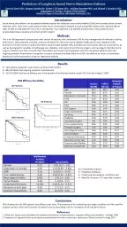
tor point stimulationof the thenar muscle (17). These researchers argued were nociceptive in patients with dorsal column dysfunction,of the median nerve. The anatomic observation and were very clear, as seen in Table 1, and had longer onsetwas evoked after repetitive stimulation; 3. MMR did not exactly share the samemechanism. Our study showed that different methodscan provoke different MMR responses. Both responses Stimulation at the wristSingle train stimulationContinuous stimulationAverageNoYesSeries of stimulihabituationpreserved responsesIntensity (mA)13.14 ( 2.00)16.20 ( 4.10)Onset latency (ms)� 60V)&#x 450; 150Sensory pathwaysmall fiberslarge fibers Receptive fieldswidespread over the body surfacethe or adjacent structures (12). Further investigation, (a reflex (apalmomental reflexes. However, our study proved thatassessed by electrophysiological methods. Median nervetrigeminal nerve stimulation. Continuous stimulation was related to the large fibers and MMRwas chiefly related to the small fibers. Based upon theresults of habituation character and latency, it was clear and MMR did not have equivalents 1.Caccia, M.R.,
8 Galimberti, V., Valla, P., Osio, M., De
Galimberti, V., Valla, P., Osio, M., Dezuanni, E. and 2.Chen, C.C., Chen, J.T., Wu, Z.A., Kao, K.P. and Liao, K.K. J. 3.Chen, C.C., Chen, J.T., Wu, Z.A., Kao, K.P. and Liao, K.K. Long 4.Dehen, H., Bathien, N. and Cambier, J. The palmomental reflex. An 5.Deuschl, G., Ludolph, A., Schenk, E. and Lucking, C.H. The 6.Guo, Y.C., Liao, K.K., Soong, B.W., Tsai, C.P., Niu, D.M., Lee, 7.Hallett, M., Berardelli, A., Delwaide, P., Freund, H.J., Kimura, J.,Central EMG and tests of motor control. Report of an IFCN 8.Herrero, J.F., Laird, J.M. and Lopez-Garcia, J.A. Wind-up of spinal 9.Miwa, H., Imamura, N., Kogahara, K., Ohori, T. and Mizuno, Y.10.Miwa, H., Nohara, C., Hotta, M., Shimo, Y. and Amemiya, K.11.Reis, D.J. The palmomental reflex. 4: 486-498, 1961.12.Rimpel, J., Geyer, D. and Hopf, H.C. Changes in the blink responses13.Shahani, B.T. Flexor reflex afferent nerve fibers in man. 14.StaroselÕtseva, N.G. and Ivanichev, G.A. The polysynaptic spine-15.Valls-Sole, J., Valldeoriola, F., Tolosa, E. and Marti, M.J. Distinc-16.Whittle, I.R., and Miller, J.D. The palmomental reflex. 17.Yamada, T., Shiga, U., Kawamura, T., Inoue, K., Ofuji, A., Had