
DifferentialdiagnosesincludeconditionssuchhypertensiveretinopathyretinalarterialmacroaneuysmCoatsdiseaseandchoroidalneovascularisation CWS Fluid in ONL HEx Blot haemorrhage DiabeticretinopathyDRret ID: 947686
Download Pdf The PPT/PDF document "PROLIFERATIVE DIABETIC RETINOPATHYAND MA..." is the property of its rightful owner. Permission is granted to download and print the materials on this web site for personal, non-commercial use only, and to display it on your personal computer provided you do not modify the materials and that you retain all copyright notices contained in the materials. By downloading content from our website, you accept the terms of this agreement.


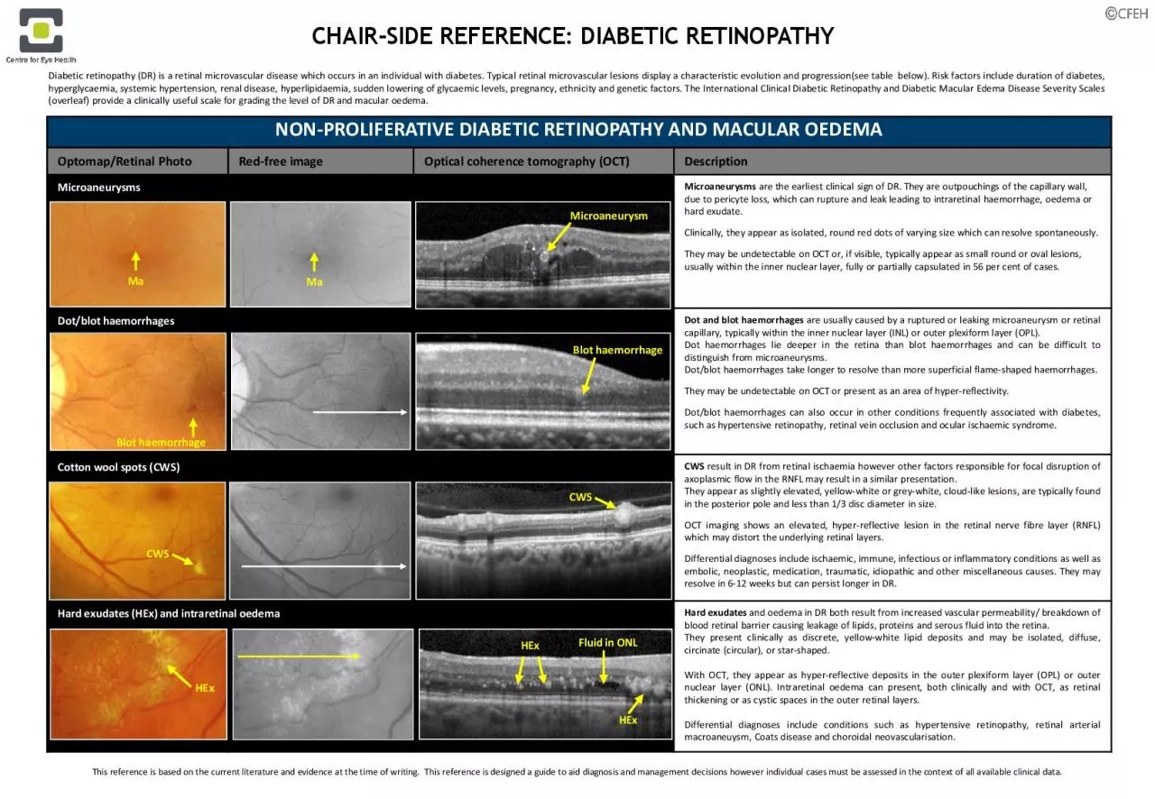
PROLIFERATIVE DIABETIC RETINOPATHYAND MACULAR OEDEMA Optomap/Retinal PhotoRedfreeimageOpticalcoherence tomography (OCT)DescriptionMicroaneurysmsMicroaneurysmsare the earliest clinicalsign of DR. They are outpouchings of the capillary wall, due to pericyteloss, which can rupture and leak leading to intraretinalhaemorrhage, oedemaor hard exudate. Differentialdiagnosesincludeconditionssuchhypertensiveretinopathy,retinalarterialmacroaneuysmCoatsdiseaseandchoroidalneovascularisation CWS Fluid in ONL HEx Blot haemorrhage Diabeticretinopathy(DR)retinalmicrovasculardiseasewhichoccursindividualwithdiabetesTypicalretinalmicrovascularlesionsdisplaycharacteristicevolutionandprogression(seetablebelow)Riskfactorsincludedurationdiabetes,hyperglycaemia,systemichypertension,renaldisease,hyperlipidaemia,suddenloweringglycaemiclevels,pregnancy,ethnicityandgeneticfactorsTheInternationalClinicalDiabeticRetinopathyandDiabeticMacularEdemaDiseaseSeverityScales(overleaf)provide Microaneurysm Ma Ma Blot haemorrhage CWSHEx HExCHAIRSIDE REFERENCE: DIABETIC RETINOPATHY This reference is based on the current literature and evidence at the time of writing.This reference is designed a guide to aid diagnosis and management decisions however individual cases must be assessed in the context of all available clinical data. OTHER NONPROLIFERATIVE DRLESIONS Venous beading (VB) colour and redfree photographyIntraretinalmicrovascular abnormalities (IRMA) colour and redfree photographyVenous Beadingis a venous calibre irregularity which occurs in areas of severe retinal hypoxia. A sausagelink appearance occurs in severe cases. Other
calibre changes include dilation, reduplication and loops.Intraretinal microvascular abnormalities (IRMA) are abnormal intraretinal shunts which appear asbranching or dilation of capillaries within the retina inareas of poor retinal perfusion. They have a similar appearance to NVbut withslightly larger vessel calibre. They are a precursor to NV which may form in close proximity PROLIFERATIVE DIABETIC RETINOPATHYOptomap/Retinal PhotoRedfreeimageOpticalcoherence tomography (OCT)DescriptionNeovascularisation (NV) and fibrousproliferation (FP) Neovascularisation(NV)appearsnewvesselswhichloopbackaroundorformdisorganisednet,distinguishingthemfromnormalcapillariesTheyarethesurfacetheinternallimitingmembrane(ILM)orposteriorhyaloidfacethevitreousandoccurtheborderbetweenhealthyretinaandareascapillarynonperfusion(retinalischaemia)Theyarepronebleeding,resultingpreretinal(PRH)vitreoushaemorrhage(VH)Dynamicinteractionbetweenandthevitreouscanleadinflammatoryresponseandsubsequentfibrousproliferation(FP)AnypreretinalorvitreoushaemorrhageshouldconsideredNVuntilprovenotherwiseNVthedisc(NVD)describesnewvesselsorwithindiscdiameterthediscopposedelsewhere(NVE)Preretinalhaemorrhage (PRH) and vitreous haemorrhage (VH)PRHorcanoccurwhennewvesselsbleedThismayoccurwhenthenewvesselsproliferatealongtheposteriorsurfacetherelativelymobilevitreous,causingtractionthenewvessels,particularlywheretherestrongadherencebetweenthevitreousandtheretinatheareaPRHmaypresentshapedboatshapedhaemorrhagetrappedbetweentheILMandtheposteriorhyaloidfacethevitreous,althoughtheymayappearlinear,blotlikearcuatewillappearreddishorgreyishareahazeobscuri
ngtheunderlyingretinaldetailOCTassistsidentifyingthelocationthehaemorrhage(whichappearshyperreflective)Tractional retinal detachment (TRD)Retinal folds or tractional retinal detachment (TRD) can occur if the vitreous is adherent to the retina in an area of fibrovascularscar formation.Thesechanges aremore likely to occur along the major vascular arcades. TRDs are concave and usually progress slowly, however a hole can form in the detached retina leading to a combined TRD and rhegmatogenousretinal detachment. Clinically, TRD will be associated with NV and FP and appear elevated. NV on posterior vitreous face VH PRH Small TRD NV on retinal surface Posterior vitreous IRMA VB VB IRMA PRH FP and NV adjacent to disc IRMA IRMACHAIRSIDE REFERENCE: DIABETIC RETINOPATHY DIABETIC RETINOPATHY FEATURES DETECTABLE ON OCT ANGIOGRAPHYRedfreeimageOCT Angiography RedfreeimageOCT Angiography Early macular ischaemiaSevere Macular ischaemiafreephoto 3x3 Retina (Cirrus AngioplexOCTangiographynoninvasivetechnologythatdetectsthemovementredbloodcellsthroughtheretinalandvesselsproduceimagethevasculaturewithouttheinjectiondyediabetes,OCTangiographycanshowfeaturesmacularorretinalischaemiaincludingfovealavascularzone(FAZ)enlargement,parafovealcapillarydropout,IRMAandneovascularisationMicroaneurysmscandetectedsomecases,butnototherswheretherepoorbloodflowthroughthemicroaneurysmAnincreasedFAZhasbeenshownpredictortheseverityandprogressiondiabeticretinopathy,Parafoveal capillary dropoutEnlargement and irregularity of the foveal avascular zone Enlargement & irregularity of FAZ 3x3 Retina (Cirrus Angioplex Parafoveal capillary dr
opout Microaneurysm 6x6 Retina & Vitreoretinal interface (Cirrus AngioplexRetinal neovascularisation IRMARetinal neovascularisationCapillary dropout IRMA IRMA 3x3 Retina cropped (Cirrus Angioplex IRMA CHAIRSIDE REFERENCE: DIABETIC RETINOPATHY free photofree photofree photofree photo International Clinical Diabetic Retinopathy Disease Severity Scale DIABETIC RETINOPATHYSTAGEOPHTHALMOSCOPYFINDINGS No apparent retinopathyNo abnormalitiesMild NPDRMicroaneurysms onlyModerate NPDRMore than just microaneurysmsbut less than severe NPDRSevere NPDR Any one of the following (and NO signs of PDR): More than 20 intraretinal haemorrhages in each of 4 quadrantsDefinite VB in 2+ quadrantsProminent IRMA in 1+ quadrantProliferative DR One of the following:Neovascularisation, vitreous/preretinal haemorrhage International Clinical Diabetic Macular EdemaDisease Severity ScaleMACULAR OEDEMASTAGE OPHTHALMOSCOPYFINDINGSAbsent No retinal thickening or hard exudates in the posterior pole Mild (noncentre involving*)Can occur at any level of DRome retinal thickening or hard exudates in posterior pole but distant from the maculaModerate (centreapproaching*) Can occur at any level of DRetinal thickening or hard exudates approaching the centre of the macula but not involving the centreSevere (centre involving*) Can occur at any level of DRetinal thickening or hard exudates involving centre of the macula*Modified by CFEH to reflect International Council ofOphthalmology guidelines (2017) which define centreinvolved macular oedema as thickening within the central 1000µm of the maculaCHAIRSIDE REFERENCE: DIABETIC RETINOPATHY