
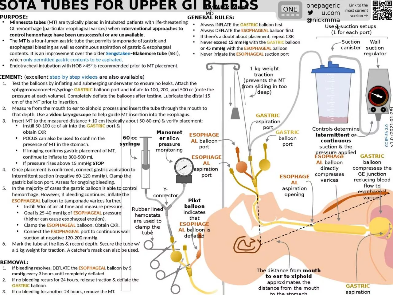
tubes for upper gi bleeds onepagericucom nickmmark Link to the most current version ONE PURPOSE Minnesota tubes MT are typically placed in intubated patients with lifethreatening GI hemorrhage particular esophageal varices when ID: 1039155
Download Presentation The PPT/PDF document "by Nick Mark MD minnesota" is the property of its rightful owner. Permission is granted to download and print the materials on this web site for personal, non-commercial use only, and to display it on your personal computer provided you do not modify the materials and that you retain all copyright notices contained in the materials. By downloading content from our website, you accept the terms of this agreement.


1. by Nick Mark MDminnesota tubes for upper gi bleedsonepagericu.com@nickmmarkLink to the most current version →ONEPURPOSE:Minnesota tubes (MT) are typically placed in intubated patients with life-threatening GI hemorrhage (particular esophageal varices) when interventional approaches to control hemorrhage have been unsuccessful or are unavailable.The MT is a four-lumen gastric tube that permits tamponade of gastric and esophageal bleeding as well as continuous aspiration of gastric & esophageal contents. It is an improvement over the older Sengstaken–Blakemore tube (SBT), which only permitted gastric contents to be aspirated.Endotracheal intubation with HOB >45° is recommended prior to MT placement. ESOPHAGEAL balloon portSuction canisterGASTRIC aspiration portGASTRIC balloon portESOPHAGEAL aspiration openingESOPHAGEAL balloon directly compresses varicesGASTRIC balloon compresses the GE junction reducing blood flow to esophageal varicesGASTRIC aspiration openings200400600800100012001400PLACEMENT: (excellent step by step videos are also available)1 kg weight traction (prevents the MT from sliding in too deep)Test the balloons by inflating and submerging underwater to ensure no leaks. Attach the sphygmomanometer/syringe GASTRIC balloon port and inflate to 100, 200, and 500 cc (note the pressure at each volume). Completely deflate the balloons after testing. Lubricate the distal 15 cm of the MT prior to insertion.Measure from the mouth to ear to xiphoid process and insert the tube through the mouth to that depth. Use a video laryngoscope to help guide MT insertion into the esophagus.Insert MT to the measured distance + 10 cm (typically about 50-60 cm) & verify placement:GENERAL RULES:ESOPHAGEAL aspiration portInstill 50-100 cc of air into the GASTRIC port & obtain CXRPOCUS can also be used to confirm the presence of MT in the stomach. If imaging confirms gastric placement of MT, continue to inflate to 300-500 mLIf pressure rises above 15 mmHg STOPOnce placement is confirmed, connect gastric aspiration to intermittent suction (negative 60-120 mmHg). Clamp the gastric balloon port. Assess for ongoing bleeding.In the majority of cases the gastric balloon is able to control hemorrhage. However, if bleeding continues, inflate the ESOPHAGEAL balloon to tamponade varices further.Always INFLATE the GASTRIC balloon firstAlways DEFLATE the ESOPHAGEAL balloon firstIf there’s a doubt about placement, repeat CXRNever exceed 15 mmHg with the GASTRIC balloon or 45 mmHg with the ESOPHAGEAL balloonNever irrigate the ESOPHAGEAL suction portInstill 50cc of air at time and measure pressure. Goal is 25-40 mmHg of ESOPHAGEAL pressure (higher can cause esophageal erosion).Clamp the ESOPHAGEAL balloon. Obtain CXR.Connect the ESOPHAGEAL port to continuous wall suction at negative 120-200 mmHg.60 cc syringeManometer allow pressure monitoringY-connectorRubber lined hemostats are used to clamp the tubeOFFINTCONWall suction regulatorPilot balloon indicates that ESOPHAGEAL balloon is deflatedControls determine intermittent or continuous suction & the pressure appliedCC BY-SA 3.0 v1.0 (2021-01-29)The distance from mouth to ear to xiphoid approximates the distance from the mouth to the stomachMark the tube at the lips & record depth. Secure the tube w/ a 1 kg weight for traction. A catcher’s mask can also be used.If bleeding resolves, DEFLATE the ESOPHAGEAL balloon by 5 mmHg every 3 hours until completely deflated. If no bleeding recurs for 24 hours, release traction & deflate the GASTRIC balloon. If no bleeding for another 24 hours, remove the MT.REMOVAL:Use 2 suction setups (1 for each port)