
Squamous Cell Carcinoma Jeremy Price MD PhD Faculty Advisor Yvonne Mowery MD PhD Duke University Durham NC USA Case History HPI 81F presents with left facial swelling difficulty ch ID: 938912
Download Pdf The PPT/PDF document "Oral Cavity" is the property of its rightful owner. Permission is granted to download and print the materials on this web site for personal, non-commercial use only, and to display it on your personal computer provided you do not modify the materials and that you retain all copyright notices contained in the materials. By downloading content from our website, you accept the terms of this agreement.


Oral Cavity Squamous Cell Carcinoma Jeremy Price, MD, PhD Faculty Advisor: Yvonne Mowery, MD, PhD Duke University, Durham, NC, USA Case: History ⢠HPI : 81F presents with left facial swelling , difficulty chewing , dentures no longer fitting , & weight loss x 3 months . 30 PY smoking hx . ⢠PE : â ECOG: 2 , dementia but independent in ADLs â HEENT: Edentulous, white left intraoral mass , exophytic and firm arising from left buccal mucosa . â Lymphatic: No
cervical, submandibular, submental, or supraclavicular lymphadenopathy Common Presentations: ⢠Oral cavity pain ⢠Facial swelling ⢠Dysgeusia, tongue immobility/deviation ⢠Dysphagia ⢠Trismus ⢠Speech changes ⢠Loose teeth ⢠Poorly fitting dentures ⢠Hx of alcohol, tobacco, betel nut use ⢠Cranial nerve deficits: â CN V2 - V3 â Trismus, impaired sensation of middle and/or lower third of face, paresthesias, impaired muscles of mastication Case: Imaging
+ Biopsy ⢠CT neck w/ contrast: 4.6 x 3.2 x 3.9 cm left inner cheek heterogeneously enhancing mass eroding into the left mandible ⢠CT chest: No metastatic disease ⢠ENT performed incisional biopsy: well - differentiated, keratinizing invasive SCC Workup & Evaluation ⢠H&P: complete H&N exam, FOL as clinically indicated ( eg , BOT involvement), tobacco/ EtOH use ⢠CT neck with contrast and/or MRI ⢠CT chest; consider PET systemic staging ⢠Core or incisional bx
of primary tumor vs FNA of palpable nodes; consider exam under anesthesia ⢠Dental, speech therapy, nutrition evaluations ⢠Multidisciplinary consultation: ENT, oral surgery, radiation oncology, medical oncology, nutrition ⢠Tobacco cessation counseling ⢠Psychosocial evaluation ⢠HPV etiology rare and not typically tested Oral Cavity Cancer: Epidemiology ⢠Oral cavity cases: 51,540 (2018) in US ⢠Deaths: 10,030 in US ⢠80 % due to tobacco and EtOH â Tobacco:
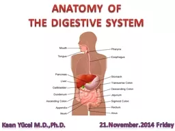
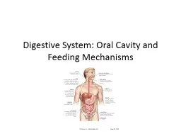
3x higher risk â EtOH + tobacco: 10 - 15x higher risk ⢠Most commonly oral tongue in US (40 - 50%) ⢠Buccal cancer common in Asia due to betel/tobacco chewing ⢠Median age at diagnosis: 62 â Most cases at age � 50 ⢠Median age at death: 68 Anatomy: Oral Cavity ⢠Oral cavity = Cavity bounded by alveolar margins of maxilla and mandible ⢠Roof: hard palate anteriorly and soft palate posteriorly ⢠Floor: mylohyoid muscle; anterior 2/3 of tongue on floor Anat
omy: Oral Cavity ⢠See also for radiographic anatomy: https://radiopaedia.org/cases/tongue - and - floor - of - mouth - neoplasm Oral Cavity Cancer: T Staging T stage, AJCC 8 th edition T0 No evidence of primary tumor T1 2 cm size AND * 5mm depth T2 2 cm size AND depth � 5 mm but 10 mm OR � 2 cm but 4 cm with depth 10 mm T3 Tumor � 4 cm OR � 10 mm depth T4 Locally advanced disease T4a Moderately advanced local disease (e.g. invades through
cortical bone, inferior alveolar nerve, FOM/intrinsic tongue muscles, skin of face, maxillary sinus) T4b Very advanced local disease (e.g. invades masticator space, pterygoid plates/space, skull base, encases internal carotid artery *AJCC 8th edition includes depth of invasion (DOI) ⢠Depth of invasion (DOI) versus Tumor Thickness â DOI = perpendicular distance from the basement membrane region to the deepest point of the infiltrative front of the tumor â Tumor Thi
ckness = perpendicular distance between the highest point of the tumor surface to the deepest point of the infiltrative front of the tumor Oral Cavity Cancer: T Staging Oral Cavity Cancer: N Staging N stage, AJCC 8 th edition N0 No regional lymph node metastasis N1 Metastasis in a single ipsilateral lymph node, 3 cm, ENE - N2 Single ipsilateral �LN ( 3 cm but 6 cm) or multiple LN ( 6 cm) N2a Metastasis in single ipsilateral lymph node �( 3 cm but 6 cm) N2b
Metastasis in multiple ipsilateral lymph nodes (all 6 cm) N2c Metastasis in bilateral or contralateral lymph nodes (all 6 cm) N3* Metastasis in a lymph node � 6 cm and ENE - OR clinically overt ENE+ N3a Metastasis in a lymph node � 6 cm and ENE - N3b Clinically overt ENE+ *N3 in AJCC 8th edition is now N3a and N3b AJCC 8 th Edition Stage Grouping 0 Tis N0 M0 I T1 N0 M0 II T2 N0 M0 III T3 N0 or N1 M0 T1 or T2 N1 M0 IVA T4a N0, N1, or N2 M0 T1, T2, or T3 N2 M0 IVB
Any T N3 M0 T4b Any N M0 IVC Any T Any N M1a or M1b Oral Cavity Cancer: Stage Grouping Case: Management ⢠Early stage lesions (T1 - 2, N0 - 1) â Surgery preferred â Definitive (chemo)radiation therapy for inoperable patients ⢠Locally advanced lesions (T3 - 4, N+) â Surgical resection +/ - radiation ⢠Add concurrent chemotherapy for ECE or + Margins â Definitive chemoradiation if unresectable Case: Surgical Management ⢠Stage IVA disease (cT4aN0M0) based on mand
ibular cortical bone invasion ⢠Surgery: â Radical resection of left buccal mucosa, FOM, and segmental mandibulectomy with sLND (IA, left IB - III, 0/22 LN ) with indeterminate margins, 14 mm depth of invasion ⢠Neck dissection routinely includes 1 st echelon nodes in level I - III ; levels IV - V may be dissected if nodal disease discovered in surgery ⢠Ipsilateral dissection for well cN + lateralized primary sites; for midline primary or invasion of a midline OAR
consider bilateral neck dissection ⢠Neck dissection when cN + â Consider for N0 patients if DOI � 3mm Oral Cavity Cancer: Surgical Management ⢠DâCruz et al, NEJM 2015: ⢠n=496 patients with T1 - T2, cN0 SCC ⢠Randomized: ⢠Oral excision �( 5 mm margin) w/ modified neck dissection (levels 1 â 4) if nodal relapse ⢠Oral excision + ipsilateral selective neck dissection (levels 1 â 3) + included levels 4 â 5 if LN+ during surgery â
¢ PORT as clinically indicated ⢠Should patients with oral cavity cancer have up front neck dissection? Improved OS with planned neck dissection Case: Post - Operative Management ⢠What is the role of adjuvant chemoradiation vs RT alone for this patient? â Bernier, Cooper et al. Head and Neck 2005: Pooled analysis of EORTC 22931 and RTOG 9501: OS improved with RT + concurrent chemo (RCT) vs RT alone for patients with +Margin and/or +ENE Case: Post - Operative Ma
nagement ⢠What is the role of chemoradiation vs RT alone for this patient? â MACH - NC Meta - Analysis ( Pignon et al, Radiother Oncol 2009): Significant OS benefit with addition of chemotherapy per MACH - NC. Absolute difference at 5 years = 5.1% Case: Post - Operative Management ⢠What is the role of chemoradiation vs RT alone for this patient? â HOWEVER: ⢠Poor performance status (ECOG 2) ⢠Advanced age (81) ⢠Comorbid dementia ⢠Therefore, post - operati
ve RT alone was utilized despite indeterminate margin ⢠Typical PORT Indications : â + Margins / Gross residual disease â T3/T4 â LVSI â PNI â �1 - 3 LNs â DOI � 3mm Radiation Technique ⢠Post - operative RT with a composite IMRT/VMAT plan ⢠Prescription: â 50 Gy in 2.5 Gy /fraction to the high risk CTV (tumor bed with margin) â 40 Gy in 2.5 Gy /fraction to low risk CTV (tumor bed with additional margin) + undissected right level IB â¢
Conventional fractionation ( most commonly utilized ) dose prescription = 60 - 66 Gy to high/intermediate risk CTVs and 54 Gy to low risk CTV at 2 Gy /fraction ⢠Alternative techniques include protons & brachytherapy Radiation Simulation ⢠Position: Supine in short thermoplastic mask, bite block â If treating neck, utilize long thermoplastic mask ⢠CT with IV contrast ⢠Consider wiring scars, bolus, based on high risk features ⢠Fuse pre - op CT/MRI to delineate
initial tumor bed for CTV design ⢠Goal : Deliver accelerated treatment without chemotherapy to provide maximal local control benefit with minimized acute toxicity â hypofractionation ⢠Common at our institution for smaller PTV volume and pN0 disease ⢠Langendijk JA et al, IJROBP 2003. ⢠46 â 50 Gy at 2 Gy / fx + boost at 2.5 Gy / fx without chemotherapy â Total 55 Gy (neg. margin) â Total 62.5 Gy (pos. margin) ⢠3 year LRC = â 87% intermediate risk
â 66% high risk ⢠Hypofractionation experiences with concurrent chemotherapy: â Jacinto et al, BMC Cancer 2018 & Sanghera et al, IJROBP 2007 Intermediate risk ECE or Microscopic + margin + 1 risk factor High risk ECE and Microscopic + margin; or ECE or +margin and � 2 risk factors Radiation Technique: Hypofractionation ⢠Pathologic nodal disease by T Stage and site for cN0 neck â Byers et al. Head Neck Surg 1988 ⢠Should the undissected level IV be inclu
ded in RT fields? â Warshavsky et al, JAMA OHNS 2019 â Rate of level IV involvement in cN0 neck is 2.53% in fixed - effects model â therefore omitted in this patient Radiation Technique: Elective Nodal Coverage Site Tx - T1 - T2 T3 - T4 Total Oral tongue (n=48) 18.6% 31.6% 25% FOM (n=62) 18.6% 26.3% 21% Lower gum (n=41) 11.5% 13.3% 12.2% Buccal mucosa (n=10) 0% 0% 0% Retromolar trigone (n=23) 36.4% 33% 34.8% Radiation Contouring ⢠GTV - Preop = initial site of gross disease
as estimated by preoperative imaging ⢠CTV50 (high risk) = Pre - op GTV / tumor bed with margin (5 - 10 mm) ⢠CTV40 (low risk) = CTV50 + 5 - 10 mm expansion based on clinical suspicion and uncertainty due to post - op anatomical changes and encompassing surgical clips + undissected right IB ⢠PTV = CTV + 3mm margin, based on daily image guidance and immobilization ⢠For contouring/dose guidelines with conventional fractionation/SIB see: https://econtour.org/cases/2
8 Case: Target Volumes GTV â Pre - op CTV40 â Low risk CTV 50 High risk Case: Target Volumes GTV â Pre - op CTV40 â Low risk CTV 50 High risk Case: Target Volumes GTV â Pre - op CTV40 â Low risk CTV 50 High risk Case: Target Volumes GTV â Pre - op CTV40 â Low risk CTV 50 High risk Key Dose Constraints: Hypofractionation Constraints OARs Dose Rationale? Rt Parotid 5 - 8 Gy Median Preserve major salivary gland Oral cavity 30 - 35 Gy Median M ucositis , pain L
arynx 15 Gy Median Wound healing around tracheostomy, hoarseness Pharynx 25 - 30 Gy Median Mucositis , pain Mandible 56 Gy Max Osteoradionecrosis Cochlea, left 30 Gy Max Ototoxicity Left eye 30 Gy Max Vision loss Pharyngeal constrictors 25 - 30Gy Median Post - op swallowing Rt SMG No constraint In PTV 40 target volume Case: Dose Volume Histogram OAR % Contoured Structure 100 80 60 40 20 R SMG L Parotid Oral Cavity Mandible Pharynx Larynx Cord L Cochlea L Eye Brainstem 0
10 20 30 40 50 Dose ( Gy ) Case: On Treatment Management Surgical ⢠Tracheostomy â A irway protection post - op ⢠Dobhoff /NGT or PEG â Allow intra - oral healing post - op and with RT induced mucositis â Speech & swallow evaluation for aspiration risk ⢠Wound healing â Ensure flap is well healed prior to RT to minimize wound complication and RT breaks Ra
diation / Chemoradiation ⢠Pain management â Secondary to post - op pain and RT - induced mucositis ⢠Xerostomia â Rx xylitol, flax seed oil, copious fluid intake to minimize ⢠Dysgeusia â Counseling patient to avoid minimized PO intake (if no PEG/NGT) ⢠Dermatitis â Ensure proper skin/wound care and analgesia ⢠Labs Abnormalities â Monitor electrolytes if decreased PO intake and/or chemotherapy. Monitor CBC if chemotherapy. Case: Follow - Up ⢠H&P +
complete H&N physical exam +/ - FOL â q1 - 3 months for year 1 â q2 - 6 months for year 2 â q4 - 8 months for years 3 â 5 â Yearly for years � 5 ⢠Baseline post - op CT at 3 months adjuvant treatment â Additional imaging practices vary per institution or as indicated by symptoms/exam ⢠TSH yearly ( if thyroid irradiated) ⢠Speech/swallowing/dental/hearing evaluations ⢠Smoking cessation ⢠Depression screening Oral Cavity Cancer: Clinical Pearl
s ⢠What is the ideal regimen of concurrent cisplatin? ⢠Noronha et al, JCO 2018: ⢠n=300 patients with LAHNC (87% oral cavity, 93% PORT) ⢠Randomized: ⢠Weekly cisplatin 30 mg/m 2 ⢠Q3 Week 100 mg/m 2 ⢠As administered with RT delivered by opposed portals ⢠Caveats: Treatment and patient characteristics differ versus typical USA patients/practices; suboptimal dosing for weekly cisplatin (30 mg/m 2 rather than 40 mg/m 2 ) Improved LRC with Q3 week cisplati
n but with more toxicity ; no difference in OS ⢠OCAT Trial ( Laskar et al, ASCO 2016 ): Conventional RT (56 - 60 Gy /6 wk ) vs CRT (56 - 60 Gy /6 wk + weekly cisplatin 30 mg/m 2 ) vs Accelerated RT (56 - 60 Gy /5 wk ) for high - risk oral cavity patients â showed similar locoregional control for all three arms References 1. Brady LW, Wazer DE, Perez CA. Perez & Bradyâs Principles and Practice of Radiation Oncology . Lippincott Williams & Wilkins; 2013. 2.
NCCN: Clinical Practice Guideline Head and Neck Cancers. 3. Siegel RL, Miller KD, Jemal A. Cancer statistics, 2018. CA Cancer J. Clin . 2018;68:7 â 30. 4. Chinn SB, Myers JN. Oral Cavity Carcinoma: Current Management, Controversies, and Future Directions. J. Clin . Oncol . 2015;33:3269 â 3276 . 5. Dirven R, Ebrahimi A, Moeckelmann N, Palme CE, Gupta R, Clark J. Tumor thickness versus depth of invasion â Analyiss of the 8 th edition AJCC Staging for oral cancer. Oral
Oncology . 2017. 6. Bernier J, Cooper JS, Pajak TF, et al. Defining risk levels in locally advanced head and neck cancers: a comparative analysis of concurrent postoperative radiation plus chemotherapy trials of the EORTC (#22931) and RTOG (# 9501). Head Neck . 2005;27:843 â 850. 7. Pignon J - P, le Maître A, Maillard E, et al. Meta - analysis of chemotherapy in head and neck cancer (MACH - NC): an update on 93 randomised trials and 17,346 patients. Radiother . Oncol .
2009;92:4 â 14. 8. Langendijk JA, de Jong MA, Leemans CR, et al. Postoperative radiotherapy in squamous cell carcinoma of the oral cavity: the importance of the overall treatment time. Int. J. Radiat . Oncol . Biol. Phys. 2003;57:693 â 700. 9. Jacinto AA, Batalha Filho ES, Viana L de S, et al. Feasibility of concomitant cisplatin with hypofractionated radiotherapy for locally advanced head and neck squamous cell carcinoma. BMC Cancer . 2018;18:1026. 10. Sanghera P
, McConkey C, Ho K - F, et al. Hypofractionated accelerated radiotherapy with concurrent chemotherapy for locally advanced squamous cell carcinoma of the head and neck. Int. J. Radiat . Oncol . Biol. Phys. 2007;67:1342 â 1351. 11. Warshavsky A, Rosen R, Nard - Carmel N, et al. Assessment of the Rate of Skip Metastasis to Neck Level IV in Patients With Clinically Node - Negative Neck Oral Cavity Squamous Cell Carcinoma: A Systematic Review and Meta - analysis. JAMA Otolary
ngol . Head Neck Surg. 2019;145:542 â 548. 12. DâCruz AK, Vaish R, Kapre N, et al. Elective versus Therapeutic Neck Dissection in Node - Negative Oral Cancer. N. Engl. J. Med. 2015;373:521 â 529. 13. Noronha V, Joshi A, Patil VM, et al. Once - a - Week Versus Once - Every - 3 - Weeks Cisplatin Chemoradiation for Locally Advanced Head and Neck Cancer: A Phase III Randomized Noninferiority Trial. J. Clin . Oncol . 2018;36:1064 â 1072 . 14. Laskar S, Chaukar D, De
shpande M, et al. Phase III randomized trial of surgery followed by conventional radiotherapy (5 fr / Wk ) (Arm A) vs concurrent chemoradiotherapy (Arm B) vs accelerated radiotherapy (6fr/ Wk ) (Arm C) in locally advanced, stage III and IV, resectable , squamous cell carcinoma of oral cavity - oral cavity adjuvant therapy (OCAT): Final results (NCT00193843 ). J. Clin . Oncol . 2016; 34(15 sup):6004. Please provide feedback regarding this case or other ARROcases to arrocas