
Additional Professor Department of Orthopaedics Learning Objectives Atraumatic Traumatic AVN Understand the pathology of AVN Progression of disease Classification of Hip AVN Principles of treatment ID: 906161
Download The PPT/PDF document "Avascular Necrosis Dr R B Kalia," is the property of its rightful owner. Permission is granted to download and print the materials on this web site for personal, non-commercial use only, and to display it on your personal computer provided you do not modify the materials and that you retain all copyright notices contained in the materials. By downloading content from our website, you accept the terms of this agreement.


Slide1
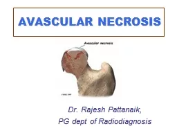
Avascular Necrosis
Dr R B Kalia,
Additional Professor,
Department of Orthopaedics.
Slide2Learning Objectives
Atraumatic /Traumatic AVN
Understand the pathology of AVN
Progression of disease
Classification of Hip AVN
Principles of treatment
Slide327 years old ,pain right groin
Non smoker, occasional alcohol intake, no medical history
On examination.
No Tenderness
Restricted internal rotation ,painful
No limp
CBC Normal, Rheumatoid factor –
ve
, ESR 18mm,CRP -6
Slide4Radiographs
Slide5Q1 Which of the following is an inappropriate diagnosis?
Early
Sero
-Negative Rheumatoid arthritis
Avascular necrosis right hip
Tubercular arthritis right hip
Torn acetabular
labrum
Slide6Q 2 The most appropriate investigation for him is?
Tc 99 bone scan
MRI scan
MARS MRI
Contrast enhanced CT Scan
Slide7Q3 This is classifiable as Ficat-Arlet-
Stage 1
Stage 2
Stage 3
Stage 4
Slide8Q 4 The most appropriate treatment ?
Non operative with follow up MRI at 6 weeks
Non weight bearing and analgesics
Bed rest, traction and analgesics
Core decompression
Slide9Q 5 The patient comes after 2 years and has severe pain in his hip, decreased ROM and a pronounced limp. Radiographs reveal collapse of the head with decreased joint space suggestive of secondary OA. The appropriate management is-
Arthroscopic debridement and irrigation
Osteochondral grafting
Bipolar hip arthroplasty
Total hip arthroplasty
Slide10Overview
AVN is a major cause of hip pain
Traumatic/Atraumatic in
etiology
Evaluation is difficult
Treatment is hip salvage/Replacement
Other common areas are –Humerus, scaphoid, talus and distal femur
Slide11Avascular necrosis- Traumatic
Femoral neck fractures- severance of the blood supply to the femoral head.
The capitulum
Femoral condyles
Proximal parts of the scaphoid and talus.
Distant parts of the bone’s vascular territory
Largely enclosed by cartilage- restricted access to local blood vessels
.
Slide12TRAUMATIC OSTEONECROSIS
Fractures and dislocations of the hip
Tear of retinacular vessels supplying the femoral head
Displaced fractures of the femoral neck – AVN 20%.
Slide13fractures of the scaphoid and talus- AVN
Principal vessels enter their distal ends
Intraosseous course from distal to proximal.
Slide14Closed compartment-Bone
Vascular sinusoids- no adventitial layer
Patency - volume and pressure of the marrow tissue
Marrow is encased in unyielding bone.
One element can expand-other gets compressed
Slide15NON-TRAUMATIC OSTEONECROSIS
Intravascular stasis
Thrombosis
Extravascular swelling and capillary compression.
Slide16Ischaemia- Multifactorial
Severance of the local blood supply
Venous stasis and retrograde arteriolar stoppage
Intravascular thrombosis
Compression of capillaries and sinusoids by marrow swelling.
Slide17Nontraumatic -AVN
Perthes’ disease,
Caisson disease
Gaucher’s disease
systemic lupus erythematosus
Slide18Arteriolar occlusion
Marrow
edema
Vascular stasis
Sinusoidal compression
Gaucher’s disease
Tuberculosis
Cortisone/alcohol
Dysbaric
ischaemia
Sickle-cell disease
Dysbaric
ischaemia
Thrombocytopenia
Fat embolism
Slide19Normal head femur
Slide20Pathology and natural history
Prolonged anoxia – Osteocyte death
Gross appearance remains unaltered
Striking histological changes in the marrow
Loss of fat cell outlines
Inflammatory cell infiltration
Marrow oedema
Replacement of necrotic marrow- mesenchymal tissue.
Slide21Bone repair?
New blood vessels and osteoblastic proliferation at the interface between ischaemic and live bone.
Vascular granulation tissue advances from the surviving trabeculae
New bone is laid down upon the dead- creeping substitution
Increase in mineral mass - increased density or ‘sclerosis’.
Slide22Further progress
Reparative formation proceeds slowly
Advances 8–10 mm into the necrotic zone.
Structural failure begins- most heavily stressed part of the necrotic segment.
Linear tangential fracture close to the articular surface.
Slide23Stage of arthritis
Articular cartilage retains its thickness and viability for a long time.
In the final stages- fragmentation collapse of the necrotic bone
Progressive deformity and destruction of the joint surface.
Slide24Clinical features
The earliest stage of bone death is asymptomatic
Patient presents with pain - lesion is usually well advanced.
Pain in or near a joint
Few complain of a ‘click’ in the joint- due to snapping or catching of a loose articular fragment.
Slide25Stage of arthritis
Joint becomes stiff and deformed.
Local tenderness may be present
Superficial joints- effusion.
Movements –may be restricted
Advanced cases- fixed deformities.
Slide26Radiographs
The early signs of ischaemia -bone marrow and cannot be detected.
3 months after the onset of ischaemia- first sign
Reactive new bone formation at the boundary of the ischaemic area -sclerosis
Slide27Thin tangential fracture line just below the articular surface –the
‘crescent sign’
.
Slide28Late -collapse and distortion of the articular surface
Slide29MRI Scan
The most sensitive modality – marrow changes are
discernable
The size of the necrotic segment-hypo-intense band in the T1,MRI
Slide30AVN –Distal femur
Slide31AVN Talus
Slide32AVN -Capitulum
Slide33Radionuclide scanning
99mTcsulphur colloid- taken up in myeloid tissue.
Useful in traumatic avascular necrosis- large segment of bone is involved.
Sickle-cell disease - ‘cold’ area contrasts significantly with the generally high nuclide uptake due to increased erythroblastic activity.
Slide34Staging the lesion
Ficat
and
Arlet
(1980) introduced the concept of
radiographic staging
for osteonecrosis of the hip
Early (pre-symptomatic) signs- sclerosis, crescent sign.
Later features- progressive demarcation and collapse of the necrotic segment in the femoral head.
Slide35Stage 1 – No radiological changes
Diagnosis was based on measurement of raised intraosseous pressure
Histological features of bone biopsy
MRI
Slide36Stage II
The femoral head contour was still normal
Early signs of reactive change in the subchondral area
Slide37Stage 3
Signs of osteonecrosis with evidence of structural damage and distortion of the bone outline.
Collapse of the necrotic segment
Slide38Stage 4
Collapse of the articular surface and signs of secondary OA.
Slide39Diagnosis of the underlying disorder
Episode of trauma- obvious
Occupation- deep-sea diving or working under compressed air
Family background of Gaucher’s disease or sickle-cell disease.
High-dosage corticosteroid administration; renal transplantation.
Low dose use –quacks, inappropriate use
Alcohol abuse is often difficult to determine
SLE- antiphospholipid antibodies may be measured.
Slide40EARLY OSTEONECROSIS
Bone contour is intact- structural failure can be prevented.
Some lesions heal spontaneously and with minimal deformity;
Non-weightbearing joints
Superomedial part of the femoral head
Non- weight bearing surfaces of the femoral condyles and talus.
Slide41Weight bearing joints
Poor prognosis-l probably end in structural failure
Simple measures like non weight bearing- reduce loading.
If the bone contour is still
intact,an
‘unloading’ osteotomy
Help to preserve the anatomy while remodelling proceeds.
Medullary decompression and bone grafting may have a place
Slide42Stage II- Core decompression B/L
Slide43LATE STAGE OSTEONECROSIS
Destruction of the articular surface may be give rise to pain and severe loss of function.
non-
operative
management,
concentrating
on pain control, modification of daily activities and appropriate, splintage of the joint
Arthrodesis of the joint, e.g. the ankle or wrist
Total joint replacement- shoulder, hip and knee.
Slide44Slide45Q1 Which of the following is an inappropriate diagnosis?
Early
Sero
-Negative Rheumatoid arthritis
Avascular necrosis right hip
Tubercular arthritis right hip
Torn acetabular
labrum
Slide46Q 2 The most appropriate investigation for him is?
Tc 99 bone scan
MRI scan
MARS MRI
Contrast enhanced CT Scan
Slide47Q3 This is classifiable as Ficat-Arlet-
Stage 1
Stage 2
Stage 3
Stage 4
Slide48Q 4 The most appropriate treatment ?
Non operative with follow up MRI at 6 weeks
Non weight bearing and analgesics
Bed rest, traction and analgesics
Core decompression
Slide49Q 5 The patient comes after 2 years and has severe pain in his hip, decreased ROM and a pronounced limp. Radiographs reveal collapse of the head with decreased joint space suggestive of secondary OA. The appropriate management is-
Arthroscopic debridement and irrigation
Osteochondral grafting
Bipolar hip arthroplasty
Total hip arthroplasty
Slide50Conclusion
AVN is difficult to diagnose early-
High degree of suspicion
Radiographs in early stages are normal-
Trust your findings more!
Best modality for early diagnosis –
MRI
Salvage can be tried for Stage I,II
Advanced stages require –
Total hip arthroplasty
Slide51Questions?