
55 plasma 45 cells 99 RBCs lt 1 WBCs and platelets Formed Elements of Blood Red blood cells erythrocytes White blood cells leukocytes granular leukocytes neutrophils eosinophils ID: 1014779
Download Presentation The PPT/PDF document "Components of Blood Hematocrit" is the property of its rightful owner. Permission is granted to download and print the materials on this web site for personal, non-commercial use only, and to display it on your personal computer provided you do not modify the materials and that you retain all copyright notices contained in the materials. By downloading content from our website, you accept the terms of this agreement.


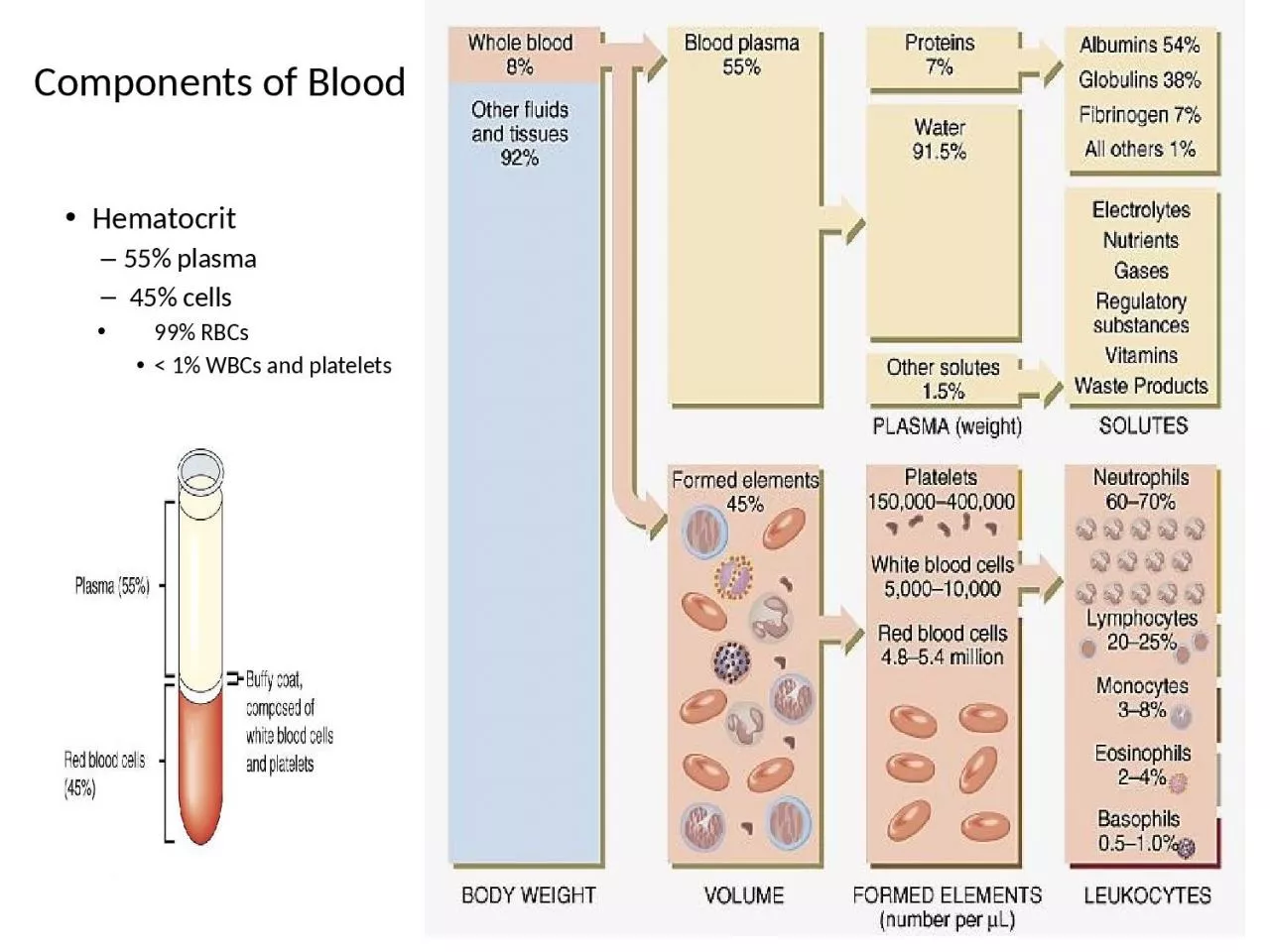
1. Components of BloodHematocrit55% plasma 45% cells 99% RBCs< 1% WBCs and platelets
2. Formed Elements of BloodRed blood cells ( erythrocytes )White blood cells ( leukocytes )granular leukocytesneutrophilseosinophilsbasophilsagranular leukocyteslymphocytes = T cells, B cells, and natural killer cellsmonocytesPlatelets (special cell fragments)Tortora & Grabowski 9/e 2000 JWS 19-2
3. Formation of blood cells: The majority of RBCs, platelets, and most of the WBCs are formed in the red marrow while only a few of them are formed in the yellow marrow. Everyone needs acontinuous production cycle of blood cells from our bone marrow throughout our lives since each blood cell has a set life expectancy. Healthy bone marrow produces as many cells as your body needs. Red cell production is increased when the body needs additional oxygen, platelets increase when bleeding occurs, and white cells increase when infection threatens.
4. Blood cells are made in the bone marrow. The bone marrow is the spongy material in the center of the bones that produces about 95 percent of the body's blood cells. There are two types of bone marrow: red marrow and yellow marrow. Yellow marrow has a much higher amount of fat cells than red marrow. Both types of marrow contain blood vessels. There are other organs and systems in our bodies that help regulate blood cells. The lymph nodes, spleen, and liver help regulate the production, destruction, and differentiation (developing a specific function) of cells. The production and development of new cells is a process called hematopoiesis.
5.
6. Leukocytes:The number of leukocytes in the blood is often an indicator of disease. There are normally between 4×10⁹ and 1.1×10¹⁰ white blood cells in a litre of blood, and ranging from 7 and 21 micrometres in diameter, they make up approximately 1% of blood in a healthy adult. An increase in the number of leukocytes over the upper limits is called leukocytosis, and a decrease below the lower limit is called leukopenia. The physical properties of leukocytes, such as volume, conductivity, and granularity, may change due to activation, the presence of immature cells, or the presence of malignant leukocytes in leukemia.
7. Types of immunity:1-Innate Immunity:The innate immunity system is what we are born with and it is nonspecific; all antigens are attacked pretty much equally. It is genetically based and we pass it on to our offspring.Surface Barriers or Mucosal Immunity Skin : The skin cannot be penetrated by most organisms unless it already has an opening, such as a nick, scratch, or cut. Mechanically, pathogens are expelled from the lungs by ciliary action as the tiny hairs move in an upward motion; coughing and sneezing abruptly eject both living and nonliving things from the respiratory system.
8. 3-Sticky mucus in respiratory and gastrointestinal tracts traps many microorganisms.4-Saliva, tears, nasal secretions, and perspiration contain lysozyme, an enzyme that destroys Gram positive bacterial cell walls causing cell lysis5- the stomach: its mucosa secrete hydrochloric acid (0.9 < pH < 3.0, very acidic) and protein-digesting enzymes that kill many pathogens. The stomach can even destroy drugs and other chemicals.
9. 2-Adaptive or Acquired ImmunityLymphocytes come in two major types: B cells and T cells. The peripheral blood contains 20–50% of circulating lymphocytes; the rest move in the lymph system. Roughly 80% of them are T cells, 15% B cells and remainder are null or undifferentiated cells called cytotoxic or killer T cell. Parts of the immune system are changeable and can adapt to better attack the invading antigen. There are two fundamental adaptive mechanisms: humoral immunity and cell-mediated immunity .
10. Humoral immunity:Immature B-lymphocyte is stimulated to maturity when an antigen binds to its surface receptors and there is a T helper cell nearby (to release a cytokine). This sensitizes or primes the B cell and it undergoes clonal selection, which means it reproduces asexually by mitosis. Most of the family of clones become plasma cells. These cells, after an initial lag, produce highly specific antibodies at a rate of as many as 2000 molecules per second for four to five days. The other B cells become long-lived memory cells.
11. Antibodies, also called immunoglobulins or Igs , constitute the gamma globulin part of the blood proteins. They are soluble proteins secreted by the plasma offspring (clones) of primed B cells. The antibodies inactivate antigens by, (a) complement fixation (proteins attach to antigen surface and cause holes to cause cell lysis), (b)neutralization(binding to specific sites to prevent attachment—this is the same as taking their parking space), (c) agglutination (clumping), (d) precipitation(forcing insolubility and settling out of solution).
12.
13. Cell mediated immunity:are non antibody-producing lymphocytes which are also produced in the bone marrow but sensitized in the thymus and constitute the basis of cell-mediated immunity. Macrophages engulf antigens, process them internally, then display parts of them on their surface together with some of their own proteins. This sensitizes the T cells to recognize these antigens.
14. Types of T lymphocyte:Cytotoxic or killer T cells(CD8+) do their work by releasing lymphotoxins, which cause cell lysis. Helper T cells(CD4+) serve as managers, directing the immune response. They secrete chemicals called lymphokines that stimulate cytotoxic T cells and B cells to grow and divide, attract neutrophils, and enhance the ability of macrophages to engulf and destroy microbes. Suppressor T cells inhibit the production of cytotoxic T cells once they are unneeded. Memory T cells are programmed to recognize and respond to a pathogen once it has invaded and been repelled.
15. Red blood cell, also called erythrocyte, cellular component of blood, millions of which in the circulation of vertebrates give the blood its characteristic colour and carry oxygen from the lungs to the tissues. The mature human red blood cell is small, round, and biconcave; it appears dumbbel-shaped in profile. The cell is flexible and assumes a bell shape as it passes through extremely small blood vessels. It is covered with a membrane composed of lipids and proteins, lacks a nucleus, and contains hemoglobin—a red, iron-rich protein that binds oxygen
16. What is anemia: Anemia is a medical condition in which the red blood cell count or hemoglobin is less than normal. Causes of common types of anemiaCommon types of anemia and their causes include:Iron deficiency anemia. is caused by a shortage of the element iron in the body. the bone marrow needs iron to make hemoglobin. Without adequate iron, the body can't produce enough hemoglobin for red blood cells.
17. Vitamin deficiency anemia. In addition to iron, the body needs folate and vitamin B-12 to produce sufficient numbers of healthy red blood cells. A diet lacking in these and other key nutrients can cause decreased red blood cell production. Additionally, some people may eat enough B-12, but their bodies aren't able to process the vitamin. This can lead to vitamin deficiency anemia.Anemia of chronic disease. Certain chronic diseases — such as cancer, HIV/AIDS, rheumatoid arthritis, and other chronic inflammatory diseases — can interfere with the production of red blood cells, resulting in chronic anemia. Kidney failure also can cause anemia.
18. Aplastic anemia. This very rare life-threatening anemia is caused by a decrease in the bone marrow's ability to produce red blood cells. Causes of aplastic anemia include infections, drugs and autoimmune diseases.Anemias associated with bone marrow disease. A variety of diseases, such as leukemia and myelodysplasia, can cause anemia by affecting blood production in bone marrow. The effects of these types of cancer and cancer-like disorders vary from a mild alteration in blood production to a complete life-threatening shutdown of the blood-making process. Other cancers of the blood or bone marrow — such as multiple myeloma, myeloproliferative disorders and lymphoma — also can cause anemia.
19. Hemolytic anemias. This group of anemias develops when red blood cells are destroyed faster than bone marrow can replace them. Certain blood diseases can cause increased red blood cell destruction. Hemolytic anemias can be inherited, or the body can develop them later in life.Sickle cell anemia. This inherited and sometimes serious anemia is caused by a defective form of hemoglobin that forces red blood cells to assume an abnormal crescent (sickle) shape. These irregular-shaped red blood cells die prematurely, resulting in a chronic shortage of red blood cells.
20. Platelet (thrombocyte) : the smallest of the formed elements in blood, a disk-shaped, non-nucleated blood element with a fragile membrane, formed in the red bone marrow by fragmentation of megakaryocytes. Platelets tend to adhere to uneven or damaged surfaces, and there are an average of about 250,000 per mm3 of blood. The bone marrow produces from 30,000 to 50,000 platelets per mm3 of blood daily, which means that in any ten-day period all the platelets in the body are completely replaced.
21. HemostasisThere are 3 mechanisms that work together to stop the flow of blood. They are 1-Vasoconstriction 2-Platelet plug formation 3-Clotting of blood
22. 1. Vasoconstriction of a damaged blood vessel slows the flow of blood and thus helps to limit blood loss. This process is mediated by:Local controls. Vasoconstrictors such as thromboxane are released at the site of the injury. Systemic control. Epinephrine released by the adrenal glands stimulates general vasoconstriction. 2. Formation of a Platelet Plug.When a blood vessel is damaged, the blood is exposed to collagen fibers in the basement membrane of the vessel . Platelets stick to collagen and become activated
23. Activated platelets release chemicals such as ADP, and thromboxane, that cause the aggregation of more platelets to the site of injury. Platelet aggregation results in the formation of a platelet plug which acts to stem the flow of blood from the broken vessel. It is essential that platelets become activated only at the site of a broken vessel. Otherwise activated platelets would form plugs and induce clots in inapropriate places. Healthy vessels secrete an enzyme called prostacyclin that functions to inhibit platelet activation and aggregation.
24.
25. 3. Clotting of BloodThe blood contains about a dozen clotting factors. These factors are proteins that exist in the blood in an inactive state, but can be called into action when tissues or blood vessels are damaged. The activation of clotting factors occurs in a sequential manner. The first factor in the sequence activates the second factor, which activates the third factor and so on. This series of reactions is called the clotting cascade.
26. Blood clotting is the transformation of liquid blood into a semisolid gel. Clots are made from fibers (polymers) of a protein called fibrin. Fibrin monomers come from an inactive precursor called fibrinogen. The body of the fibrinogen molecule has caps on its ends that mask fibrin-to-fibrin binding sites. If the caps are removed then fibrin monomers polymerize to form fibrin polymers. This process requires thrombin, the enzyme that converts fibrinogen to fibrin. This process also requires calcium, which acts as a kind of glue to hold the fibrin monomers to each other to form the polymeric fiber. The fibrin fibers form a loose meshwork that is stabilized by clotting factor XIII. The stabilized meshwork of fibrin fibers traps erythrocytes, thus forming a clot that stops the flow of blood.
27.
28. Blood clotting tests: Before you have surgery your doctor may order blood tests to determine how quickly your blood clots. This group of tests is known as a coagulation study, individually these tests are commonly referred to as a PT (Prothrombin Time), PTT(Partial Thromboplastin Time). During some surgeries it is important that the blood not clot as quickly as normal, and medications may be given to slow the clotting time.
29. . Drugs commonly used to slow clotting have a variety of names, but Heparin, Coumadin, Lovenox and Warfarin are among the most common. In other cases, the patient may not clot quickly enough, and steps may be taken to make the blood to clot more quickly. Prothrombin Time Blood Test-PT This test is done to evaluate the blood for its ability to clot. It is often done before surgery to evaluate how likely the patient is to have a bleeding or clotting problem during or after surgery. Normal PT Values: 10-12 seconds (this can vary slightly from lab to lab)
30. Partial Thromboplastin Time Blood Test-PTT This test is performed primarily to determine if heparin (blood thinning) therapy is effective. It can also be used to detect the presence of a clotting disorder. Normal PTT Values: 30 to 45 seconds (this can value slightly from lab to lab)
31. The Lymph SystemLymph is an alkaline (pH > 7.0) fluid that is usually clear, transparent, and colorless. It flows in the lymphatic vessels and bathes tissues and organs in its protective covering. There are no RBCs in lymph and it has a lower protein content than blood. Like blood, it is slightly heavier than water (density = 1.019 ± .003). Lymph carries lipids and lipid-soluble vitamins absorbed from the gastrointestinal (GI) tract. Since there is no active pump in the lymph system, there is no back-pressure produced. The lymphatic vessels, like veins, have one-way valves that prevent backflow. Additionally, along these vessels there are small bean-shaped lymph nodes that serve as filters of the lymphatic fluid. It is in the lymph nodes where antigen is usually presented to the immune system.
32. The human lymphoid system has the following:· primary organs: bone marrow (in the hollow center of bones) and the thymus gland (located behind the breastbone above the heart), and · secondary organs at or near possible portals of entry for pathogens: adenoids, tonsils, spleen (located at the upper left of the abdomen), lymph nodes (along the lymphatic vessels with concentrations in the neck, abdomen, Peyer's patches (within the intestines), and the appendix.
33.