
The tectum which is dorsal to the cerebral aqueduct The tegmentum which is ventral to the cerebral aqueduct The crus cerebri or the cerebral peduncle which are ventral to tegmentum At the tectal ID: 1000526
Download Presentation The PPT/PDF document "Mid brain The regions of the mid brain:" is the property of its rightful owner. Permission is granted to download and print the materials on this web site for personal, non-commercial use only, and to display it on your personal computer provided you do not modify the materials and that you retain all copyright notices contained in the materials. By downloading content from our website, you accept the terms of this agreement.


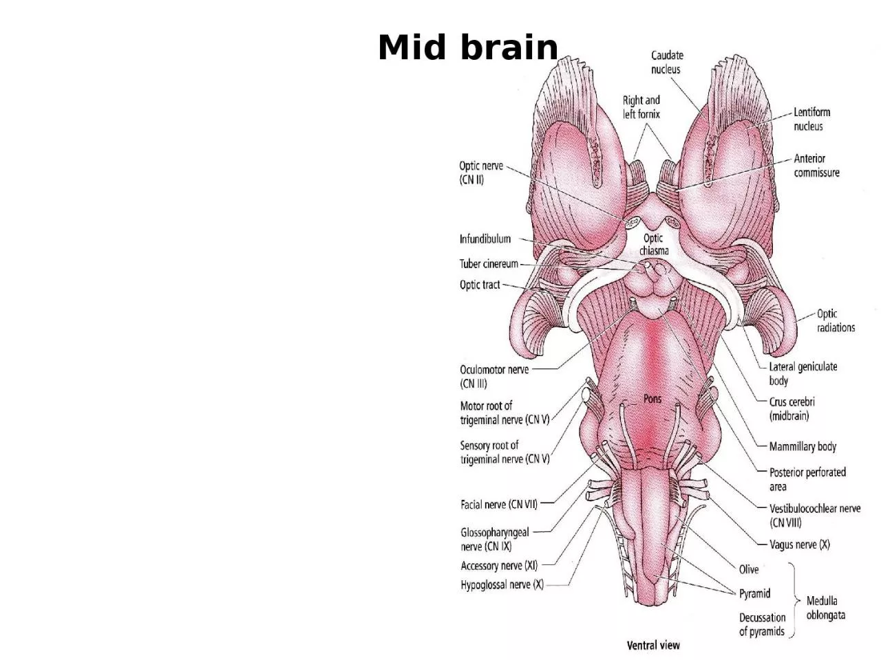
1. Mid brain
2. The regions of the mid brain:The tectum which is dorsal to the cerebral aqueduct.The tegmentum which is ventral to the cerebral aqueduct.The crus cerebri or the cerebral peduncle which are ventral to tegmentum. At the tectal region of the mid brain, there are two pairs of rounded structures which are called the colliculi. Two superior and two inferior colliculi.
3. The cross section of the mid brain at level of the inferior colliculus: 1- There are two rounded swellings which are called the inferior colliculi. They are considering as the reflex center for the auditory stimuli. They receive input from the lateral lemniscus and give output to the medial geniculate body of the thalamus. This inferior colliculus will direct the attention to the unexpected sound.
4. 2- At the level of the inferior colliculus, in the tegmentum, there is the nucleus of the trochlear nerve (4th cranial nerve) that supplies the superior oblique muscle of the eyeball, the fibers of this nerve will decussate before leaving the mid brain and leave through the posterior aspect of the mid brain (all the cranial nerves leave through the lateral or ventral parts of the brain stem except the 4th cranial nerve).
5. 3- At this level, in the tegmentum, there is large decussation of the superior cerebellar peduncle (between the mid brain and cerebellum).
6. 4- The substantia nigra is very clear structure in the section of the mid brain and has a characteristic black color due to melanin pigmentation. The substantia nigra is an important motor center. The substantia nigra, is one of the five nuclei that constitute the basal ganglia circuit, which controls voluntary movements. It is divided into the pars compacta and the pars reticulata, which mainly contain dopaminergic and GABAergic neurotransmitter producing cells respectively.
7. The connections of the substantia nigra: The input to the substantia nigra is from the cerebral cortex through subthalamic nucleus, and gives output (project) to the thalamus, and there is reciprocal connection to the corpus striatum. The pathway between the substantia nigra to the corpus striatum is an important pathway contains the neurotransmitter called the dopamine. The low amount of dopamine will affect this pathway that lead to Parkinson's disease.
8. The connections of the substantia nigra: The input to the substantia nigra is from the cerebral cortex through subthalamic nucleus, and gives output (project) to the thalamus, and there is reciprocal connection to the corpus striatum. The pathway between the substantia nigra to the corpus striatum is an important pathway contains the neurotransmitter called the dopamine. The low amount of dopamine will affect this pathway that lead to Parkinson's disease.
9. The second cross section of the mid brain is at the level of the superior colliculus:At the tectal region, there is the superior colliculus which is considered as an important reflex center of the visual stimuli.
10. The connection of the superior colliculus: The input is from the lateral geniculate body of the thalamus, and the output to cranial nerve nuclei of the 3rd, 4th and 6th cranial nerves. The reciprocal connection is with the spinal cord. The cerebral cortex is controlling strongly the reflex activity of the superior colliculus.
11. The functions of the superior colliculus of the mid brain:Direct the attention or direction of the gaze toward the target by rapid shifting of the direction of the gaze reflexly that strongly controlled or affected by the cerebral cortex input to the superior colliculus.Responsible for follow up of the object in the visual field.It directs the attention or the direction of the head toward the source of the cutaneous stimuli.
12. 2. At this level, the tegmentum contains cranial nerve nucleus of the oculomotor (3rd) nerve. This nucleus has two important groups:General somatic nucleus for innervation of the extra ocular muscles except lateral rectus and superior oblique.Parasympathetic nucleus for supplying two muscles inside the eyeball; these are the sphincter pupillae and ciliary muscle (that responsible for accommodation). This nucleus also is called Edinger- Westphal nucleus or called accessory oculomotor nucleus.
13. 3. Red nucleus which has red color in fresh section. It has the following functions:Alternative pathway for corticospinal tract.Coordination of the movement through its connection with the cerebellum.
14. 4. Ventral tegmental area of Tasi which is situated between the red nucleus and the substantia nigra. This Tasi area contains dopamine. Some studies were showed an increase in the dopamine level in this area in schizophrenic patients.
15. 5. Substantia nigra also presents at this level.
16. The crus cerebri or the cerebral peduncle: It is situated ventral to tegmental region and it is composed of large bundles of white matter. These fibers have the following arrangements:The lateral 1/5th of fibers is occupied by the temporopontine (corticopontine) fibers.The middle 3/5th of fibers is made up of corticonuclear and corticospinal fibers (pyramidal fibers).The medial 1/5th of fibers is made up of frontopontine (corticopontine) fibers.