
Dr Anita Rani Anatomy Lecture 17 th December 2016 Lesson Plan Introduction Skin amp Superficial fascia Deep Fascia Layers Muscles Nerves amp Arteries Bones Arches Applied Anatomy ID: 915710
Download Presentation The PPT/PDF document "Sole and Arches of Foot" is the property of its rightful owner. Permission is granted to download and print the materials on this web site for personal, non-commercial use only, and to display it on your personal computer provided you do not modify the materials and that you retain all copyright notices contained in the materials. By downloading content from our website, you accept the terms of this agreement.

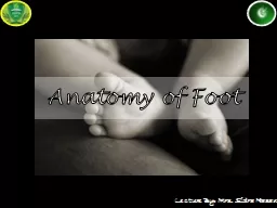


Slide1
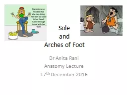
Sole andArches of Foot
Dr Anita RaniAnatomy Lecture 17th December 2016
Slide2Lesson PlanIntroduction
Skin & Superficial fasciaDeep FasciaLayersMusclesNerves & ArteriesBones: ArchesApplied Anatomy
Slide3Homologous to palm BUT
Is an organ of support and locomotionGreat toe : lost its power of mobility & prehensionLesser four toes markedly reduced in sizeTarsal bones and Ist Metatarsal forms broad base for better support
Introduction
Slide4Skin
Thick for protectionFirmly adherent to underlying plantar aponeurosisCreased
Slide5Innervation
Slide6Plantar ReflexBabinski’s Sign
Slide7Superficial fascia
Fibrous and denseFibrous bands bind the skin to plantar aponeuposisDivide the fat in to tight compartments: water cushionReinforce spring effect to archesContains superficial nerve and vessels
Forms superficial transverse metatarsal ligament
Slide8Slide9Superficial and deep transverse metatarsal ligaments
Slide10Deep FasciaPlantar
aponeurosisDeep transverse metatarsal ligamentFibrous flexor sheath of toes
Slide11Muscles of sole of foot
18 intrinsic + 4 Extrinsic musclesArranged in 4 layersFirst layer : 3 [2 small abductors
& 1 small flexor]Second layer: 7
(
5 +
2
) [
2 long tendons of toes – FHL & FDL] + FD
Accessorius
& 4 lumbricals
]Third Layer: 3 [ 2 small flexors & 1 adductor]Fourth layer : 9 ( 7+2) [2 long tendons of TP & PL + 3 PI + 4 DI]
Slide12Slide13First layerAbductor Hallucis
FDBAbductor digiti minimi
Slide14Second layer
Slide15Third Layer
2 small flexors & 1 adductorAdductor HallucisFHB
FDMB
Slide16Fourth Layer
Slide17Slide18Nerves of sole
Slide19Slide20Arteries of Sole
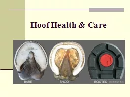
Slide21Slide22Arches of foot
Foot Support the body weight & Serve as a lever to propel the bodySegmented Arches help to sustain stresses of thrusts and weights.
Slide23Arches of foot serve as elastic springs for efficient walking
Slide24Identify the bony components….
Slide25Slide26Slide27Slide28Slide29Maintenance of ArchesBony factor
Intersegmental tiesTie beams/ bow stringsSlings
Slide30Slide31Slide32Pes Planus & Pes Cavus
Slide33Claw foot
Slide34Talipes deformities
Slide35Club Foot
Slide36