
CONGENITA DR SAKTI PRASAD DAS ASST PROFESSOR Arthrogryposis multiplex congenita is a collective term applied to a very large number of different syndromes characterised by non progressive multip ID: 941174
Download Pdf The PPT/PDF document "ARTHROGRYPOSIS MULTIPLEX" is the property of its rightful owner. Permission is granted to download and print the materials on this web site for personal, non-commercial use only, and to display it on your personal computer provided you do not modify the materials and that you retain all copyright notices contained in the materials. By downloading content from our website, you accept the terms of this agreement.


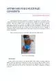
ARTHROGRYPOSIS MULTIPLEX CONGENITA DR SAKTI PRASAD DAS, ASST. PROFESSOR Arthrogryposis multiplex congenita is a collective term applied to a very large number of different syndromes characterised by non - progressive, multiple joint contractures present at birth. 1,2 The joints usually develop normally in early embryonic life but, as gestation progresses, movements are required to facilitate normal development. Where there are abnormalities that prev ent this from occurring, such as neurological or connective - tissue disorders or physical restriction, the condition forms. The muscles involved are partially or completely replaced by fat and fibrous tissue. The most common form, accounting for 40% of case s, is amyoplasia. Other secondary problems are associated with generalised fetal akinesia. Fig1 . Case of Amyoplasia Epidemiology It may occur to some extent in 1 in 3,000 live births. The condition is usually detected at birth or before by ultrasound examination. It is often secondary to other conditions. If they are X - linked this will produce a male preponderance but otherwise there is an equal sex incidence. It has been found to be more common in some isolated communities in Finland and Israel. 3 Aetiology Over 150 different syndromes may cause this symptom complex; therefore, careful history and examination are important to try to elucidate the underlying cause. The basic cause is fetal akinesia (reduced fetal movements). The underlying cause can be environmental (lack of ability to move) or genetic (single gene conditions). The underlying cause may be part of a more complex disorder affecting muscles, nerves or connective tissue. 4,5 A variety of maternal illnesses may result in arthrogryposis . This varied aetiology has contributed to a more comp
lex classification of the symptom complexes. Amyoplasia is the most common type of arthrogryposis and this accounts for about one third of cases. The conditions may be classified as below and broadly un der the following headings: Disorders characterised mainly by limb involvement. Disorders that involve the limbs and other body parts. Disorders with limb involvement and central nervous system dysfunction. Other associated syndromes and conditions. Fig 2 . A case of whistle face of syndrome. ( Freeman - Sheldon syndrome) Classification A - Disorders with mainly limb involvement : o Amyoplasia. Sporadic condition. Most common type of arthrogryposis (accounting for 1/3 of cases). Distinct appearance of limbs and joints (including internally rotated, adducted shoulders, fixed extended elbows, pronated forearms, flexed wrists and fingers and bilateral talipes equinovarus. Normal IQ. o Oddly, 80% have midline facial capillary haemangi oma B - Disorders with involvement of limbs and other body parts : o Multiple pterygium syndrome (pterygium meaning 'wing' and referring to triangular membranes affecting the neck, knees, elbows, ankles, etc.): Autosomal recessive: multiple joint contractures w ith marked pterygia (and dysmorphic facies and cervical vertebral anomalies). Autosomal dominant: multiple pterygia (with or without mental retardation). o Other syndromes: Freeman - Sheldon syndrome. Osteochondrodysplasias. Chromosomal disorders. Cerebro - ocu lo - facial skeletal syndrome. Presentation History History can be examined in terms of family history, pregnancy and delivery. A bout family history of : o Other affected children or members of the family. o Consanguinity in
creases the risk of rare recessive disorders. o Increasing parental age may increase risk - both mother and father. o A parent may have a mild form or have had infantile contractures. o Ask about miscarriages, possibly with fetal abnormalities. A bout maternal disease . This may include myotonica dystrophica that can produce a very severe condition but also myasthenia gravis and multiple sclerosis. A bout the pregnancy and delivery: o Infection with some viruses, including rubella and coxsackie , can cause neuropathy. o Prolonged maternal pyrexia can produce contractures due to abnormal nerve growth and migration. This can also be produced by very hot baths, hot tubs and saunas in pregnancy. o Drugs, including phenytoin and alcohol, can impair fetal movements. o Oligohydramnios reduces fetal movement s. A septate uterus or large fibroids can do the same. o An abnormal lie is common and this will complicate delivery. o There may have been amniotic bands or placental abnormality. A short cord or one wrapped around a limb reduced mobility. o Multiple pregnancy restricts room to move. Examination Classical presentation of amyoplasia shows involvement of both upper and lower extremities with the lower extremities typically m ore involved. Abnormalities include: Shoulders (adduction, internal rotation). Elbows (extension or fixed flexion). Wrists (flex ion). Deformity of thumb and palm and rigid interphalangeal joints. Hips (with dislocation of one or both sides). - dislocation and subluxation Knees (fixed extension or flexion). Rigid bilateral clubfeet/vertical t alus . Other characteristic features include: o Thin subcutaneous tissue and absent skin creases. o Symmetrical deformities (becoming more sever
e distally). o Rigid joints. o Congenital dislocation of the hips (and sometimes the knees). o Atrophy or absence of groups of muscles. o Normal sensation. o Contractures (especially of distal joints). o Pterygia (these are winglike triangular membranes occurring typically in the neck, knees, elbows, ankles or fingers). o Other deformities of the limbs (including shortening, webs, com pression often due to cord wrapping, absent patellae, dislocated radial heads, and dimples). o Deformities of face and jaw (including asymmetry, flat nasal bridge, haemangiomata, micrognathia and trismus . o Scoliosis , genital deformity and umbilical or ingui nal herniae are common. o There may be many other malformations of the skeleton, respiratory tract, urinary system and nervous system. Fig 3 – A case with bilateral knee dislocation Fig 4 . A case with bilateral knee subluxation and bilateral club foot Differential diagnosis There is a wide and varied range of rare conditions to be considered in the diagnosis. 3 Conditions causing arthrogryposis include . 6 A Fetal abnormality : o Neurogenic disorders . Myelomeningocele Sacral agenesis Spinal muscular atrophy (anterior horn cell disease of prenatal origin (SMA 0), not Werdnig - Hoffman (SMA 1). Congenital contracture syndrome (lethal). Cerebro - oculo - facial syndrome. Marden - Walker syndrome. Pena - Shokeir syndrome. o Myopathic disorders . Congenital myopathies Congenital muscular dystrophy. Myasthenic syndromes. Intrauterine viral myositis Mitochondrial disorders. o Connective tissue disorders Diastrophic dysplasia. Osteochondroplasia. Met at
ropic dwarfism. o Mechanical limitations to movement . Oligohydramnios as in Potter's syndrome and multiple births. B. Maternal disorders : o Maternal infection (including rubella, Coxsackie and enterovirus infections). o Drugs (including for example alcohol, phenytoin and methocarbamol ). o Trauma . o Intrauterine vascular abnormalities/compromise . o Other maternal illnesses (including myotonic dystrophy , myasthenia gravis and multiple sclerosis). Investigations X - rays of all joints may show bony abnormalities, including missing bones, skeletal dysplasia , scoliosis, ankylosis and fractures arising from difficult delivery. X - ray of spine and pelvis should always be included. Ultrasound, CT and MRI scans may all be useful to assess muscle mass and abnormalities in other tissues like the central nervous system. If muscles are very flaccid, a blood test for creatine kinase may be revealing. Antiviral antibody may also show a cause. Cytogenetic studies may be required Management This is likely to involve several different specialists and therapists. Ideally, management should be co - ordinated by a key specialist (often a paediatrician) who is part of a team looking after affected patients. Broadly speaking, management can be divided into medical, surgical, social and psychological care. Medical care Physical therapy to improve the range of motion and stretch surrounding tissues is very useful, especially in amyoplasia and distal arthrogryposis, although in diastrophic dysplasia it may lead to ankylosis instead. It should be started early . 7 Splinting between times can correct deformity, especially in the hands and wrists. Serial casting after physical therapy has achieved maximum usefulness with weekly changes of cast and gentle
manipulati on. 8 Surgical care Surgery is often needed to correct soft - tissue contractures and joint deformities. It can also reduce and stabilize dislocated hips, correct foot deformities and stabilize spinal deformities. Anesthesia can be a problem, as some patients with some forms of arthrogryposis are at risk of malignant . Social and psychological care This encompasses some fundamental care needs which will be associated with the condition. The extent of the need for care will var y depending on the individual, their circumstances and the impact of the condition on the particular individual. Information is important and support groups provide useful information and advice. 9 The new system of social care and support is called Self Directed Support and this should allow for the varied and complex care needs of this particular condition. Prognosis Prognosis depends on the underlying cause, but most have a normal life span. If, however, there is a central nervous system problem in addition, about 50% of patients die in the first year. Scoliosis is common and can appear at any age but needs correction before it becomes severe. The long - term prognosis in terms of dependency is poor in many cases but for amyoplasia it is quite reasonab le. 10 Of those with amyoplasia, 2/3 are ambulant by age 5. Most achieve independent life and can attend mainstream school. However, most require serial casting or surgery. Prevention Genetic advice may be essential to prevent arthrogryposis. Extrinsicall y derived contractures have a low recurrence risk, but the recurrence risk for intrinsically derived contractures depends on aetiology and ranges from 3% to 50%. 3 Ambulation Regarding ambulation it has been studied that functional ambulation in these patients required hip
motion to within 30[degrees] of full extension and knee motion to within 20[degrees] of full extension. Ambulation also required hip extensor strength of goo d (grade 4), quadriceps strength of fair (grade 3), or crutchable upper extremities and orthotic substitutes. Foot and spine deformities also interfered with ambulatory ability. Progressive spine deformity is the common cause of decreased ambulation. Pati ents not satisfying these critetria should have emphasis on sitting and activities of daily living. 11 R eferences 1. Hall JG ; Genetic aspects of arthrogryposis. Clin Orthop Relat Res. 1985 Apr;(194):44 - 53. 2. Hall JG ; Arthrogryposis multiplex congenita: et iology, genetics, classification, diagnostic approach, and general aspects. J Pediatr Orthop B. 1997 Jul;6(3):159 - 66. 3. Chen H ; Arthrogryposis, Medscape, Jul 2009 4. Gordon N ; Arthrogryposis multiplex congenita. Brain Dev. 1998 Oct;20(7):507 - 11. 5. Banker BQ ; Neuropathologic aspects of arthrogryposis multiplex congenita. Clin Orthop Relat Res. 1985 Apr;(194):30 - 43. 6. Clinical Guideline ; UCL and Great Ormond Street Hospital; Clinical Guideline Arthrogryposis 7. Bernstein RM ; Arthrogryposis and amyoplasia. J Am Acad Orthop Surg. 2002 Nov - Dec;10(6):417 - 24. 8. Smith DW, Drennan JC ; Arthrogryposis wrist deformities: results of infantile serial casting. J Pediatr Orthop. 2002 Jan - Feb;22(1):44 - 7. 9. Arthrogryposis information and support , The Arthrogryposis Group 10. Sells JM, Jaffe KM, Hall JG ; Amyoplasia, the most common type of arthrogryposis: the potential for good outcome. Pediatrics. 1996 Feb;97(2):225 - 3 1. 11. M Mark Hoffer, Susan swank, fred estman, douglas Clark, Robert Tietz. Ambulation in severe arthrogryposis. Journal of paediatrics orthopaedics. 1983,3, :293 - 2