
The Serbian trial on blood GSTP as novel biomarker of dialysis adequacy and dosage Sanja Simi ć Ogrizovi ć Clinic of nephrology CCS Medical faculty University of Belgrade Belgrade Serbia ID: 926846
Download Presentation The PPT/PDF document "GLUTATHIONE TRANSFERASE P1 AS THE MARKER..." is the property of its rightful owner. Permission is granted to download and print the materials on this web site for personal, non-commercial use only, and to display it on your personal computer provided you do not modify the materials and that you retain all copyright notices contained in the materials. By downloading content from our website, you accept the terms of this agreement.


Slide1
GLUTATHIONE TRANSFERASE P1 AS THE MARKER OF DIALYSIS ADEQUACY The Serbian trial on blood GSTP as novel biomarker of dialysis adequacy and dosage
Sanja Simić-OgrizovićClinic of nephrology CCS Medical faculty, University of Belgrade Belgrade, Serbia
Clinical Center of Serbia
Slide2In order to quantify the removal of uraemic toxins and the treatment time, proposed a mathematical model based on the dialytic removal of urea
The concept of Kt/V urea, Product of the dialyser clearance (K)Multiplied by the duration of treatment (t)divided by the estimated volume of distribution (V) of ureaSargent John and Gotch Frank 1975. They introduced the concept of Kt/V urea in assessment of dialysis adequacy which is related to MHD patient’s mortality
Slide3spKt/V
urea index > 1.3 (KDOQI) spKt/Vurea index >1.4 (EBPG) ??!25 years later Kt/V > 1.0/session = adequate dialysis
Slide41846 pts
HEMO study Kt/V= 1.32Kt/V = 1.72p >0.05Majority of MHD patients achieve the recommended target Kt/V urea, however the survival of these patients remains disappointing
No significant differences in mortality or hospitalization between the dosage groups
Slide5a.Kt/V urea is an ‘urea-centric’ mathematic model adopted to quantify the detoxification from a single compound in a single dialytic session
Slide6b. Kt/V represents only the actions of small-solute removal, not predictive of the kinetic performance of other classes of uremic toxins, such as middle molecules or protein-bound solutes
Slide7Removal of large solutes is enhanced with HDF and HF (convection therapies-large volumes of substitution fluid)
Larger convective transportBetter correction of uremia vascular damages CV morbidity mortality
?
Slide8Cardiovascular diseases are main cause of mortality of
hemodialysis patientsCVD mortality by age, race, and gender in the general population
and in dialysis patients
Data from the general population are from the National Center for Health Statistics multiple cause of mortality files 1993. Data from dialysis
patientsinclude
hemodialysis
and peritoneal dialysis combined from USRDS 1994-1996.
Reprinted with permission from
Am J Kidney
Dis
32[
Suppl
3]: S115, 1998.
Slide9The complicated puzzle of uremic CVD
Stenivnkel et al Emerging Biomarkers for Evaluating Cardiovascular Risk in he Chronic Kidney Disease Patient: How Do New Pieces Fitinto the Uremic Puzzle?Clin J Am Soc Nephrol 2008; 3: 505-521EUTox Work Group suggests that most of the molecules involved in the vascular damage were protein-bound and/or middle moleculesand urea does not necessarily belong to toxic solutes
Slide10OS in ESRD patients cornerstone of atherosclerotic process
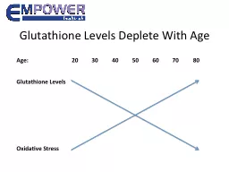
1.Carotid artery IMT in chronic HD patients correlates with lipid peroxidation byproduct2.S MDA is a strong predictor of prevalent cardiovascular disease in HD patientsStenivnkel et al Clin J Am Soc Nephrol 2008; 3: 505-521Kao MP, Ang DS, Pall A et al. Oxidative stress in renal dysfunction: mechanisms, clinical sequelae and therapeutic options. J Hum Hypertens 2010; 24: 1–8.
Slide11What is
GLUTATHIONE TRANSFERASE P1 ???
Slide12Members of the glutathione transferase (GST) enzyme superfamily (cytosolic
, mitochondrial and microsomal proteins) in detoxification and antioxidation The family of cytosolic GSTs comprises different classes: 1.Alpha (GSTA), 2. Mu (GSTM), 3. Pi (GSTP)4. Theta (GSTT)
Slide13GSTP1 enzyme in dialysis patients Role in detoxification accumulated
uraemic toxins and posses strong antioxidant activity towards reactive oxygen species (ROS) and peroxides. Mimic-Oka J, Simic T, Djukanovic Lj et al. Glutathione and its associated enzymes in peripheral blood cells in different stages of renal insufficiency. Amino Acids 1992; 2: 215–224.Lin YS, Hung SC, Wei YH et al. GST M1 polymorphism associates with DNA oxidative damage and mortality among hemodialysis patients. J Am Soc Nephrol 2009; 20: 405–415.Influenced by the genetic polymorphism
Slide14New role of erythrocyte member of cytosolic GST family (e-GST) dialysis patients e-GST is
overexpressed in uremic patients in response to increased OS and uremic conditionsThe enzyme is compartmentalized within the RBC (non-dialyzable)Ideal for new dialysis adequacy biomarker
Slide1582 healthy subjects
44 patients on bic. HD 59 patients on online HDFeGST not as a classical biomarker,but more suitably an endogenous biosensor of blood toxicityPilot/cross section study
Slide16a.
eGST is being naturally overexpressed when the toxin level increases;
Slide17(b)
eGST expression is reasonably linked not to the quantitative level of blood toxins nor to their size, but probably to their own specific toxicity
Slide18(c) Contrary to the Kt/
Vurea e-GST reflects the adequacy of multiple dialytic sessions within 1–2 months of life span of circulating erythrocytes
Slide19Inflammation Oxidative stressVitB12-Folic acid
HyperHcy HyperPTH Inadequate dialysisMalnutritionANEMIA rHEpo resistance rHEpo resistance CVD
Slide20GLUTATHIONE TRANSFERASE P1 AS THE MARKER OF DIALYSIS ADEQUACY
Prospective multicentre study with a follow-up period of 3 years
Slide21STUDY PARTICIPANTSPrincipal Investigator: Prof dr
T.Simic Co-Investigator(s): Prof dr S.Simic Ogrizovic and Prof dr F.Galli Laboratory project leader: Ass prof M. Pljesa Ercegovac Clinical project leader: Dr J. Maslovaric Cost benefit analysis: Prof dr N. Bogavac Laboratory research: V. Coric Technician: S.Zivotic
Slide22Main objectivesTo verify the potential of e-GST as a biomarker of dialysis adequacy, complementary to the Kt/V urea, in different dialysis settings (standard bicarbonate
hemodialysis (bHD) and HDF). To verify potential of e-GST as reflection of “average” adequacy of multiple dialyses session and its association with EPO resistance will also be assessed.
Slide23Main objectives3. To examine whether glutathione S-transferase P1 (GSTP1) A, B, C and
D genotypes are associated with differences in detoxification potential mediated by e-GST in MHD patients. Secondary objectivesTo assess prognostic significance of e-GST in terms of CV morbidity and mortality in HD patients. To compare cost-effectiveness of e-GST and Kt/V methods.
Slide24Subjects, groups and centersPatients undergoing HD (640) and HDF (160) will be recruited from several Serbian HD centers: Clinic of nephrology, CCS, Belgrade;
Clinical Hospital Zemun, Belgrade; Clinical of nephrology, Military academy, Belgrade;Special hospital for BEN, Lazarevac; Special hospital for internal medicine, Mladenovac; Center of hemodialysis Obrenovac, Clinic of nephrology, Clinical Center Nis. The laboratory part of the study will be carried out in the Laboratory for Kidney Research, Institute of Medical and Clinical Biochemistry, Faculty of Medicine University of Belgrade, and University of Perugia.
Slide25Study population In prevalent HD patients: any history of HTA, hypotension, ischemic vascular disease (MI, AP, cerebral stroke), heart failure, DM and smoking will be obtained by interview, physical examination and analysis of medical records as well for BMI and EPO doses
. Subject inclusion criteria: Older than 18Dialysis vintage over 6 months More than 3 months on the same dialyses modality Having obtained his/her or his/her legal representative’s informed consent Subject exclusion criteriaHistory of hepatitis B or C Active infection and cancer
Slide26BIOCHEMICAL PARAMETERSSampling: b
lood will be sampled on a midweek day as EDTA blood for plasma, buffy coat and erythrocytes. Analysis: B.parameters: s.concentration of Hb, urea, albumin, iron, ferritin, hs-CRP will be analyzed using automated methods. Polymorphism of GSTP1 DNA will be isolated from the collected buffy coat by modified protocol using QIAamp DNA Blood kit. PCR will be performed using the primers PiF2306/PiR2721 for exon 5 (Ile105Val), and PiF3402/PiR3800 for exon 6 (Ala114Val). (Prof T.Simic, Belgrade)e-GST activity will be determined by a spectrophotometric assay at 340 nm (37⁰C), using a Modular P800 (Roche, Basel, Switzerland) automated apparatus (Prof F. Galli, Peruga).
Slide27Clinical parameters at baselineDemographic parameters (age, sex, dialysis vintage,
origine kidney disease), co-morbidities (HTA,DM, ischaemic cardiopathy, peripheral arteriopathy and a previous transient ischaemic attack) and type of vascular accesses will be recorded. Pre- and post-session BW, BP and HR will be recorded together with the dialysis parameters, including filter type, blood flow, dialysis time and total infusion. For all examined MHD patients the following data will be calculated: ESA resistance will be expressed as an ESA index: the weekly weight adjusted ESA dose (DDD) divided by the haematocrit (Hct) ; Total weekly kT/V; Body weight and BMI after dialysis session.
Slide28Clinical parameters during the study: Information regarding death and causes of death will be obtained from hospital records and other relevant documents. Causes of death will be classified as cardiovascular death, if myocardial infarction and/or stroke occurred.
Myocardial infarction will be diagnosed by cardiologist on the basis of clinical presentation, ECG parameters and dynamic of enzyme activities. Stroke will be diagnosed by neurologist according to clinical presentation and CT scan.
Slide29D
ialysis adequacy Kt/VeGST
Slide30??????????
costbenefits