/
MYCETOMA BY : YASSER MAGHRABY MYCETOMA BY : YASSER MAGHRABY

MYCETOMA BY : YASSER MAGHRABY - PowerPoint Presentation

evans
evans . @evans
Follow
67 views
Uploaded On 2023-11-18

MYCETOMA BY : YASSER MAGHRABY - PPT Presentation

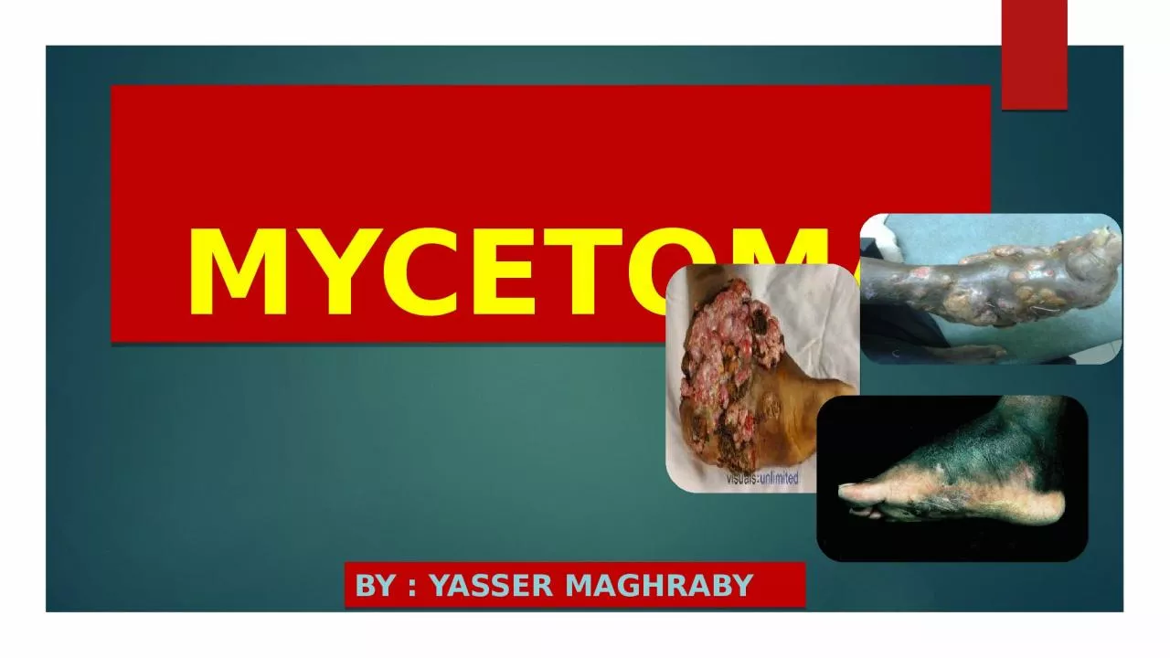
Mycetoma Definition Chronic granulomatous progressive inflammatory disease caused by various species of fungi or bacteria actinomycetes and characterized by the formation of aggregates ID: 1032845

clinical mycetoma occure actinomycetes mycetoma clinical actinomycetes occure patients multiple grains actinomycetoma causative bones bacterial pus fungi skin species

Share:

Link:

Embed:

Download Presentation from below link

Download Presentation The PPT/PDF document "MYCETOMA BY : YASSER MAGHRABY" is the property of its rightful owner. Permission is granted to download and print the materials on this web site for personal, non-commercial use only, and to display it on your personal computer provided you do not modify the materials and that you retain all copyright notices contained in the materials. By downloading content from our website, you accept the terms of this agreement.


Presentation Transcript

1. MYCETOMABY : YASSER MAGHRABY

2. Mycetoma

3. Definition Chronic granulomatous progressive inflammatory disease caused by various species of fungi or bacteria (actinomycetes), and characterized by the formation of aggregates of the causative organisms (grains) within abscesses. This results in severe damage to skin, subcutaneous tissues and bones of the feet, hands and other parts of the body, and grains are discharged to the surface through draining sinuses.

4. EtiologyMycetoma may be caused by various species of fungi (eumycetoma) and aerobic actinomycetes (actinomycetoma), which occur as saprophytes in soil or on plants.From these sources, they are implanted subcutaneously, usually after a penetrating injury.The disease is largely confined to tropical and subtropical climates, usually among agricultural workers.

5. Main aetiological agents of mycetomaActinomycetesFungiActinomadura maduraeActinomadura pelletieriStreptomyces somaliensisNocardia brasiliensisNocardia otitidis-caviarumNocardia asteroidesDark grainMadurella mycetomatisMadurella grisea Leptosphaeria senegalensisCurvularia lunataPale grainScedosporium apiospermumNeotestudina rosatiiAcremonium spp.Fusarium spp.

6. PathogenesisThe causative organism enter through the sites of local traumaA neutrophilic response is initially occurs which may be followed by a granulomatous reactions Spread occure through skin facial planes and can involve the bones Heamatogenic or lymphatic spread are un common

7. EpidemiologyIt occurs commonly in in tropical and subtropical areas espicialy in adult males who work barfooted.About 90% of the patients are males between 20 -50 years

8. Epidemiology

9. Epidemiology

10. Clinical featuresHistoryThe earliest sign is painless subcutaneous swelling /lump Some patients have a history of penetrating injury at the siteAfter some years massive swelling of the area , with indurations ,skin ruptures , and sinus tract formationThere may be little pain ,tenderness , feverPatients may complain of deep itching sensations

11. Clinical features the clinical features are the same whatever it eumocytoma or actinomycetes The most common lesions are the foot or the lower leg , but uncommon to occure any where on the body as arms head shoulder thigh and buttoks The earliest stage is firm painless nodule , development of other nodules produce irregular lump appearance .Some nodules are breake down and discharge pus containing grains.Extention to underlying bonesand joints gives rise to periostitis , osteomyelitis ,and arthritis

12. Clinical presentations

13. Clinical featuresIn advanced cases destructions of bones within an infected area may be almost complete and gross deformity may result

14. Clinical featuresPain occure in about 20% pf the patients ,and usally is due to secondary bacterial infections . in General Eumocytomas are more circumscribed and progress slower than Actinomycetomas

15. Investigations Direct KOH of pus : color of granules determine there is fungal or actinomycetes .Culture in sabouraud,s agar:Fungi (chloramphenicol only) at room temp . Growth 5- 10 daysActinomycetes (cycloheximide only) at room temp growth 2-3 days

16. Investigations Mycetoma can be accurately diagnosed by Fine Needle Aspiration (FNA) cytology. Mycetoma lesion has a distinct appearance in a cytology smear characterised by the presence of polymorphous inflammatory cells consisting of an admixture of neutrophils, lymphocytes, plasma cells

17. DermatopathologyThe causal organism produce a chronic granulomatous reaction leading to dense scar tissue and local endarteritis changes .Colonies which form white or red or black or pale granules according to etiological agent may be discharged in pus through multiple sinuses .The causal agent can be stained better in biopsy samples with gram stain in actinomycetoma ,, or with Gomori methenamine silver or PAS stain for eumocytoma

18.

19. Investigation (Radiological study)Cortical thining is due to compression from outside by the mycetomaCortical hypertrophy may presentas a sunray appearance and acodman triangle .Multiple lytic lesion or cavities may be larg ,few in number and with well defined margin in Eumocytoma , or small ,numerous and with ill defined margin in Actinomycetoma Disuse osteoporosis may occure in late mycetoma

20. Radiological study

21. InvestigationCT scan can allows better definition of the changes observed in in bone x ray filmsMagnetic resonance imaging (MRI) provides a better assessment of the degree of bone involvementUltrasonography single or multiple thick walledcavities with hyperreflective echos in mycetoma

22. Differential diagnosisChronic osteomyelitis of bacterial or tuberculous aetiology may resemble mycetoma, particularly in the early stages.Elephantiasis there is no sinus tracts .Laboratory tests will be distinguish between the causative organisms.

23. TreatmentLocalized lesions that can be excised without residual disability are best so treated .Infection by species of actinomycetes may be susceptible to chemotherapeutic agents. The combination of dapsone with streptomycin or co-trimoxazole plus streptomycin has been reported to give good results.An alternative second drug is rifampicin. Amikacin may also be used in recalcitrant Nocardia infections

24. TreatmentAmong the fungal causes of mycetoma, M. mycetomatis is the most sensitive to therapy, as it responds to ketoconazole in some cases.Itraconazol 200mg/d until cure The minimum treatment duration is about 10 months.Radical surgery usually amputation should be considerd carefuly

25. Prognosis

26. PrognosisActinomycetoma can be cured with the appropriate antibiotic therapy but eumycetoma has a high rate of recurrence and can require amputation.A high incidence of secondary bacterial infection in mycetoma lesions has been reported. This can cause increased pain and disability as well as septicaemia, which may be fatal if untreated.

27. شكراً للحـــــــضور