
Songserm T Amonsin A Jamon R SaeHeng N Meemak N Pariyothorn N et al Avian Influenza H5N1 in Naturally Infected Domestic Cat Emerg Infect Dis 2006124681683 httpsdoiorg103201eid1204051396 ID: 1041608
Download Presentation The PPT/PDF document "Figure 1 Figure 1. Microscopic ..." is the property of its rightful owner. Permission is granted to download and print the materials on this web site for personal, non-commercial use only, and to display it on your personal computer provided you do not modify the materials and that you retain all copyright notices contained in the materials. By downloading content from our website, you accept the terms of this agreement.


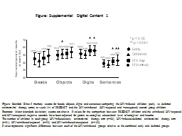


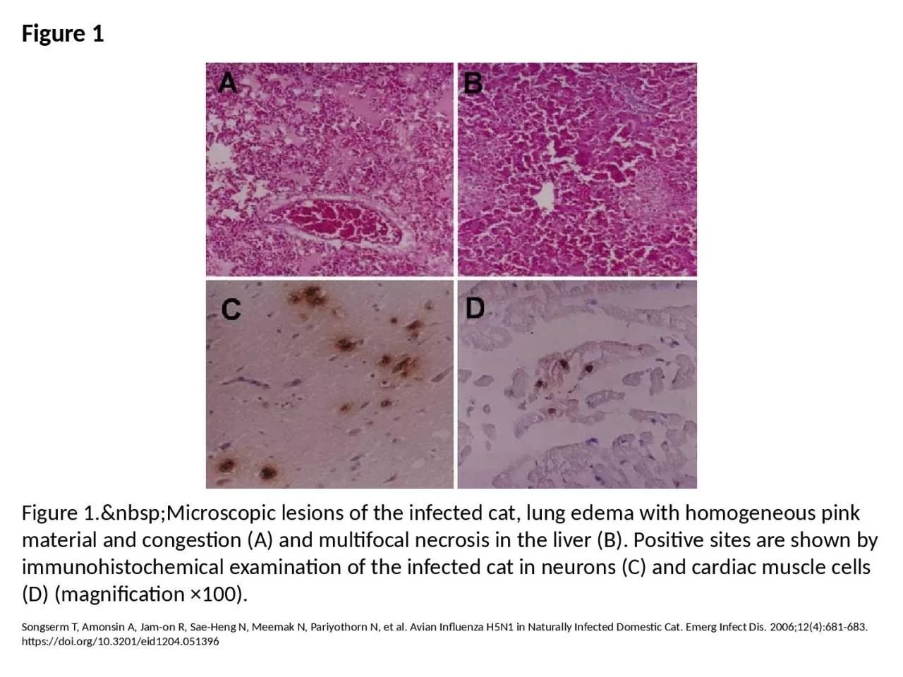
1. Figure 1Figure 1. Microscopic lesions of the infected cat, lung edema with homogeneous pink material and congestion (A) and multifocal necrosis in the liver (B). Positive sites are shown by immunohistochemical examination of the infected cat in neurons (C) and cardiac muscle cells (D) (magnification ×100).Songserm T, Amonsin A, Jam-on R, Sae-Heng N, Meemak N, Pariyothorn N, et al. Avian Influenza H5N1 in Naturally Infected Domestic Cat. Emerg Infect Dis. 2006;12(4):681-683. https://doi.org/10.3201/eid1204.051396