
D Litt et Phil Health Studies D N Ed M Art et Scien B A Cur Dip Occupational Health Dip Genetic Counselling Dip Audiometry and Noise Measurement Diagnostic Radiographer Medical Ethicist ID: 938828
Download Pdf The PPT/PDF document "Researched and Authored by Prof Michael ..." is the property of its rightful owner. Permission is granted to download and print the materials on this web site for personal, non-commercial use only, and to display it on your personal computer provided you do not modify the materials and that you retain all copyright notices contained in the materials. By downloading content from our website, you accept the terms of this agreement.


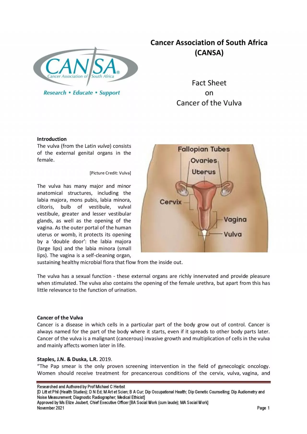
Researched and Authored by Prof Michael C Herbst [D Litt et Phil (Health Studies); D N Ed; M Art et Scien; B A Cur; Dip Occupational Health ; Dip Genetic Counselling; Dip Audiometry and Noise Measurement; Diagnostic Radiographer; Medical Ethicist ] Approved by Ms Elize Joubert, Chief Executive Officer [BA Social Work (cum laude); MA Social Work] November 20 2 1 Page 1 Cancer Association of South Africa (CANSA) Fact Sheet o n Cancer of the Vulva Introduction The vulva (from the Latin vulva ) consists of the external genital organs in the female . The vulva has many major and minor anatomical structu res, including the labia majora , mons pubis , labia minora , clitoris , bulb of vestibule , vulval vestibule glands, a s well as the opening of the vagina . As the outer portal of the human uterus or womb, it protects its opening by a âdouble doo râ: the labia majora (large lips) and the labia minora (small lips). The vagina is a self - cleaning organ, sustaining healthy microbial flora that flow from the inside out. The vulva has a sexual function - these external organs are richly innervated and provide pleasure when stimulated. The vulva also contains the opening of the female urethra , but apart from this ha s little relevance to the function of urination. Cancer of the Vulva Cancer is a disease in which cells in a particular part of the body grow out of control. Cancer is always named for the part of the body where it starts, even if it spreads to other body parts later. Cancer of the vulva is a malignant (cancerous) invasive growth and multiplication of cells in the vulva and mainly aff Staples, J.N. & Duska, L.R. 2019. âThe Pap smear is the only proven screening intervention in the field of gynecologic oncology. Women should receive treatment for precancerous conditions of the cervix, vulva, vagina, and Researched and Authored by Prof Michael C Herbst [D Litt et Phil (Health Studies); D N Ed; M Art et Scien; B A Cur; Dip Occupational Health ; Dip Genetic Counselling; Dip Audiometry and Noise Measurement; Diagnostic Radiographer; Medical Ethicist ] Approved by Ms Elize Joubert, Chief Executive Officer [BA Social Work (cum laude); MA Social Work] November 20 2 1 Page 2 endometrial lining. Women with inherited conditions should consider having a risk - reducing surgery once they have finished childbearing. The human papil loma virus vaccination should be offered to all girls and boys aged 11 to 12 years, and can also be given as early as age 9 and through 26 years of age.â Tumour Grade and Tumour Stage Tumour grade and stage are terms used to describe the severity of a tumour, w hile tumo u r grade describes the appearance of cancerous cells in the tissue by examining them under a microscope . T umo u r stage encompasses:
⢠The location of the tumo u r. ⢠The size and/or extent of the original tumo u r. ⢠Whether cancer cells have spread to lymph nodes or anywhere else in the body. ⢠The number of tumo u rs present. Doctors use tumo u r grade, cancer stage, and a patientâs age and general health to decide the course of treatment for the patient and determine prognosis. Prognosis describes all factors including the disease course, cure rate, chances of survival, and risk of rec urrence of cancer . What are the cancer stages? Different systems of cancer staging are used to describe the types of cancer. Below is a common method in which stages are ranged from 0 to IV. ⢠Stage 0: The tumo u r is confined to its place of origin (in situ) and has not spread to nearby tissue. ⢠Stage I: The tumo u r is located only in the original organ, is small, and has not spread. ⢠Stage II: The size of the tumo u r is large but has not spread. ⢠Stage III: The tumo u r has become larger and may have spread to surrounding tissues and/or lymph nodes. ⢠Stage IV: The tumo u r has spread to other distant organs of the body, which is known as the metastasis stage. TNM staging Another common staging method used for cancer is the TNM system, which stands for tumo u r, node (which means spread of the tumo u r to lymph nodes), and metastasis. When a patientâs cancer is staged using the TNM system, a number will be present along with the letter. This number signifies the extent of the disease in each category - tumo u r, node, and metastases. Another system of cancer staging divides cancer into five stages, which include: ⢠In situ: Abnormal cells are present but have not spread to nearby tissue. ⢠Localized: Cancer is located only in the original organ and shows no sign of its s pread. ⢠Regional: Cancer has spread to nearby lymph nodes, tissues, or organs. ⢠Distant: Cancer has spread to distant parts of the body. ⢠Unknown: The stage cannot be figured out due to a lack of enough information. Researched and Authored by Prof Michael C Herbst [D Litt et Phil (Health Studies); D N Ed; M Art et Scien; B A Cur; Dip Occupational Health ; Dip Genetic Counselling; Dip Audiometry and Noise Measurement; Diagnostic Radiographer; Medical Ethicist ] Approved by Ms Elize Joubert, Chief Executive Officer [BA Social Work (cum laude); MA Social Work] November 20 2 1 Page 3 What are the cancer grades? Cancer grades are based on examination of the suspected tissue sample under a microscope. This involves surgically removing a piece of the suspected cancerous tissue and sending it to the lab for analysis. The entire procedure is known as a biopsy. A doctor who special izes in diagnostic tests (pathologist) examines the cells of the tissue and determines whether they are har
mless (benign or noncancerous) or harmful ( malignant or cancerous). They describe the microscopic appearance of the cells and assign a numerical âgra deâ to most cancers . Generally, a lower grade indicates slow - growing cancer and a higher grade indicates fast - growing cancer. The most commonly used grading system is as follows: ⢠Grade I: Cancer cells that look like normal cells but are not growing rapidly. ⢠Grade II: Cancer cells that don't look like normal cells with their growth being faster than normal cells. ⢠Grade III: Cancer cells that look abnormal and have the potential to grow rapidly or spread more aggressively. Sometimes, the following system can be used: ⢠GX: Grade cannot be assessed (undetermined grade) ⢠G1: Well - differentiated (low grade) ⢠G2: Moderately differentiated (intermediate grade) ⢠G3: Poorly differentiated (high grade) ⢠G4: Undifferentiated (high grade) Incidence of Cancer of the Vulva in South Africa According to the outdated National Cancer Registry (20 1 7 ) , known for under reporting, the following number of cancer of the vulva cases w as histologically diagnosed in South Africa during 20 1 7 : Group - Females 20 1 7 Actual No of Cases Estimated Lifetime Risk Percentage of All Cancers All females 5 41 1: 5 80 1 , 3 0 % Asian females 1 2 1: 667 0 , 93 % Black females 4 39 1: 57 3 2 , 35 % Coloured females 2 9 1: 906 0, 6 4 % White females 61 1: 607 0, 36 % The frequency of histologically diagnosed cases of cancer of the vulva in South Africa for 20 1 7 w as as follows (National Cancer Registry, 20 1 7 ): Group - Females 20 1 7 0 â 19 Years 20 â 29 Years 30 â 39 Years 40 â 49 Years 50 â 59 Years 60 â 69 Years 70 â 79 Years 80+ Years All females 5 3 4 1 86 1 50 63 57 28 18 Asian females 0 0 1 0 1 2 6 2 Black females 4 2 8 1 75 1 33 51 33 9 6 Coloured females 0 1 7 7 3 5 5 2 Researched and Authored by Prof Michael C Herbst [D Litt et Phil (Health Studies); D N Ed; M Art et Scien; B A Cur; Dip Occupational Health ; Dip Genetic Counselling; Dip Audiometry and Noise Measurement; Diagnostic Radiographer; Medical Ethicist ] Approved by Ms Elize Joubert, Chief Executive Officer [BA Social Work (cum laude); MA Social Work] November 20 2 1 Page 4 White females 1 5 3 1 1 8 1 7 8 8 N.B. In the event that the totals in any of the above tables do not tally, this may be the result of uncertainties as to the age, race or sex of the individual. The totals for âall malesâ and âall femalesâ, however, always reflect the correct totals. According to Bruni, et al ., (2019), the burden of cervical cancer for
South Africa for 2018 is estimated as: ⢠Annual number of cases of cancer of the vulva 2 262 ⢠Annual number of deaths due to cancer of the vulva 210 Causes and Risk Factors for Cancer of the Vulva Several risk factors for cancer of the vulva have been identified. Researchers have made a lot of progress in understanding how certain changes in DNA can cause normal cells to become cancerous. DNA is the chemical that carries the instructions for nearly everything the bodyâs cells do. [Picture Credit: Cancer of Vulva] Certain genes that promote cell division are called oncogenes . Others that slow down cell division o r cause cells to die at the right time are called tumour suppressor genes . Cancers can be caused by DNA mutations (defects) that turn on oncogenes or turn off tumour suppressor genes. Usually DNA mutations related to cancers of the vulva occur during life rather than having been inherited before birth. Acquired mutations may re sult from cancer - causing chemicals in tobacco smoke. Sometimes they occur for no apparent reason. Studies suggest that squamous cell cancer of the vulva (the most common type) can develop in at least 2 ways. In up to half of cases, human papilloma virus (HPV) infection appears to have an important role. The second process by which vulvar cancers develop does not involve HPV infection. Vulvar cancers not linked to HPV infection (the keratinising subtype) are usually diagnosed in older women (over age 55 ). These women often show mutations of the p53 tumour suppressor gene. The p53 gene is important in preventing cells from becoming cancerous. When this gene has undergone mutation, it is easier for cancer to develop. [Picture Credit: Lichen Sclerosus] Researched and Authored by Prof Michael C Herbst [D Litt et Phil (Health Studies); D N Ed; M Art et Scien; B A Cur; Dip Occupational Health ; Dip Genetic Counselling; Dip Audiometry and Noise Measurement; Diagnostic Radiographer; Medical Ethicist ] Approved by Ms Elize Joubert, Chief Executive Officer [BA Social Work (cum laude); MA Social Work] November 20 2 1 Page 5 Lichen Sclerosus Lichen sclerosus appears in: o w omen (often after menopause) o m en (uncommon) o c hildren (rare) Early in the disease, small white spots appear on the skin. The spots are usually shiny and smooth. Later, the spots grow into bigger patches. The skin on the patches becomes thin and crinkled. Then the skin tears easily, and bright red or purple bruises are common. Sometimes, the skin becomes scarred. If the disease is a mild case, there may be no symptoms. Other symptoms are: ⢠i tching (very common) ⢠d iscomfort or pain ⢠b leeding ⢠b listers Signs and Symptoms of Cancer of the Vulva Signs and symptoms of cancer o f the vulva may include:
⢠i tching that does n o t go away ⢠p ain and tenderness ⢠b leeding that is n o t from menstruation ⢠s kin changes, such as colour changes or thickening ⢠a lump, wart - like bumps or an open sore (ulcer) ⢠a bnormal bleeding ⢠b urning ⢠p ainful urination ⢠w art - like growths (similar to genital warts) ⢠c hange in the appearance of an existing mole (specific to vulvar melanoma) ⢠i tching ⢠l ocal pain Diagnosis of Cancer of the Vulva If someone suspects that she may have cancer of the vulva she should visit a medical pra c titioner that specialises in womenâs cancers (gynaecological cancer specialist). Apart from a close examination of the vulval area, the doctor will also do a general medical examination of the patient to determine her general condition of health. Researched and Authored by Prof Michael C Herbst [D Litt et Phil (Health Studies); D N Ed; M Art et Scien; B A Cur; Dip Occupational Health ; Dip Genetic Counselling; Dip Audiometry and Noise Measurement; Diagnostic Radiographer; Medical Ethicist ] Approved by Ms Elize Joubert, Chief Executive Officer [BA Social Work (cum laude); MA Social Work] November 20 2 1 Page 6 The d octor may use a bright light and a magnifier to examine the vulva, so that the skin can be seen more clearly. He/she may then take small samples of tissue (biopsies) from any areas that look unusual. This may be done under a local anaesthetic. The doctor will also usually do an internal examination to check the vagina, cervix and the neck of the womb for any abnormality. The doctor will use a speculum (a plastic or metal instrument) to hold the vaginal walls open. He/she may also take a cervical smear tes t (a small sample of cells taken from the cervix). The doctor may also examine the back passage (anus). Michalski, B.M., Pfeifer, J.D., Mutch, D. & Council, M.L. 2020. Background: Vulvar cancers, although rare, are becoming an increasingly serious threat to women's health. Cancer of the vulva accounted for 0.3% of all new cancers in the United States in 2019, with 6,070 newly diagnosed cases. This review details the epidemiology, pathogenesis, diagnosis, staging, and treatment of vulvar mali gnancies. Objective: To review cancer entities of the vulva, including vulvar intraepithelial neoplasms, squamous cell carcinoma (SCC), malignant melanoma, basal cell carcinoma, neuroendocrine tumors, and adenocarcinomas. Materials and methods: Literature review using PubMed search for articles related to cancer of the vulva. Results: Vulvar intraepithelial neoplasms represent premalignant precursors to SCC of the vulva. There are several different histopathologic subtypes of SCC, and treatment is dependent on characteristics of primary tumor and lymph node involvement. Melanoma is the second most common can
cer to affect the vulva, and staging is based on tumor, node, and metastatic spread. Conclusion: Vulvar malignancies are rare, and diagnosis is dependent on biopsy and pathologic evaluation. Treatment for vulvar malignancies depends on histopathologic diagnosis but ranges from wide local excision with or without lymph node biopsy or dissection to radiation therapy with chemo - or immunotherapy. Overall surv ival varies by diagnosis. Tan, A., Bieber, A.K., Stein, J.A. & Pomeranz, M.K. 2019. â Vulvar malignancies represent a serious gynecologic health concern, especially given the increasing incidence over the past several decades. Squamous cell carcinoma and melanoma are common subtypes, although other neoplasms, such as basal cell carcinoma and Paget disease of the vulva, might be seen. Many v ulvar cancers are initially misdiagnosed as inflammatory conditions, delaying diagnosis and worsening prognosis. It is essential that dermatologists are familiar with characteristic findings for each malignancy to ensure appropriate diagnosis and managemen t. Herein, we review the unique epidemiologic and clinical characteristics of each major vulvar malignancy, as well as discuss their respective prognoses and current management recommendations. â Treatment of Cancer of the Vulva Treatment options for can cer of the vulva depend on the type and stage of the cancer as well as the personâs overall health and preferences , and may include: Surgery Operations used to treat cancer of the vulva include: ⢠Removing the cancer and a margin of healthy tissue (excision). ⢠Removing a portion of the vulva (partial vulvectomy). ⢠Removing the entire vulva (radical vulvectomy). Researched and Authored by Prof Michael C Herbst [D Litt et Phil (Health Studies); D N Ed; M Art et Scien; B A Cur; Dip Occupational Health ; Dip Genetic Counselling; Dip Audiometry and Noise Measurement; Diagnostic Radiographer; Medical Ethicist ] Approved by Ms Elize Joubert, Chief Executive Officer [BA Social Work (cum laude); MA Social Work] November 20 2 1 Page 7 ⢠Extensive surgery for advanced cancer. ⢠Reconstructive surgery. ⢠Possible s urgery to remove nearby lymph nodes . Michalski, B.M., Pfeifer, J.D., Mutch, D. & Council, M.L. 2020. Background: Vulvar cancers, although rare, are becoming an increasingly serious threat to women's health. Cancer of the vulva accounted for 0.3% of all new cancers in the United States in 2019, with 6,070 newly diagnosed cases. This review details the epidemiology, pathogenesis, diagnosis, staging, and treatment of vulvar malignancies. Objective: To review cancer entities of the vulva, including vulvar intraepithelial neoplasms, squamous ce ll carcinoma (SCC), malignant melanoma, basal cell carcinoma, neuroendocrine tumors, and adenocarcinomas. Materials and methods:
Literature review using PubMed search for articles related to cancer of the vulva. Results: Vulvar intraepithelial neoplasms re present premalignant precursors to SCC of the vulva. There are several different histopathologic subtypes of SCC, and treatment is dependent on characteristics of primary tumor and lymph node involvement. Melanoma is the second most common cancer to affect the vulva, and staging is based on tumor, node, and metastatic spread. Conclusion: Vulvar malignancies are rare, and diagnosis is dependent on biopsy and pathologic evaluation. Treatment for vulvar malignancies depends on histopathologic diagnosis but ran ges from wide local excision with or without lymph node biopsy or dissection to radiation therapy with chemo - or immunotherapy. Overall survival varies by diagnosis. Frøding, L.P., Zobbe, V., Fokdal, L.U., Niemann, I.C.S., Nøtrup, T.J., Kristensen, E., Ch ristiansen, A.P., Loft, A. & Schnack, T.H. 2019. âThis review summarises the current guidelines for vulva cancer in Denmark. Vulva cancer is a rare disease. The diagnosis is often delayed, which results in large tumours and regional spread. The most impor tant prognostic factor is inguinal lymph node metastases. Staging and treatment is centralised to two hospitals. Primary treatment is wide local excision combined with removal of either inguinal sentinel nodes or lymphadenectomy. Treatment is associated wi th considerable morbidity, and supportive care is often necessary. Local curable recurrences are common. Relapses in the groin are associated with a poor prognosis. Thus, long term follow - up is essential. *) On behalf of Dansk Gynækologisk Cancer Gruppe fo r vulvacancer. â Radiation therapy Radiation therapy uses high - powered energy beams, such as X - rays, to kill cancer cells. Radiation therapy for cancer of the vulva is usually administered by a machine that moves around oneâs body and directs radiation to precise points on the skin (external beam radiation). Chemotherapy Chemotherapy is a drug treatment that uses chemicals to kill cancer cells. Chemotherapy drugs are typically administered through a vein in the arm or by mouth. Singareddy, R., Bajwa, H.K., Alluri, K.R., R aju, K.V.V.N., Rao, T.S. & Rajeev, G. 2019. AIM: The aim of this study was to evaluate the patterns of recurrence and outcomes in patients with vulvar cancer treated with surgery, with or without postoperative radiotherapy (RT) or RT alone. Researched and Authored by Prof Michael C Herbst [D Litt et Phil (Health Studies); D N Ed; M Art et Scien; B A Cur; Dip Occupational Health ; Dip Genetic Counselling; Dip Audiometry and Noise Measurement; Diagnostic Radiographer; Medical Ethicist ] Approved by Ms Elize Joubert, Chief Executive Officer [BA Social Work (cum laude); MA Social Work] November 20 2 1 Page 8 MATERIALS AND METHODS: The hospita
l records were retrospectively analyzed from February 2007 to May 2016. All patients that had biopsy - proven carcinoma vulva were included for analysis. Surgery was done by simple or radical vulvectomy with or without lymph node dissecti on. Radical RT dose was 60 Gy (photons alone or with electron boost). Adjuvant RT was administered in case of node positivity or positive margins. RESULTS: A total of 76 patients were diagnosed as squamous cell carcinoma of vulva. The median age was 59 yea rs. About 59 patients were treated by surgery and 17 patients were treated by radical RT. The most common stage was IB. Adjuvant RT was administered in 10 patients. About 13 patients (22%) underwent inguinal lymphadenectomy. At a median follow - up of 35 mon ths, 49 patients (64.4%) were alive, 27 died, and 6 patients were lost to follow - up. Locoregional recurrence rate was 24.5% in radical surgery group, 12% in surgery plus adjuvant RT group, and 47% in radical RT group. Distant metastasis rate was 4%. The pr ogression - free survival at 3 years was 45.3% and 35.5% in the surgery group and radical RT group, respectively. CONCLUSION: Surgery resulted in favorable local control rates in early - stage carcinoma vulva. Adjuvant RT is indicated in case of nodal positivi ty or positive margins. Radical RT can be considered as an alternative in patients with comorbidities. Nandwani, M., Barmon, D., Begum, D., Liegise, H. & Kataki, A.C. 2019. An overview of vulvar cancer: a single - center study from Northeast India. J Obstet Gynaecol India . 2019 Dec;69(6):541 - 545. doi: 10.1007/s13224 - 019 - 01261 - z. Epub 2019 Aug 16. STUDY: Carcinoma vulva is a rare cancer of the female genital tract. It mostly presents in postmenopausal women. The treatment of vulvar cancer is surgery, chemoradiation, radiotherapy or a combination of all modalities. Here, we present a study of 33 cases of carcinoma vulva over a period of 2 years at a Northeast India regional cancer institute describing its demographic features and treatment outcomes. MET HODOLOGY: A retrospective cohort study of vulvar cancer diagnosed at Northeast India regional cancer institute from January 2017 to December 2018. RESULTS: A total of 33 cases of biopsy proven carcinoma (Ca) vulva were studied. Maximum number of cases belo nged to the age group: 60 - 69 years (39.4%). 66.67% cases had palpable inguinal lymph nodes at presentation, and 100% had squamous cell carcinoma on histopathology. Maximum number of cases belonged to stage III (44.8%), and least number of cases belonged to stage IV (10.3%) of FIGO 2009 staging of Ca vulva. 87.9% cases underwent treatment, and 12.1% were lost to follow - up. Out of the cases who underwent treatment, 55.2% cases were taken up for primary surgery and 44.8% cases for primary radiotherapy. 75% cas es who underwent surgery received
adjuvant radiotherapy. No complication was seen in patients post - radiation. But, 6.25% patients post - surgery developed lymphocyst and 18.75% patients developed wound necrosis ( p â>â0.05). CONCLUSION: Vulvar cancer is not a common malignancy of the female genital tract that presents in sixth and seventh decades of life and often with palpable inguinal lymph nodes. Though early stages of Ca vulva are treated by surgery, the incidence of immediate postoperative complications in our study was more as compared to post - radiotherapy. Also, maximum patients in the present study post - surgery received adjuvant radiotherapy. Thus, radiotherapy can be considered as the primary treatment modality for patients with early as well as advance d vulvar carcinoma. Follow - up T ests after T reatment After completing treatment for cancer of the vulva, the doctor may recommend periodic follow - up exam inations to look for a cancer recurrence. Researched and Authored by Prof Michael C Herbst [D Litt et Phil (Health Studies); D N Ed; M Art et Scien; B A Cur; Dip Occupational Health ; Dip Genetic Counselling; Dip Audiometry and Noise Measurement; Diagnostic Radiographer; Medical Ethicist ] Approved by Ms Elize Joubert, Chief Executive Officer [BA Social Work (cum laude); MA Social Work] November 20 2 1 Page 9 About Clinical Trials Clinical trials are research studies that involve people. They are conducted under controlled conditions. Only about 10% of all drugs started in human clinical trials become an approved drug. Clinical trials include: ⢠Trials to test effectiveness of new treatments ⢠Trials to test new ways of using current treatments ⢠Tests new interventions that may lower the risk of developing certain types of cancers ⢠Tests to find new ways of screening for cancer The South African National Clinical Trials Register provides the public with updated information on clinical trials on human participants being conducted in South Africa. The Register provides information on the purpose of the clinical trial; who can participate, where the trial is located, and contact details. For additional information, please visit: www.sanctr.gov.za/ Medical Disclaimer This Fact Sheet is intended to provide general information only and, as such, should not be considered as a substitute for advice, medically or otherwi se, covering any specific situation. Users should seek appropriate advice before taking or refraining from taking any action in reliance on any information contained in this Fact Sheet. So far as permissible by law, the Cancer Association of South Africa ( CANSA) does not accept any liability to any person (or his/her dependants/ estate/ heirs ) relating to the use of any information contained in this Fact Sheet. Whilst the Cancer Association of South Africa (
CANSA) has taken every precaution in compiling this Fact Sheet, neither it, nor any contributor(s) to this Fact Sheet can be held responsible for any action (or the lack thereof) taken by any person or organisation wherever they shall be based, as a result, direct or otherwise, of information contained in, or accessed through, this Fact Sheet. Sources and References Consulted or Utilised American Cancer Society http://www.cancer.org/cancer/vulvarcancer/detailedguide/vulvar - cancer - what - causes http://www.cancer.org/cancer/vulvarcancer/detailedguide/vulvar - cancer - staging Bruni, L., Albero, G., Serrano, B., Mena, M., Gómez, D., Muñoz, J., Bosch, F.X.& de Sanjosé, S. 2019. ICO/IARC Information Centre on HPV and Cancer ( HPV Information Centre ). Human Papillomavirus and Related Diseases in South Africa. Summary Report 17 June 2019. [Date Accessed] Cancer.Net http://www.cancer.net/cancer - types/vulvar - cancer/symptoms - and - signs Researched and Authored by Prof Michael C Herbst [D Litt et Phil (Health Studies); D N Ed; M Art et Scien; B A Cur; Dip Occupational Health ; Dip Genetic Counselling; Dip Audiometry and Noise Measurement; Diagnostic Radiographer; Medical Ethicist ] Approved by Ms Elize Joubert, Chief Executive Officer [BA Social Work (cum laude); MA Social Work] November 20 2 1 Page 10 Cancer of Vulva https://www.google.co.za/search?q=vulvar+cancer+pictures&source=lnms&tbm=isch&sa=X&ei=uOgBUvujJ4mZhQfujoG4A Q&ved=0CAcQ_AUoAQ&biw=1366&bih =614#facrc=_&imgdii=_&imgrc=Jv6wg7AHBHEjCM%3A%3BrHAAxmj8DDLJeM%3Bh ttp%253A%252F%252Fwww.uptomed.ir%252FDigimed.ir%252Fcecil%252FCecil%252FHTML%252Ff4 - u1.0 - B978 - 1 - 4160 - 2805 - 5..50214 - 7..gr2.jpg%3Bhttp%253A%252F%252Fwww.uptomed.ir%252FDigimed.ir%252Fcecil%252 FCecil%252FHTML%252F770.htm %3B592%3B600 Family Practice Notebook http://www.fpnotebook.com/Gyn/Hemeonc/VlvrCncr.htm Frøding, L.P., Zobbe, V., Fokdal, L.U., Niemann, I.C.S., Nøtrup, T.J., Kristensen, E., Christiansen, A.P., Loft, A. & Schnack , T.H. 2019. Workup, treatment and follow - up of vulva cancer. Ugeskr Laeger , 2019 Aug 19;181(34):V04190216. Lichen Sclerosus https://www.google.co.za/search?q=lichen+sclerosus&source=lnms&tbm=isch&sa=X&ei=xekBUqa6EIOohAfnx4CgBA&sqi= 2&ved=0CAcQ_AUoAQ&biw=1366&bih=614#facrc=_&imgdii=_&imgrc=Z2CNFoyzwumlLM%3A%3BWr7PAzCrrP8I6M%3Bht tp%253A%252F%252Fdermatlas.med.jhmi.edu%252Fdata%252 Fimages%252Flichen_ruber_moniliformis_2_040626.jpg%3 Bhttp %253A%252F%252Fkootation.com%252Flichen - plaque.html%3B1280%3B960 MacMillan Cancer Support http://www.google.co.za/url?sa=t&rct=j&q=&esrc=s&frm=1&source=web&cd=3&ved=0CDsQFjAC&url=http%3A%2F%2Fw ww.ma cmillan.org.uk%2FCancerinformation%2FCancertypes%2FVulva%2FSymptomsdiagnosis%2FDiagnosis.aspx&ei=dzzM UbqCCeWP7AaeyICoDw&usg=AFQjCNHFGLgC - GHjUJL75IqhwqZI - R - o2Q&sig2=wX6IEvvLDQJF7N7PXeAXAA http://www.macmillan.org
.uk/Cancerinformation/Cancertypes/Vulva/Treat ingvulvalcancer/Treatmentoverview.aspx Mayo Clinic http://www.mayoclinic.com/health/vulvar - cancer/DS00768/DSECTION=symptoms http://www.mayoclinic.com/health/vulvar - cancer/DS00768/DSECTION=treatments - and - drugs Medscape Today http://www.medscape.com/viewarticle/722721 Michalski, B.M., Pfeifer, J.D., Mutch, D. & Council, M.L. 2020. Cancer of the vulva: a review. Dermatol Surg . 2020 Sep 15. Micheletti, L. & Preti, M. 2014. Surgery of the vulva in vulvar cancer. Best Pract Res Clin Obstet Gynaecol . 2014 Oct; 28:1074 - 87. Doi: 10.1016/j. bpobgyn.2014.07.011. Epub 2014 Jul 22 National Cancer Institute http://www.cancer.gov/clinical trials/learningabout/what - are - clinical - trials Nandwani, M., Barmon, D., Begum, D., Liegise, H. & Kataki, A.C. 2019. An overview of vulvar cancer: a single - center study from Northeast India. J Obstet Gynaecol India . 2019 Dec;69(6):541 - 545. doi: 10.1007/s13224 - 019 - 01261 - z. Epub 2019 Aug 16. National Institute of Arthritis and Musculoskeletal and Skin Diseases http://www.niams.nih.gov/Health_Info/Lichen_Sclerosus/ Rajaram, S. & Gupta, B. 2015. Management of Vulvar Cancer. Rev Recent Clin Trials . 2015; 10(4):282 - 8. Singareddy, R., Bajwa, H.K., Alluri, K.R., Raju, K.V.V.N., Rao, T.S. & Rajeev, G. 2019. Carcinoma vulva patterns of recurrence and treatment outcomes: a single - institution experience. J Cancer Res Ther . 2019 Oct - Dec;15(6):1328 - 1331. doi: 10.4103/jcrt.JCRT_44_18. Staples, J.N. & Duska, L.R. 2019. Cancer screening and prevention highlig hts in gynecologic cancer. Obstet Gynecaol Clin North Am . 2019 Mar;46(1):19 - 36. doi: 10.1016/j.ogc.2018.09.002. Tan, A., Bieber, A.K., Stein, J.A. & Pomeranz, M.K. 2019. Diagnosis ana management of vulvar cancer: a review. J Am Acad Dermatol. 2019 Dec;81(6):1387 - 1396. doi: 10.1016/j.jaad.2019.07.055. Epub 2019 Jul 23. Researched and Authored by Prof Michael C Herbst [D Litt et Phil (Health Studies); D N Ed; M Art et Scien; B A Cur; Dip Occupational Health ; Dip Genetic Counselling; Dip Audiometry and Noise Measurement; Diagnostic Radiographer; Medical Ethicist ] Approved by Ms Elize Joubert, Chief Executive Officer [BA Social Work (cum laude); MA Social Work] November 20 2 1 Page 11 Tumour Grade and Tumour Stage https://www.medicinenet.com/cancer_101_pictures_slideshow/article.htm Vulva https://www.google.co.za/search?q=vulva&source=lnms&tbm=isch&sa=X&ei=dfzLUeKOG6Sr7AaXyoDQDw&sqi=2&ved=0C AcQ_AUoAQ&biw=942&bih=464#facrc=_&imgdii=_&imgrc=OY5RECi O6MOp0M%3A%3BescM9pygEvj5aM%3Bhttp%253A %252F%252Fwww.news - medical.net%252Fimage.axd%253Fpicture%253D2010%25252F4%25252Ffemale - genitals.jpg%3Bhttp%253A%252F%252Fwww.news - medical.net%252Fhealth%252FWhat - is - the - Vulva.aspx%3B438%3B349 Wikipedia http://en.wiki pedia.org/wiki/Vu