
Nov 12 2018 Click me The brain is divided into four main regions Cerebrum Diancephalon aka interbrain Brain stem Cerebellum Regions of the Human Brain Largest region Responsible for all higher brain functions ID: 1042563
Download Presentation The PPT/PDF document "Brain Structure and Function" is the property of its rightful owner. Permission is granted to download and print the materials on this web site for personal, non-commercial use only, and to display it on your personal computer provided you do not modify the materials and that you retain all copyright notices contained in the materials. By downloading content from our website, you accept the terms of this agreement.


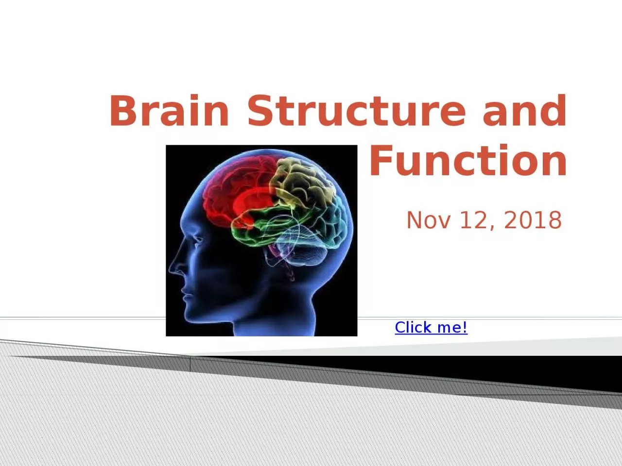
1. Brain Structure and FunctionNov 12, 2018Click me!
2. The brain is divided into four main regions: CerebrumDiancephalon (aka interbrain)Brain stemCerebellumRegions of the Human Brain
3. Largest regionResponsible for all ‘higher’ brain functions:speechmemoryreasoningemotionconsciousnessinterpretation of sensationvoluntary movementCerebrum
4. Divided into left and right hemispheresWrinkled texture Raised areas called gyriShallow grooves called sulciDeep groves called fissuresCerebrum features
5. The outer part of the cerebrum – called the cerebral cortex -- is composed of gray matter Gray matter = cell bodies and unmyelinated nerve fibersThis is where thought, sensation, etc. occurThe inner part of the cerebrum is mostly white matterWhite matter = mylinated nerve fibersThis is the connection between different regions of the brainInternal anatomy of cerebrum
6. The corpus callosum is one especially important area of white matter – it’s the connection between the two cerebral hemispheresInternal anatomy of cerebrum
7. JFF: Functional regions of cerebrum
8. JFF: Functional regions of cerebrumDamage to one of these regions can cause loss of specific functions (e.g. partial paralysis due to stroke)BUTThe brain does have some ability to heal itself – to form new connections or to have one hemisphere take over the job of the other hemisphere - this is called brain plasticityJody’s storyBut also see Phineas Gage’s story
9. The diencephalon consists of three parts:thalamusepithalamus (completely enclosed by thalamus)hypothalamusDiencephalon structure
10. structures that are part of the diencephalonDiencephalon structure
11. Regulation of autonomic functions, includingBody temperatureWater balanceMetabolismControl of endocrine system Limbic system – emotional / visceral brain (sex, food, thirst, pain, pleasure)Mostly, these functions are done by the hypothalamusDiencephalon Functions
12. The brain stem consists of three partsMidbrainPonsMedulla oblongataBrain Stem
13. Controls many autonomic functions, including:BreathingBlood pressureHeart rateAlso allows passage of nerves fibers between brain and spinal cordBrain Stem Functions
14. Back of the brain, beneath cerebrumLike cerebrum, it hasTwo hemispheresWrinkled surfaceOuter cortex made of gray matter outside and inner region of white matterCerebellumFunctions: BalanceCoordination of muscles
15. Do the diagrams and questions on your guided notesYou Do:
16. Following a stroke, a person develops the symptoms listed below. Which part of the brain was damaged? ataxia (an inability to coordinate muscular movement) cerebellumDrooping left side of face right cerebral hemisphereQuestion
17. Fluid that surrounds and protects brain and spinal cord Produced by the choroid plexuses from blood plasma, and drains back into the bloodCerebrospinal Fluid (CSF)Some diseases can be diagnosed by examining CSF collected during a spinal tapWhich glial cells are involved with CSF? What do they do?
18. Fluid that surrounds and protects brain and spinal cord Produced by the choroid plexuses from blood plasma, and drains back into the bloodCerebrospinal Fluid (CSF)Some diseases can be diagnosed by examining CSF collected during a spinal tapWhich glial cells are involved with CSF? ependymalWhat do they do? Circulate CSF
19. Protects the brain from toxins, most drugs, and fluctuations in other chemicals such as ionsTwo major barriersRelatively impermeable brain capillariesAstrocytes which control flow of materials from blood vessels to neuronsBypassing the blood brain barrier with sound wavesBlood Brain Barrier
20. Why is the blood brain barrier necessary?Does the blood brain barrierblock the passage of alcohol? Blood Brain Barrier
21. Why is the blood brain barrier necessary?Without a controlled environment, uncontrolled neural activity might occurDoes the blood brain barrierblock the passage of alcohol?No – you can tell by the effectsBlood Brain Barrier
22. What were our objectives, and what did you learn about them. What was our learner profile trait and how did we exemplify it?How does what we did today address our unit question?Closure
23. For the following questions, A = cerebrum, B= diencephalonC = brain stem, D = cerebellumThe thalamus, hypothalamus, and epithalamus constitute the _________________.Control of temperature, endocrine activity, and thirst are associated with the ____________.The area of the brain that controls breathing and heart rate is the _________________.Exit Ticket
24. A shallow groove on the surface of the cortex is called a A) fissure B) gyrus C) furrow D) sulcusThe three major parts of the brain stem are the A) cerebrum, cerebellum, and diencephalon B) thalamus, epithalamus, and hypothalamus C) midbrain, pons, and medulla oblongata D) basal nuclei, pineal body, and choroid plexusExit Ticket
25. Exit Ticket678910EC 1EC 2