
Lama AlAbdi Supervised by Dr Reem AlAjmi Generay It is an instrument used to examine and monitor objects we cant see with the naked eye It has many applications Microscope There ID: 1044522
Download Presentation The PPT/PDF document "Lab#1 Microscopy & Epithelial Tissue" is the property of its rightful owner. Permission is granted to download and print the materials on this web site for personal, non-commercial use only, and to display it on your personal computer provided you do not modify the materials and that you retain all copyright notices contained in the materials. By downloading content from our website, you accept the terms of this agreement.


1. Lab#1Microscopy&Epithelial TissueLama AlAbdiSupervised by: Dr. Reem AlAjmi
2. Genera!y, It is an instrument used to examine andmonitor objects we can’t see with the naked eye.It has many applications.Microscope
3. There are different classes and types of microscopes dependingon the magnification power, their applications, and how theyare operated:Transmission electron microscope (TEM).Scanning electron microscope (SEM).Inverted microscope.Light (compound) microscope.Types of microscopes
4. The compoundmicroscope is made of 3systems:The mounting andmovement System.The magnificationSystem.The i!umination System.Compound Microscope
5. The mounting and movement systemThe body tube:carries the ocular lensesThe arm:Supports and connects theupper part of the microscopeThe coarse focusing knob:for stage movementThe fine focusing knob:for image sharpnessThe nose piece:Carries the objective lenses andmove them accordingly above thestageThe stage:Horizontal platform upon whichthe slide of interest restThe base:Supports the microscope
6. The ocular lenses:5X, 10X, and 15XThe objective lenses:Scanning lens: 3.5X or 4X or 5XLow power lens: 10XHigh power lens: 40XOil immersion lens: 100XTHE MAGNIFICATION SYSTEMHow to calculate the magnification power?Magnification power = ocular lens xobjective lens.(e.g.) 10X x 40X = 400X hint: Don’t forget the unit
7. THE ILLUMINATION SYSTEMThe illuminator:light sourceThe iris diaphragm:controls the amount of lightreaching the slideThe condenser:collects and concentrate the light
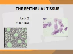
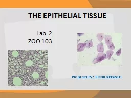
8. THE EPITHELIAL TISSUE Prepared by : Reem AldossariLab 2ZOO 103
9. Types of Epithelial Tissue Epithelial tissue can be divided into two groups depending on the number of layers of which it is composes. simple epithelium: which consist of only one layersquamous epitheliumcuboidal epitheliumcolumnar epitheliumStomachIntestineCollecting tubuleThyroid glandLining of MouthTop viewBowman’s capsuleSide viewstratified epithelium: which consist of more than one layer( Squamous,cuboidal,columnar)Skin of Toad( Frog )
10. Covering “surface” epithelium. Glandular epitheliumNeuro_epithium Types of Epithelial Tissue The Simple.Stratified.epithelium of cells specialized to produce secretion. (Goblet cell)Epithelial tissue can be divided into two groups depending on their function of which it is composes.
11. Squamous epithelium
12. Simple Cuboidal Epithelium
13. Simple Columnar Epithelium
14. 12Glandular Epithelium
15. Stratified sequamous epithelium in skin keratinized and non keratinized
16. Stratified cuboidal epithelium in sweat gland
17. Thank you for your attention