
Localized arrangement of adjacent amino acids formed as the polypeptide chain folds The regions of ordered structures formed by interaction of hydrogen bond donor and hydrogen bond acceptor residues of the repeating peptide unit ID: 1006353
Download Presentation The PPT/PDF document "Structure of Protein-II SECONDARY STRUCT..." is the property of its rightful owner. Permission is granted to download and print the materials on this web site for personal, non-commercial use only, and to display it on your personal computer provided you do not modify the materials and that you retain all copyright notices contained in the materials. By downloading content from our website, you accept the terms of this agreement.


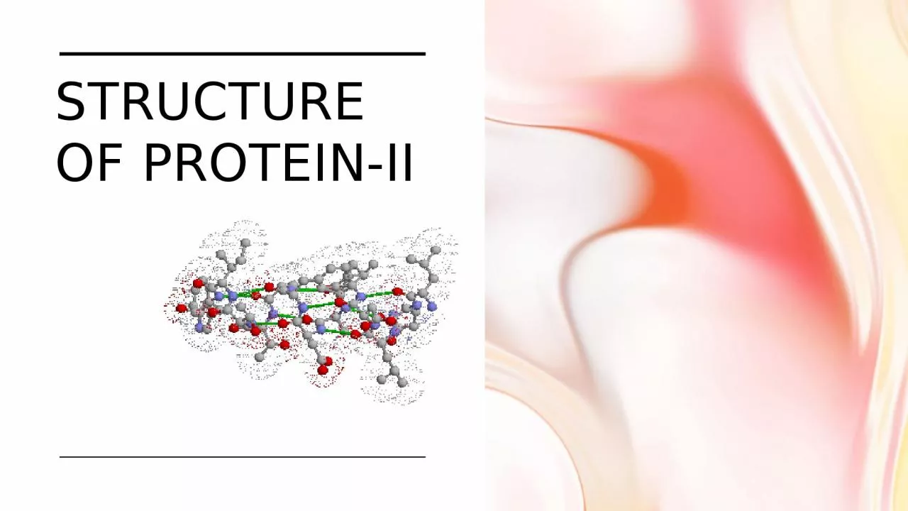
1. Structure of Protein-II
2. SECONDARY STRUCTURELocalized arrangement of adjacent amino acids formed as the polypeptide chain folds.The regions of ordered structures formed by interaction of hydrogen bond donor and hydrogen bond acceptor residues of the repeating peptide unitTypesα-helixβ-pleated sheetβ-bendsβ-barrels coiled-coilsNon repetitive structures Super secondary structures
3. ALPHA HELIXSpiral structureTightly packed, coiled polypeptide backbone core.Side chain extend outwardsStabilized by H bonding b/w carbonyl oxygen and amide hydrogen.The distance between two amino acid residues is 1.5 ÅAmino acids per turn – 3.6Pitch is 5.4 Å and width is 5.0 Å Each peptide bond participates in the hydrogen bonding. This gives maximum stability to α-helixAlpha helical segments are found in many globular proteins like myoglobins, troponin- C etc
4. Amino acids that prefer to adopt α-helical conformations in proteins include methionine, alanine, leucine, glutamate and lysine, as their small side chains minimize the steric hindrance in helix formationOther less common helices found in proteins are 3(10)-helix which is more stretched than ideal α – helix and π – helix which is more compact and extremely rare
5. Types of helix310 -helixVery tightly coiled H- bonding pattern n+3 rarely found in natureπ-helixVery loosely coiled H- bonding pattern n + 5Rarely found in nature.α-helixAlso called the 3.612
6. BETA PLEATED SHEETThe peptide chain is extended into a zigzag arrangement resembling a series of pleats, with the peptide bonds organized in planes of alternating slopes (alternating ascending and descending direction)Formed when 2 or more polypeptides line up side by side (different segment of same peptide or two peptide chain)
7. The characteristics of the β – pleated sheet includeEach peptide is planar and has a trans conformation.The C=O and N – H groups of the peptide bonds from adjacent chains point towards each other and are in the same plane so that hydrogen bonding is possible between them.All R- groups on any one chain alternate, first above, then below the plane of the sheet.
8. Types 2 typesParallel and Anti-parallel In parallel arrangement, the C-terminus end and the N-terminus end are on the same sides for both segmentsIn anti-parallel arrangement, the C-terminus end of one segment is on the same side as the N-terminus end of the other segment. Parallel arrangement is less stable because the geometry of the individual amino acid molecules forces the hydrogen bonds to occur at an angle, making them longer and thus weaker. Anti-parallel arrangement the hydrogen bonds are aligned directly opposite each other, making for stronger and more stable bonds
9. connection between β strandsThe two major sorts of connection between β strandsa hairpin or same end connectiona crossover or opposite end connection
10. Tight turnsThere are different types of the tight turns depending on the number of atoms forming the turn
11. SUPER SECONDARY STRUCTURES (MOTIFS)Certain groupings of secondary structural elements are called motifs
12. The helix is fundamental in allowing Proteins to sit within the plasma membraneit is the foundation of many very important Proteins Visual Pigments Immune cell receptors
13. Coiled-coil2-6 alpha-helices are coiled together like the strands of a ropeCollagen triple helix
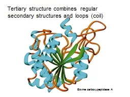
14. TERTIARY STRUCTURE3 – dimensional conformation of a polypeptide chain in its folded stateThe common features of protein tertiary structure reveal much about the biological functions of the proteins and their evolutionary originsThe function of a protein depends on its tertiary structure. If this is disrupted, it loses its activityTertiary structure is based on various types of interactions between the side-chains of the peptide chainSecondary structure and loops come together to form “Domain”, the smallest tertiary structural unit
15. DOMAINSPolypeptide chains containing more than 200 residues usually fold into two or more globular clusters known as domainsFundamental functional and 3 dimensional structure of proteins.Domains often have a specific function such as the binding of a small molecule
16. Tertiary structure stabilizationTertiary interactions are frequently stabilized by sequestration of hydrophobic amino acid residues in the protein core.Consequent enrichment of charged or hydrophilic residues on the protein's water-exposed surface
17. Types of Interaction
18. DETERMINATION OF TERTIARY STRUCTUREThe known protein structures have come to light through:X-ray crystallographic studiesNuclear Magnetic Resonance studiesThe atomic coordinates of most of these structures are deposited in a database known as the Protein Data Bank (PDB)It allows the tertiary structures of a variety of proteins to be analyzed and compared
19. QUATERNARY STRUCTUREThe biological function of some molecules is determined by multiple polypeptide chains – multimeric proteins.Arrangement of polypeptide subunit is called quaternary structureSubunits are held together by noncovalent interactions.Eg: Hemoglobin has the subunit composition a2b2Insulin is formed by two peptide chains, but since these two chains are linked by disulphide linkage, Insulin does not qualify as quaternary protein
20. importance of bonding interactions(Why?)Most important interactions in tertiary structure: van der waals and hydrogen bonding interactionsLeast important interactions: covalent and ionic bondingTwo reasons for this:More opportunity for van der waals and hydrogen bonding interactionsPresence of water surrounding the protein structure
21. There are only limited number of amino acids with residues capable of interacting with hydrogen bonds, those too are far outnumbered by number or residues capable of forming van der waal and hydrogen bondsMost stable tertiary structure has most of the hydrophilic groups on the surface so that they interact with water and most of the hydrophobic groups in core so they avoid water and interact with each other.Hydrophilic amino acids form a hydrogen bond with water and thus number of ionic and hydrogen bonds contributing to tertiary structure is reduced, this leaves van der waal and hydrophobic interactions to largely determine the three – dimensional shape of the protein