
Ankle Brachial Index Exam CDR Doug E Henry PT CWS Federal Medical Center Butner NC Ob jectives Outline the BOP Guidelines for evaluating vascular disease Identify the clinical signs of peripheral arterial diseasePAD ID: 1037690
Download Presentation The PPT/PDF document "The Clinical Impact of the" is the property of its rightful owner. Permission is granted to download and print the materials on this web site for personal, non-commercial use only, and to display it on your personal computer provided you do not modify the materials and that you retain all copyright notices contained in the materials. By downloading content from our website, you accept the terms of this agreement.


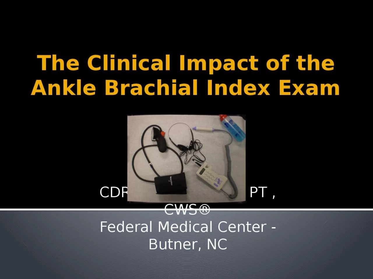
1. The Clinical Impact of theAnkle Brachial Index ExamCDR Doug E. Henry, PT , CWS®Federal Medical Center - Butner, NC
2. ObjectivesOutline the BOP Guidelines for evaluating vascular diseaseIdentify the clinical signs of peripheral arterial disease/PADSummarize the significance of the ABI examination in relationship to evaluation of PADSummarize the strengths & limitations of ABI examinationDescribe or interpret how current literature impacts clinical findings of the ABI examinationOutline which class of compression stockings are indicated or contraindicated for pnts
3. FCC Butner
4. The comparison of blood pressures in the arms (brachial) and lower extremities both (posterior-tibial) and (dorsalis pedis), using an appropriately sized blood pressure cuff and a Doppler. The highest systolic reading of the DP or PT pulse, indicated by the first audible Korotkoff sound, is divided by the highest systolic brachial reading. This gives the clinical ratio for interpretation.ABI or ABPI Procedure
5. DELAYED HEALINGTissue IschemiaPoor VascularityInstability of metabolites – Decreased H&H, Dehydration, poor glucose control… Nutrition – Low Albumin for Healing DemandHeavy Bacterial Bio-burdenInfectionOther
6. PAD risk factorsAtherosclerosisTobacco use Metabolic Syndrome: DM, HTN, Dyslipidemia,ObesityCADThromboangitis obliteransVasculitisRenal DiseaseRaynaud’s DiseaseSickle Cell DiseaseOther…
7. Risk – Hirsch et al 2006ACC/AHA 2005 Guidelines for the Management of Patients with Peripheral Arterial Disease, Journal of American College of Cardiology, vol 47, 2006AgeIndividuals at Risk for Lower Extremity PAD<50DM and one other risk factor (smoking, dislipidemia, hypertension)50-69History of smoking or DM70All patients older AnyLeg symptoms with exertion (suggestive of claudication) or ischemic rest painAnyAbnormal LE pulse examinationAnyKnown atherosclerotic coronary, carotid, or renal artery disease
8. PAD – PresentationClinical presentation: Claudication, atrophy, reduction of mobility, integumentary lesions, and delayed or unexplained slow healing wounds.Local presentation: discoloration, bruising, numbness, weakness, decreased pulses, atrophic appearance.
9. PAD - PathophysiologyDiminished blood flow resulting in tissue ischemia, ↓ O2 levels, ↓ neutrophil activity, ↓inflammatory response =↑ rates of infection… ↑ NON-healing ulcerations → necrosis → gangrene, amputation…Pathophysiology
10. Clinical Practice Guidelines
11. Committee Members Jeffrey Allen, MDMatt Hardin, DermatologistCDR Kevin Elker, RN, CWOCNCAPT Matt Taylor (ret), PT, DPT, OCS, CWSPam Baker, RN, CWOCNPatina Walker-Geer, NPCDR Christine Fallon, NP, WCCLCDR Sherrie Wheeler, RNCDR Cubie Beasley, RN ….
12. The Vascular Exam BOP Wound Care CPG
13. Basic Vascular Evaluation Skin TemperatureCapillary RefillPalpation of LE pulses DP & PTElevation PallorDependent RuborAtrophic foot
14. Subjective VascularPainIntermittent ClaudicationNocturnal painRest pain
15. Claudication Nocturnal Rest PainRuthorford, BR et al. Recommended standards for reports dealing with lower extremity ischemia: Revised version. Journal of Vascular Surgery. Sept 1997; 26. 517-38.Patients report pain with activity. Calf or leg/s feel heavy or cramping pain when they walk a specific distance each time.Relieved with about 10 minutes of rest. Occurs when the involved vessel is approximately 50% occluded.Occurs at night when the patient is in bed Relieved by placing the legs in a dependent position to increase blood flow, often over the edge of the bed.Occurs in the absence of activity with legs in a dependent positionIndicates advanced occlusive disease, typically greater than 90% of the affected vessel/s
16. ABI correlation to PADABI ValueDisease severity>0.90No disease0.69-0.90Mild to moderate intermittent claudication0.40-0.60Severe intermittent claudication0.25-0.40Rest pain and tissue loss Aronow WS. Management of peripheral arterial disease. Cardiology Review. 2005;13(2):61-68.
17. Atrophic Arterial signsIntact: cold, dry, flaking, chaffing, shiny skin, bluish, thick toenails, non-palpable pulse, chronic loss of autonomic and/or protective sensationOpen: eschar, denuded/dry wound bed.Arterial UlcerationsIf cellulitis, abscess, gangrene, or deep ulceration is present, consider immediate referral for treatment and amputation prevention
18. Elevation Pallor vs Dependent RuborWith the patient in the supine position, elevate the affected leg approximately 60 degrees. Note the color of the soles of the foot while elevatedNote the total = T/sec to pallorThe presence of a purplish-red discoloration in one or both legs is caused by the retention of de-oxygenated blood in the dilated skin capillaries of a patient with arterial disease. To differentiate from cellulitis or other mechanisms, have the patient lie in a supine position and elevate the leg approximately 60 degrees. If the discoloration fades, the most likely mechanism is dependent rubor
19. Pallor with ElevationErmer-Seltun J. Lower Extremity Assessment, Acute and Chronic Wounds. 2012, pg. 173. Pallor developing within…INDICATES25 secondsSevere arterial disease25-40 secondsModerate arterial disease40-60 secondsMild arterial disease60 secondsNo arterial diseaseInterpretation per BOP Wound Care CPG
20. Elevation Pallor vs Dependent Rubor
21. The Clinical Impact of the ABIEBM
22. ABI ValueEvaluation of PADAssessment of LE circulation pre-surgicallyAssessment of LE circulation s/p re-vascularization proceduresRisk for cardiovascular events including all causes leading to mortalitySimple, quick & non-invasive diagnosis Cost-Effective!
23. ABI – McDermottLower ankle/brachial index, as calculated by averaging the dorsalis pedis & posterior tibial arterial pressure, & association with leg functioning in peripheral arterial diseaseMcDermott et al, Journal of Vascular Surgery, Dec 2000n= 244 men & women age 55+ with & w/o PADOutcome measures = walking velocity & endurance measured with 6min/walk testReviewed 3 different methods of calculating ABI to determine the best testing method
24. ABI – McDermott cont.PAD defined as ABI less than < 0.90 Method 1: higher LE arterial pressure - 47% Method 2: lower LE arterial pressure - 59% Method 3: average of dorsalis pedis & posterior tibial pulses used to calculate ABI 52%Results: Method #2 was most closely associated with correctly predicting PAD but Method #3 predicted leg function using the outcome measures in regression analysis
25. ABI Specificity & SenstivityKhan et al. Critical Review of the Ankle Brachial Index. Current Cardiology Reviews. 2008, 4, 101-106AuthorSpecificitySensitivityConditionsSchroeder et al.99%68%Highest AP93% 89%Lowest APNiazi et al.83%69%Highest AP64%84%Lowest APLijemer96%79%Highest APStoffers et al82%79%Highest AP
26. CAT - 2013 For a 60 year old person with a positive reading less than 0.80 [0.90] what is the probability of mortality in this patient?Relationship of High and Low Ankle Brachial Index to All-Cause and Cardiovascular Disease Mortality: Resnick et al, Circulation 2004
27. CAT – Resnick et aln= 4549, age 45-74, ethnicity native, outcome = mortality, retrospective cohort, level 2BBoth abnormally low (4.9%) and high (9.2%) ABI results are linked to cardiovascular eventsAll cardiac events were associated including: CVA, TIA, MI…PAD determined by ABI <0.90
28. CAT – Resnick et al
29. ABI interpretationhttp://circ.ahajournals.org/content/126/24/2890.longAHA Measurement and Interpretation of ABI – Aboyans et al 2012ABI RatioINDICATION>/= 1.30Indicates non-compressible vessels >250mmHg. This finding is common in diabetic patients, due to increased rates of arteriosclerosis. Further diagnostic testing may be needed to evaluate whether blood flow is adequate for healing1.0 to 1.29 Normal ratio0.8 to 0.99 Blood flow should be adequate for healing.< 0.8 Patient may need to be referred for further diagnostic testing to evaluate whether blood flow is adequate for healing
30. ABI interpretationIDSA Guidelines for Diabetic Foot Infections - Lipsky et al 2012ABI RatioINDICATION>/= 1.30Poorly Compressible vessels, Arterial calcification0.9 to 1.30Normal ratio0.60 to 0.89Mild arterial obstruction0.40 to 0.59Moderate obstruction< 0.40Severe obstruction
31. ABI limitationsThe ABI is one screening toolElevated BP → erroneous resultsChronic Long Term Diabetes often N-CPerform Toe Brachial Index (TBI) in patients with non-compressible vessels Refer to vascular for advanced testingNon-invasive: LEAD studies/segmental limb pressure, doppler wave, pulse volume recorders, TcPO2, MRA, duplex angiographyInvasive: Arteriogram, CTA
32. Ankle Brachial Index (ABI)Assessment tool for arterial pathology- Compression TherapyVenousArterialComfort w/ compressionSubjective pain &/or discomfortNormally good skin turgor +Atrophic signs at footPulses normal or obscurePulse weak or absent
33. Levels of CompressionClassmmHgIndications120-30mmHg (light)Varicosities230-40mmHg (medium)Venous Insufficiency, +/- ulcer340-50 mmHg (strong)Treatment of refractory ulcer450-60 mmHg (high)LymphedemaByrant, RA, Nix, DP: Acute & Chronic Wounds, Current Management Concepts, 3rd Ed, Mosby Elsevier 2007, St. Louis, MO. Pp 299-306.
34. Compression StockingsTypeAvailableContra-indicatedIndicationsTED Hose (8-12 mmHg pressure)SupplyAmbulatoryPatients!Post-op DVT prevention, bed-bound pntsTherapeutic Stockings (20-30mmHg)PTSevere Arterial disease (PAD), cellulitis, CHF (caution)Dependent edema control, varicose veins, lymph prevention (post-mastectomy)TherapeuticStockings30-40mmHg PTmod-severe PAD, cellulitis, CHFVenous insufficiency, venous stasis, venous ulcersCustomPT same as aboveLymphedema pts, morbidly obese, anatomical variety
35. Compression StockingsAdvantages: Provide graded compression, enhance circulation for wound healing, pain relief, and prevention of DVT, variety of styles for ease of application.Disadvantages: Can be difficult to apply, require compliance, cleaning (cannot be dried in dryer)
36. Compression Stockings-InstructionsApplied before getting out of bed in the morning, removed at night or bathing ONLY (TED hose worn in bed by the non-ambulatory for prevention)Wash in washing machine, hang dryShould last 8-12 monthsD/C if develop chest pains or skin irritation, consider less compression if skin tears at dorsal crease in elderlyLower compression options=tubi-grip D, E & F
37. Take home messageThe evaluation of arterial disease is a critical step for wound healing at the LETreatment delays increase probability of failurePAD pnts have higher cardiac risks, mortality and amputation risksLimb salvage in the diabetic population is greatly improved with assertive mgmt!
38. Vascular examinationPMHSubjective ReportClinical ExaminationClinical Tests for Arterial DiseaseABI (Sensitivity & Specificity)Referral: Vascular Medicine
39. TOOLS D2HENRY@BOP.GOVBOP WOUND CARE CPGINTERPRETATION 2012 AHA2012 IDSACOMPRESSION HOSE GUIDES
40. Questions…D2HENRY@BOP.GOV
41. Referenceswww.cebm.netRothwell PM. External validity of randomised controlled trials: "to whom do the results of this trial apply?" Lancet 2005; 365(9453):82-93Montori VM, Jaeschke R, Schunemann HJ, Bhandari M, Brozek JL, Devereaux PJ et al. Users' guide to detecting misleading claims in clinical research reports BMJ 2004; 329(7474):1093-1096Amsler et al. In search of optimal compression therapy for venous leg ulcers: A meta-analysis of studies comparing divers bandages with specifically designed stockings. J Vasc Surg 2009;50:668-74.Straus, Sharon, Richardson W. Scott, Glasziou, Paul, Haynes R. Brian. Evidence-Based Medicine. How to Practice and Teach EBM. 2005 3rd Edition, Elsevier Churchill Livingstone. Byrant, RA, Nix, DP: Acute & Chronic Wounds, Current Management Concepts, 3rd Ed, Mosby Elsevier 2007, St. Louis, MO. Pp 299-306.Guyatt, Gordon, Rennie, Drummond. Users’ Guides to the Medical Literature. Essentials of Evidence-Based Clinical Practice. 2005 4TH Edition, JAMA & Archives Journals, AMA.