
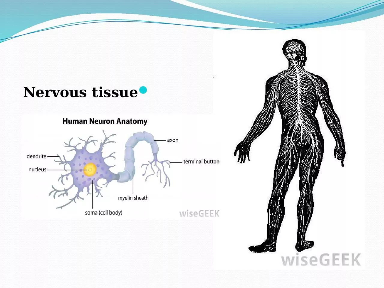
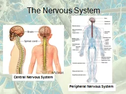
is the main component of the two parts of the nervous system the brain and spinal cord of the central nervous system CNS and the branching peripheral nerves of the peripheral nervous system ID: 916968
Download Presentation The PPT/PDF document "Nervous tissue Nervous tissue" is the property of its rightful owner. Permission is granted to download and print the materials on this web site for personal, non-commercial use only, and to display it on your personal computer provided you do not modify the materials and that you retain all copyright notices contained in the materials. By downloading content from our website, you accept the terms of this agreement.


Slide1
Nervous tissue
Slide2Nervous tissue
is the main component of the two parts of the
nervous system; the brain and
spinal cord
of the
central nervous system
(CNS), and the branching peripheral nerves of the
peripheral nervous system
(PNS), which regulates and controls bodily functions and activity. It is composed of
neurons
, or nerve cells, which receive and transmit impulses, and
neuroglia
, also known as
glial
cells, which assist the propagation of the
nerve impulse
as well as providing
nutrients
to the neuron.
Nervous tissue is made up of different types of nerve cells, all of which have an
axon
, the long stem-like part of the cell
that sends
action potential
signals to the next cell.
Slide3Functions of the nervous system are sensory input, integration, control of
muscles
and glands,
homeostasis
, and
mental activity
.
Structure
Nervous tissue is composed of
neurons
, also called nerve cells, and
neuroglial
cells
. Typically, nervous tissue is categorized into four types of tissue. In the
central nervous system
(CNS), the tissue types found are
grey matter
and
white matter
. In the
peripheral nervous system
(PNS), the tissue types are
nerves
and
ganglia
. The tissue is categorized by its neuronal and
neuroglial
components.
Slide4Components
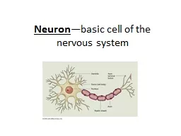
Neurons are cells with specialized features that allow them to receive and facilitate nerve impulses, or
action potentials, across their membrane to the next neuron They possess a large cell body (
soma
), with cell projections called
dendrites
and an
axon
. Dendrites are thin, branching projections that receive electrochemical signaling (
neurotransmitters
) to create a change in voltage in the cell. Axons are long projections that carry the action potential away from the cell body toward the next neuron. The bulb-like end of the axon, called the
axon terminal
, is separated from the dendrite of the following neuron by a small gap called a
synapse
. When the action potential travels to the axon terminal,
Slide5Slide6Nerve cells are functionally connected to each other at a junction known as a
synapse
, where the terminal branches of an axon and the dendrites of another neuron lie in close proximity to each other but never make direct contact.
Slide7Neurotransmitters are released across the synapse and bind to the
post-synaptic receptors, continuing the nerve impulse.
Neurons are classified both functionally and structurally.
Functional classification
:
Sensory neurons
(
afferent
): Relay sensory information in the form of an
action potential
(nerve impulse) from the PNS to the CNS
Motor neurons
(
efferent
): Relay an action potential out of the CNS to the proper
effector
(muscles, glands)
Interneurons
: Cells that form connections between neurons and whose processes are limited to a single local area in the brain or spinal cord
Slide8Structural classification
:
Multipolar
neurons
: Have 3 or more processes coming off the
soma
(cell body). They are the major neuron type in the CNS and include
interneurons
and motor neurons.
Bipolar neurons
: Sensory neurons that have two processes coming off the soma, one dendrite and one axon
Pseudounipolar
neurons
: Sensory neurons that have one process that splits into two branches, forming the axon and dendrite
Unipolar
brush cells
: Are
excitatory
glutamatergic
interneurons
that have a single short dendrite terminating in a brush-like tuft of
dendrioles
. These are found in the granular layer of the
cerebellum
Slide9Slide10cells that make up the primary immune system for the CNS.
Neuroglia
encompasses the non-neural cells in nervous tissue that provide various crucial supportive functions for neurons. They are smaller than neurons, and vary in structure according to their function. Neuroglial cells are classified as follows
Microglial
cells
: Microglia are
macrophage
They are the smallest
neuroglial
cell.
Slide11Astrocytes
: Star-shaped
macroglial cells with many processes found in the CNS. They are the most abundant cell type in the brain, and are intrinsic to a healthy CNS.
Oligodendrocytes
: CNS cells with very few processes. They form
myelin sheaths
on the axons of a neuron, which are lipid-based insulation that increases the speed at which the action potential, can travel down the axon.
Schwann cells
: The PNS equivalent of
oligodendrocytes
, they help maintain axons and form myelin sheaths in the PNS.
Satellite
glial
cell
: Line the surface of neuron cell bodies in
ganglia
(groups of nerve body cells bundled or connected together in the PNS)
Enteric
glia
: Found in the
enteric nervous system
, within the gastrointestinal tract.
Slide12Classification of Tissue
1-In the Central Nervous System: Grey matter is composed of cell bodies, dendrites,
unmyelinated
axons, protoplasmic
astrocytes
(
astrocyte
subtype), satellite
oligodendrocytes
(non-
myelinating
oligodendrocyte
subtype), microglia, and very few
myelinated
axons.
White matter
is composed of
myelinated
axons, fibrous
astrocytes
,
myelinating
oligodendrocytes
, and microglia.
2-In the Peripheral Nervous System
:
Ganglion
tissue is composed of cell bodies, dendrites, and satellite
glial
cells.
Slide13Nerves
are composed of
myelinated and unmyelinated axons, Schwann cells surrounded by
connective tissue
.
The three layers of connective tissue surrounding each nerve are:
Endoneurium
. Each nerve axon, or fiber is surrounded by the
endoneurium
, which is also called the
endoneurial
tube, channel or sheath. This is a thin, delicate, protective layer of connective tissue.
Slide14Perineurium
. Each
nerve fascicle containing one or more axons, is enclosed by the perineurium
, a connective tissue having a lamellar arrangement in seven or eight concentric layers. This plays a very important role in the protection and support of the nerve fibers and also serves to prevent the passage of large molecules from the
epineurium
into a fascicle.
Epineurium
. The
epineurium
is the outermost layer of dense connective tissue enclosing the (peripheral) nerve.
Slide15Functions of Nerve Tissue
Nervous tissue
allows an organism to sense stimuli in both the internal and external environment.
The stimuli are
analysed
and integrated to provide appropriate, co-
ordinated
responses in various organs
.
The afferent or sensory neurons
conduct nerve impulses from the sense organs and receptors to the central nervous system
.
Internuncial
or connector neurons
supply the connection
between the afferent and efferent neurons as well as different parts of the central nervous system.
Efferent or somatic motor neurons
transmit the impulse from the central nervous system to a muscle (the
effector
organ) which then react to the initial stimulus
.
Autonomic motor or efferent neurons
transmit impulses to the involuntary muscles and glands
.