
By PROF Saeed Abuel Makarem DRSANAA ALSHAARAWY OBJECTIVES At the end of the lecture the students should be able to Identify and describe the muscles of the pectoral region Pectoralis major ID: 1038846
Download Presentation The PPT/PDF document "PECTORAL REGION AND AXILLA" is the property of its rightful owner. Permission is granted to download and print the materials on this web site for personal, non-commercial use only, and to display it on your personal computer provided you do not modify the materials and that you retain all copyright notices contained in the materials. By downloading content from our website, you accept the terms of this agreement.


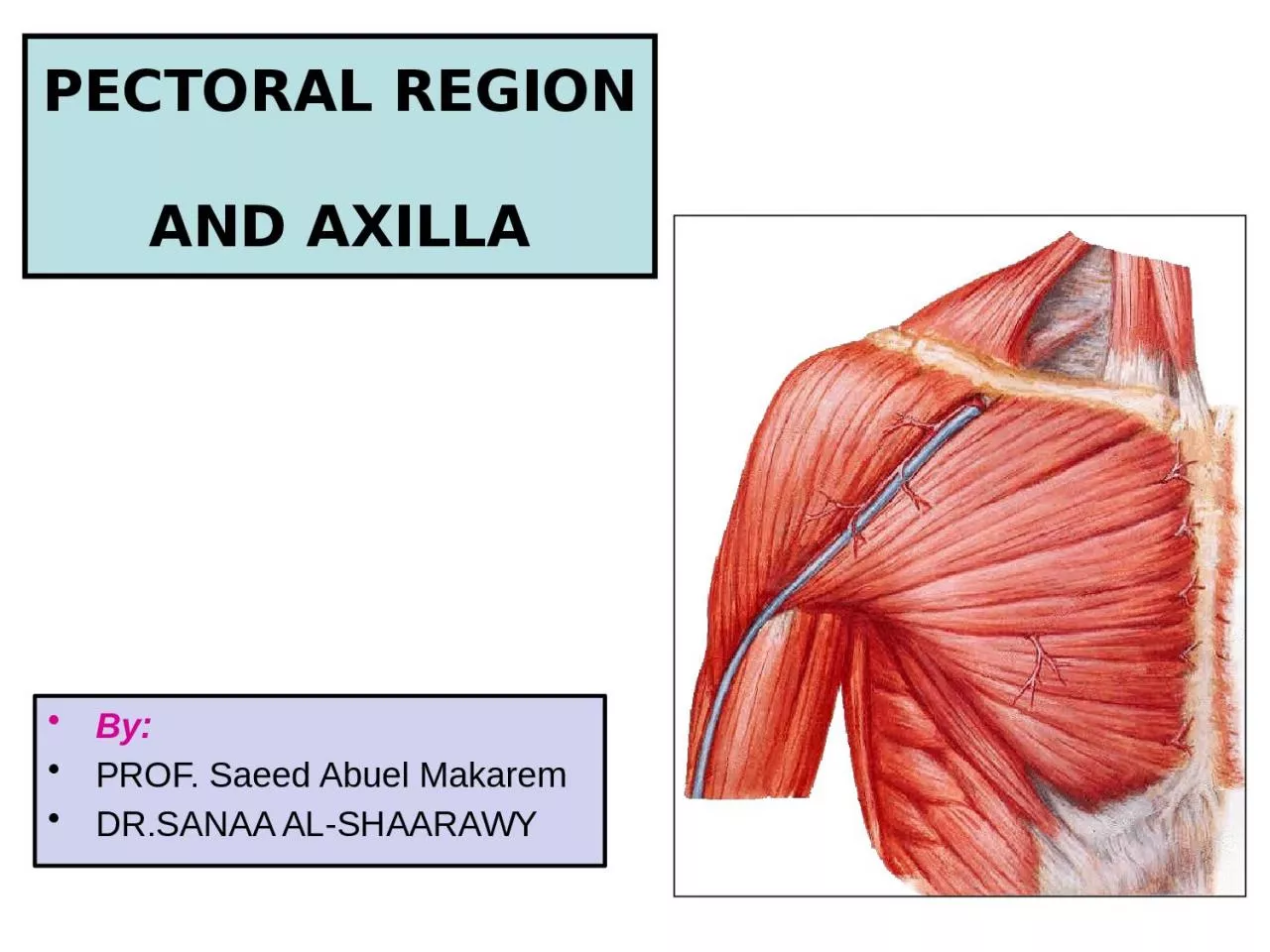
1. PECTORAL REGION AND AXILLABy:PROF. Saeed Abuel MakaremDR.SANAA AL-SHAARAWY
2. OBJECTIVESAt the end of the lecture the students should be able to :Identify and describe the muscles of the pectoral region.Pectoralis major.Pectoralis minor.Subclavius.Serratus anterior.Describe and demonstrate the boundaries and contents of the axilla.
3. Pectoralis MajorOrigin : Clavicular head: From medial ½ of the front of the clavicle.Sternocostal head: From ,Sternum.Upper 6 costal cartilages.Aponeurosis of external oblique.Insertion :Lateral lip of bicipital groove.Nerve supply : Medial & lateral pectoral nerves.Action : Adduction and medial rotation of the arm.Clavicular head helps in flexion of arm (shoulder).
4. Pectoralis MinorOrigin: from 3rd , 4th , and 5th ribs close to their costal cartilages.Insertion: coracoid process.Nerve supply: medial pectoral nerve.Action:Depression of shoulder.Draw the ribs upward and outwards during deep inspiration
5. SubclaviusOrigin: From 1st rib at the junction with its costal cartilage.Insertion: Subclavian groove at the inferior surface of middle 1/3 of clavicle.Nerve supply:Nerve to subclavius from upper trunk of brachial plexus.Action: Steadies the clavicle during movement of the shoulder joint.
6. Clavipectoral FasciaIt is a thickened membrane of deep fascia between the subclavius and pectoralis minor.It is pierced by :Lateral pectoral nerve.Thoraco-acromial arteryCephalic vein.Few lymph vessels.
7. Origin: Upper eight ribs.Insertion: Ventral aspect of medial border and inferior angle of scapula.Nerve supply: Long thoracic nerve.Action:Draws the scapula forward (protraction, in boxing).Rotates scapula outwards in raising the arm above 90 degree.Serratus anterior
8. The AxillaA pyramid-shaped space between the upper part of the arm and the side of the chest through which major neurovascular structures(Axillary vessels & nerves) pass between neck & thorax and upper extremity.Axilla has an apex, a base and four walls
9. Boundaries of the AxillaApex: Is directed upwards into the root of the neck.is bounded, by 3 bones:Clavicle anteriorly.Upper border of the scapula posteriorly.Outer border of the first rib medially.It is called cervico-axillary canal.
10. Base: Formed by skin stretching between the anterior and posterior walls.is bounded:In front by the anterior axillary fold (formed by the lower border of the Pectoralis major muscle).behind by the posterior axillary fold (formed by the tendon of latissimus dorsi and the teres major muscle).medially by the ribs and the chest wall.
11. Anterior wall:Is formed byPectoralis majorPectoralis minorSubclaviusClavipectoral fascia: Pectoralis majorPectoralis minorClavipectoral fascia
12. Posterior wall:Is formed by:SubscapularisLatissimus dorsiTeres major muscles
13. The medial wall:Is formed by:Serratus anteriorUpper 4-5 ribs & Intercostal muscles .The lateral wall:Is formed by:CoracobrachialisBiceps brachii Intertubercular groove of the humerus.
14. Contents of The Axilla Cords and braches of brachial plexus.Axillary artery and its branches.Axillary vein and its tributaries. Axillary lymph nodes. Axillary fat.Loose connective tissue.The neurovascular bundle is enclosed in connective tissue sheath, called ‘axillary sheath’Axillary a. & v.Brachial plexus
15. Location & Formation Brachial Plexus is present in the posterior triangle of the neck & axilla It is formed by the union of the anterior Rami of the C 5th, 6th, 7th & 8th and the 1st thoracic spinal nerve. What is a Brachial Plexus ?Brachial Plexus is a network of nerves that present at the root of the neck to enter the upper limb The roots of C5 & C6 unite to form Upper trunkThe root of C7 continues as the Middle trunkThe roots of C8 & T1 unite to form Lower trunk 15
16. The Plexus can be divided into 5 stages: Roots: in the posterior∆Trunks: in the posterior∆ Divisions: behind the clavicleCords: in the axillaBranches: in the axillaThe first 2 stages lie in the posterior triangle, while the last 2 sages lie in the axilla.16
17. The anterior divisions of the upper and middle trunks unite to form the Lateral cord.The anterior division of the lower trunk continues as the Medial cord.All the posterior divisions of three trunks join to form the Posterior cord.17
18. B R NCHE SLateral cordMedial cordPosterior cordLateral pectoral nerve.Medial pectoral nerve. Axillary nerve.Musculocutaneous nerve.Ulnar nerve.Radial nerve.Median nerve (lateral root).Median nerve (medial root).Upper & lower subscapular nerves.Medial cutaneous nerve of arm & forearm.Thoracodorsal N.
19. SUMMARYMuscles connecting the upper limb with anterior and lateral thoracic wall are the muscles of pectoral region, these are :Pectoralis major.Pectoralis minor.Subclavius.Serratus anterior.The axilla is a pyramidal space situated between the upper part of arm and the side of the chest, it has 4 walls (anterior, posterior, medial and lateral), base, and apex.The axilla is very important space because it contains :Axillary vessels.Cords of brachial plexus and their branches.Axillary lymph nodes.
20. superiorly: by the outer border of first rib, superior border of scapula, and posterior border of clavicle.medially: serratus anterior and by the ribcageanteriorly: by the pectoralis major, minor, and subclavius (see also anterior axillary fold). posteriorly: by the subscapularis above, and teres major and latissimus dorsi below (see also posterior axillary fold)laterally: by the intertubercular sulcus (coracobrachialis and the short head of the biceps brachii are in the axilla.)floor/base: by the skin (visible surface of armpit) Boundaries of Axilla
21. THANK YOU