
aka Forensic Serology Sources wwwcrosscuttingconceptscom Forensic Chemistry by David E Newton Infobase Publishing 2007 Take out your notebook and copy the following questions Provide a short answer ID: 1040065
Download Presentation The PPT/PDF document "Blood & Blood Spatter Analysis" is the property of its rightful owner. Permission is granted to download and print the materials on this web site for personal, non-commercial use only, and to display it on your personal computer provided you do not modify the materials and that you retain all copyright notices contained in the materials. By downloading content from our website, you accept the terms of this agreement.


1. Blood & Blood Spatter Analysisa.k.a. Forensic SerologySourceswww.crosscuttingconcepts.comForensic Chemistry by David E. NewtonInfobase Publishing; 2007
2. Take out your notebook and copy the following questions. Provide a short answer. Blood is……….The most common blood type is………Agglutination is……..A presumptive test is ……….A confirmatory test is……….
3. BLOOD as EvidenceValued as evidence especially in violent crimes such as murder and rapeCharacteristics are variable among the population making it possible to rule in or rule out a person as the perpetrator.Stains and blood spatter patterns reveal information about the crime
4. What is Blood? Components of blood: Plasma: the pale, yellowish colored liquid portion that contains proteins, enzymes, antibodies, dissolved nutrientsRed blood cells: carry oxygen to cells; carbon dioxide from cells; get the red color from hemoglobinWhite blood cells: several different types: lymphocytes, monocytes, and neutrophils; responsible for immunity and fighting infectionPlatelets: tiny fragments of cells that aid in blood clotting
5. White blood cellsRed Blood Cellsplatelets
6. Blood TypesKarl Landsteiner: Searched to explain why it is not possible to transfuse blood from any one human into any other humanWhy did some people do well when transfused blood from another person and others die? Why did the transfusion fail? Later announced that human blood can be classified into a few classes or types known today as types A, B, AB, and O.
7. Early 1970’s: forensics began using blood groupings for clues to link blood to an individual. Blood type is considered class evidence because it is not unique to an individual the way DNA and fingerprints are unique.
8. The rise of blood as evidenceThere was little research in forensic serology prior to the 20th century. A couple of tests for blood were developed in the late 1800’s: Guaic test (by van Deem) and the hydrogen peroxide test (by Schonbein). Both tests detect hemoglobin in blood. Mathieu Orfila: suggested that blood could be analyzed using a microscope.
9. The type of blood represents the antigens present on the red blood cell. Blood TypeAntigen on RBCAntibody in plasmaType AA antigen B antibodyType BB antigenA antibodyType ABA and B antigensNo antibodyType ONo A or B antigenBoth A and B antibodies
10. Using Blood type to solve a crimeFrequency of types in the populationBlood typeExpected FrequencyA positive1 in 3A negative1 in 16B positive1 in 12B negative1 in 67AB positive1 in 29AB negative1 in 167O positive1 in 3O negative1 in 15
11. TypeAfrican AmericanAsianCaucasianHIspanicO positive47%39%37%53%O negative4%1%8%4%A positive24%27%33%29%A negative2%0.5%7%2%B positive18%25%9%9%AB positive4%7%3%2%AB negative0.3%0.1%1%0.2%Correlation of Blood Type and Race
12. Blood types continued……Agglutination: the clumping of blood cells Results from an antigen-antibody reactionBlood cells containing a specific antigen will clump when mixed with blood that contains the antibody against that antigen. This was the reason behind failed transfusions in Landsteiner’s time.
13. Blood types continued…..Why failed transfusions cause death? Patients cells clump. Clumped cells block blood vessels and stop flow of blood. Clumped cells can rupture releasing contents such as free hemoglobin that is toxic to the body when it is not attached to the red blood cell.
14. Using Blood type to solve a crimeCan be used to narrow down a pool of suspects if the blood type (class evidence)Example: if the stain is type A then potential suspects that are not type A can be excluded. The presence of the Rh antigen on red blood cells) gives another criteria for inclusion or exclusion as well. There are other antigens that have also been discovered that help to further narrow the range of suspects. Ex. Duffy, Kidd, MNS, Kell, Lewis, LutheranThere are also antigens present on White Blood cells know as HLA (human lymphocyte) antigens.
15. Blood StainsFirst determine whether the stain is blood or some other red substance. Blood released from the body is bright red for about the first 3 to 5 minutes. As it dries it turns a brown to black color. Wet blood is usually easier to test than dried blood.
16. Presumptive test: a quick test that can be performed at the scene to determine if a piece of evidence is relevant and needs to be sent for further testing. Presumptive tests can save time and money.
17. Presumptive Tests for Blood……One of the earliest Adler’s or Benzidine test; mix hydrogen peroxide with benzidine and treat the blood stain. Oxidation-reduction reaction: Benzidine + Hydrogen peroxide + hemoglobin in blood diazo dye that is blue-green in color. The benzidine is reduced in the reaction. In 1973, the EPA banned benzidine as a carcinogen.
18. https://www.youtube.com/watch?v=pnH2HnB-GrIKastle-Meyer TestReagent contains: potassium hydroxide (KOH), phenolphthalein (in indicator) and zinc dustStain is treated with the reagent. The Reaction: when Kastle-Meyer reagent is combined with hydrogen peroxide and blood, the hemoglobin in the blood catalyzes the conversion of phenolphthalein causing it to change to a deep pink color. Can give false positive with vegetable materials present
19. Hemastix test: Cellulose strip with a mixture of o-toluidine and hydrogen peroxide on a tiny padMoisten strip and dip in to the sample to be testedThe reaction: Hemoglobin catalyzes the conversion of o-toluidine to a green colored product. Intensity of the green color is matched to a scale to indicate the concentration of blood present in the sample.
20. Luminol (5-amino-2,3-dihydro-1,4-phthalizinedione)First developed by Walter Specht in 1937The reaction: Luminol when treated with hydrogen peroxide in the presence of blood, one ring of the luminol breaks apart. Nitrogen gas is released and 3-aminophthlate is produced in an excited state. After a brief moment, the 3-APA gives off a photon (425 nm). A blue flourescent light is noted. Highly sensitive: detects bloodstains diluted up to 10 million; works well on old blood stains tooDoes not interfere with blood typing or DNA analysisCan give false positives with plant enzymes, oxidizers, metals, and chlorine
21. Presumptive Test SummaryPresumptive TestIndication of PositiveSituation UsedFalse PositivesPhenolphtalein (Kastle-Meyer)Bright pink coloron visible stainsVegetable material (potatoes and horseradish)Tetramethylbenzidine (TMB) / HemastixGreen to blue-green coloron visible stainsOxidizing agents, catalyst, vegetable peroxidase, cosmeticsLuminolBlue-white to yellow-green lightlatent bloodPlant enzymes, oxidizing agents, metals, chlorineFluoresceinFluoresce with UV light sourcelatent blood, vertical surfaceCopper, hypochlorite
22. Confirmatory Tests for BloodTeichmann test (1853)Reagent: mixture of glacial acetic acid and sodium chlorideThe reaction: reagent causes hemoglobin molecules in blood to split producing brown crystals of hemin (purple to almost black)
23. Confirmatory Tests for BloodTakayma Test (1912)The reaction: pyridine is added to blood causing the reduction of hemoglobin to a pyridine-hemoglobin complex that is salmon pink color.
24. Human blood or animal bloodOnce a stain has tested positive for blood, the origin (animal or human) must be determined. Ouchterlony test or precipitin test (1960)The test: based on antibody-antigen reactionExperimental animal (rabbit) is injected with sample of human blood. Rabbit makes antibodies to human blood cells. Blood is drawn from rabbit and mixed with the unknown blood sample. If the blood is human, clumping will be observed and it can be concluded that the unknown blood was human.
25. Bloodstain PatternsImportant elements of Blood pattern analysis: Type of surface on which blood fallsSmooth, hard surface results in more clearly defined dropsShapes of the individual dropsRound (circular) shape means the blood dropped verticallyDrops at an angle appear more ellipticalGreater angle = more elliptical shapePointed end always faces the direction of the source
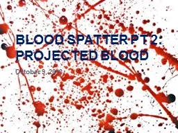
26. Spatter PatternsLow velocity: outward projection of blood drops in a relatively cohesive amoeba-shaped arrayMedium velocity: individual droplets that are about 1 millimeter or more in diameter [weapon moves with a velocity of 100 ft/sec (30 m/s)]Possible weapons: person’s fist, hammer, knife or baseball bat (blunt instruments)High velocity: droplets less than 1 millimeter in diameter (tiny drops) produced by rapidly moving object such as a bullet fired from a gun
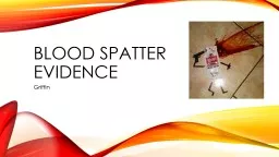
27. What can a blood spatter tell you……The source of the blood (where the victim was struck)The type of impact that produced the bloodThe number of incidents that occurred to produce the blood (how many impacts)The direction and speed that the victim was moving during the crimeThe position of the victim during the crimeWhether the victim’s arteries were cut
28. www.crosscuttingconcepts.com
29. Trigonometry and Angle of Impact To determine the angle of impact:Measure the width and length of the blood spatter. Length must be longer than width.Divide width by length.Take the arcsine of your result.This formula will not produce accurate results at extreme angles ( less than 10˚ or greater than 60 ˚). o determine the angle of impact:Measure the width and length of the blood spatter. Length must be longer than width.Divide width by length.Take the arcsine of your result.This formula will not produce accurate results at extreme angles ( less than 10˚ or greater than 60 ˚).
30. Find your blood pattern sheets from yesterday. Begin measuring each blood drop. Measure the length and width for each drop. You can write this information on the droplet sheet next to each drop. Once you have 3 or 4 consistent measurements, average them and record on your lab data sheet. Instructions to follow for what to do next.
31. Area of Convergence (2d area)After many strings have been placed, a general area of convergence will appear where the strings overlap.This can also be done on a computer with image analysis software.www.crosscuttingconcepts.com
32. Point of Origin (3d volume)The procedure for generating the point of origin is just like the area of convergence, except the angle of impact for each stain is calculated.This is a method of adding a third dimension to the 2d area of convergence calculation.n is just like the area of convergence, except the angle of impact for each stain is calculated.This is a method of adding a third dimension to the 2d area of convergence calculation.2d Solution – no angular data