
Panzera F Dujardin JP Nicolini P Caraccio MN Rose V Tellez T et al Genomic Changes of Chagas Disease Vector South America Emerg Infect Dis 2004103438446 httpsdoiorg103201eid1003020812 ID: 1009266
Download Presentation The PPT/PDF document "Figure 3 Figure 3. Gonial mitot..." is the property of its rightful owner. Permission is granted to download and print the materials on this web site for personal, non-commercial use only, and to display it on your personal computer provided you do not modify the materials and that you retain all copyright notices contained in the materials. By downloading content from our website, you accept the terms of this agreement.


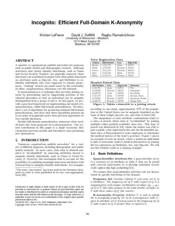

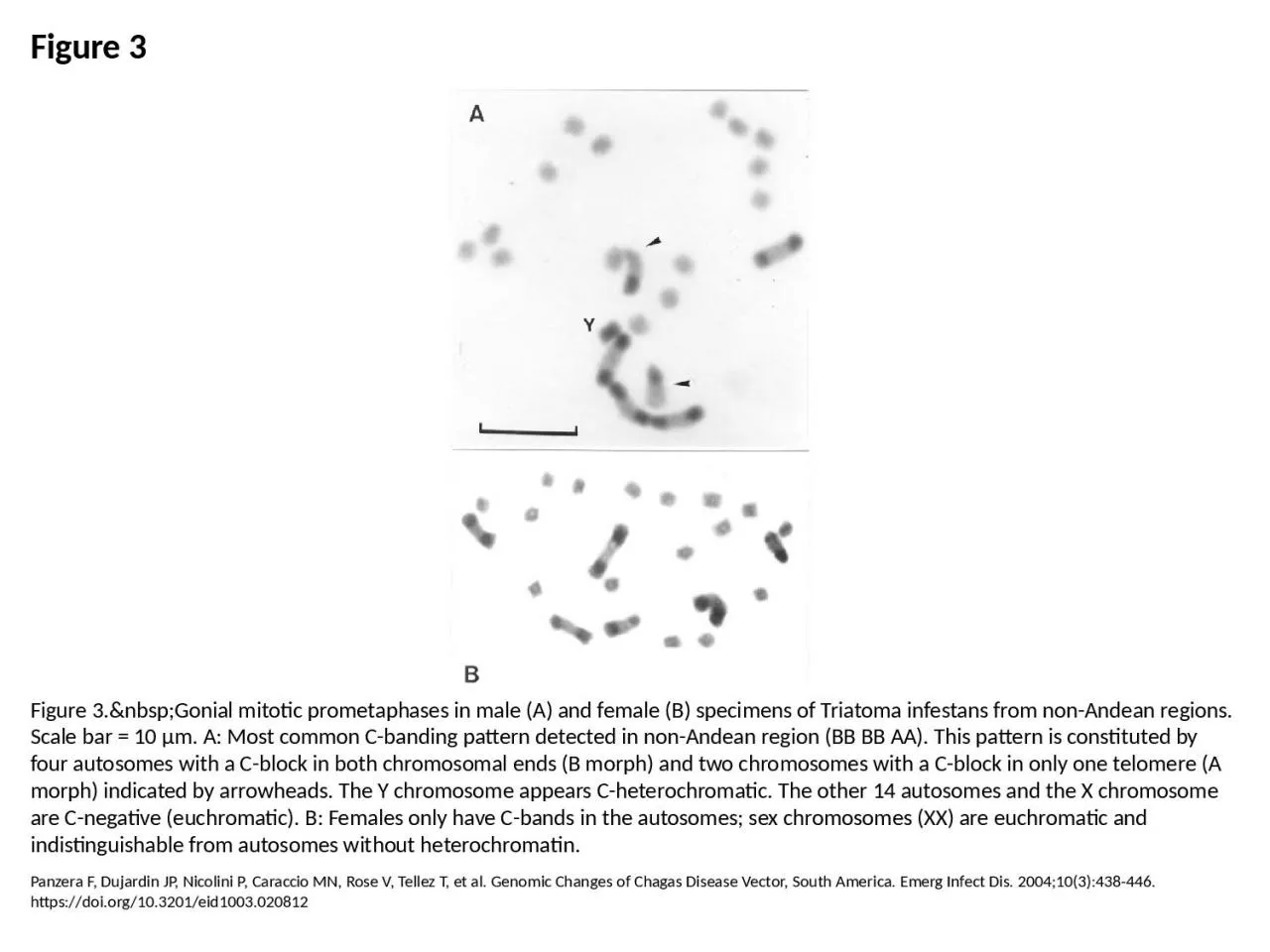
1. Figure 3Figure 3. Gonial mitotic prometaphases in male (A) and female (B) specimens of Triatoma infestans from non-Andean regions. Scale bar = 10 μm. A: Most common C-banding pattern detected in non-Andean region (BB BB AA). This pattern is constituted by four autosomes with a C-block in both chromosomal ends (B morph) and two chromosomes with a C-block in only one telomere (A morph) indicated by arrowheads. The Y chromosome appears C-heterochromatic. The other 14 autosomes and the X chromosome are C-negative (euchromatic). B: Females only have C-bands in the autosomes; sex chromosomes (XX) are euchromatic and indistinguishable from autosomes without heterochromatin.Panzera F, Dujardin JP, Nicolini P, Caraccio MN, Rose V, Tellez T, et al. Genomic Changes of Chagas Disease Vector, South America. Emerg Infect Dis. 2004;10(3):438-446. https://doi.org/10.3201/eid1003.020812