/
PROGRESSION OF CANCER  FOCUS ON METASTASIS WITH DIFFERENT ASPECTS PROGRESSION OF CANCER  FOCUS ON METASTASIS WITH DIFFERENT ASPECTS

PROGRESSION OF CANCER FOCUS ON METASTASIS WITH DIFFERENT ASPECTS - PDF document

gelbero
gelbero . @gelbero
Follow
342 views
Uploaded On 2022-09-01

PROGRESSION OF CANCER FOCUS ON METASTASIS WITH DIFFERENT ASPECTS - PPT Presentation

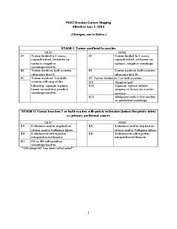
26 Fatma Tennur Genç Hacettepe Un28vers28ty Faculty of Med28c28ne Ankara TURKEY ABSTRACT 27e role of metastas28s on the poor prognos28s of cancer 28s unden28ably 28 ID: 944522

cancer cells metastas tumor cells cancer tumor metastas cell platelets factor growth tgf platelet factors spread thel ncrease mportant

Share:

Link:

Embed:

Download Presentation from below link

Download Pdf The PPT/PDF document "PROGRESSION OF CANCER FOCUS ON METASTAS..." is the property of its rightful owner. Permission is granted to download and print the materials on this web site for personal, non-commercial use only, and to display it on your personal computer provided you do not modify the materials and that you retain all copyright notices contained in the materials. By downloading content from our website, you accept the terms of this agreement.


Presentation Transcript

26 PROGRESSION OF CANCER : FOCUS ON METASTASIS WITH DIFFERENT ASPECTS Fatma Tennur Genç Hacettepe Unversty Faculty of Medcne, Ankara, TURKEY ABSTRACT e role of metastass on the poor prognoss of cancer s undenably mportant. For ths reason, revealng the role of metastass and defnng the factors contrbutng to t play a key role n the treatment of cancer. In ths revew, we amed to research varous genes, markers and factors related to metastass, the functons of platelets n metastass, - noss secton, we gave more detals on epthelal-mesenchymal transton whch has a role on the malgnancy and we examned the eect of factors on metastass. In other two sectons, we mentoned that thromboss and platelets support metastass, platelets play a role n dodgng mmune system and there are two opposte mechansms trgge - rng tumor formaton. Keywords: Metastass, epthelal-mesenchymal transton, platelet INTRODUCTION Cancer s a process relyng on unregulated cell dv - son. Cancer cells not only dvde and multply rregu - larly, but they also accumulate and form tumors. Bengn tumors do not have the ablty to spread to neghbor tssue and organs, and they are derentated, meanng that they possess smlar characterstcs lke regular cells; whereas malgn tumors are expected to spread to negh - bor tssues. Alongsde ther ablty to nfltrate and da - mage tssues, they can also spread to some other parts of the body and form secondary cancer colones, whch are called metastass. Revealng the causes of cancer and de - termnng ts course n early phases s recently one of the most mportant felds of study. Many pathways whch are partcular to lvng organsms and cannot be expla - ned n vtro, provde deas regardng the spread of cancer and determnng ts prognoss beforehand. s revew ams to approach metastass wth derent aspects and provde a general knowledge. Progresson Of Cancer t s known. It s not easy to descrbe the metastass wth expermental methods by formng mcroenvronment. Metastass s expected to show up durng late phases, for ths reason detectng factors whch can predct early me - tastass and prognoss of the dsease s very mportant snce t can aect treatment protocol and mprove the survval. HER2 and PyMT have ncreased the spread of tumor cells by early epthelal change n transgenc mce and human ductal carcnoma (1). Cytokeratn (CK) s - row under normal condtons. In breast cancer patents, a CK (+) result provdes mportant nformaton about a presence of mcrometastass and shows that there s a hgh rsk for relapse (2). e most mportant factor to determne f a malg - nant transformaton wll occur s the mcro envronment of the tumor (3). By excretng varous cytoknes, the tu - mor actvates stromal fbroblasts. Stromal cells excrete growth factors and cytoknes, thus epthelal-mesenchy - examne EMT n squamous cell carcnoma, the cancer stem cells whch express hgh level of CD44 undergo EMT wth the help of stromal factors and become can - cer stem cells. ese cells enter the blood crculaton and mgrate to a secondary ste; they undergo a mesenchy - mal-endothelal transton (MET) and jon the epthe - lal cells, causng metastass (5). e most mportant cause of EMT s exposure to TGF-ß (6). Most growth factors stmulate prolferaton and trgger carcnogene - TGF-ß excreted from human platelets show smlar bo - logc characterstcs wth growth nhbton factor exc - reted from BSC-1* (Epthelal cell orgnated from Af - rcan green monkey) cells (7, 8). An experment done

Adress for Correspondence: Fatma Tennur Genç, Hacettepe Unversty Faculty of Medcne, Ankara, TURKEY - e-mal: tennurgenc07@gmal.com Receved: 02.12.2015 - Accepted: 18.12.2015 27 wth transgenc mce reveals a deteroraton of tumor formng mechansm wth ncreased TGF-ß sgnal (9). But the fact that TGF-ß ncreases EMT and causes me - tastass by ncreasng the spread n prmary carcnomas s an mportant aspect (10). When TGF-ß antbodes were gven to mce breast cancer models wth a T-cell defcency and normal natural kller (NK) cell functons, mce spleen NK actvty was mproved and tumor cell prolferaton was regressed (11). An ncrease on TGF-ß nduces metastass and angogeness n prostate cancer, whle weakenng the mmune system (12). TGF-ß plays an mportant role on cell prolferaton and extracellular matrx confguraton. In patents wth non-metastatc prmary leson or lymph node metastass, TGF-ß levels measured n perpheral vens and tumor dranng ven s closely related to nvason and metastass of gastrc car - cnoma (13). In a research n whch nvason and metastass were analyzed by measurng IGF bndng protens, IGFBP-2 was ncreased n prostate cancer patents when com - pared to healthy ndvduals. However, IGFBP-3 s df - ferent. In patents whose preoperatve plasma has low amounts of IGFBP-3, prostate cancer has a more aggres - sve course (14). Spread Of Tumor Cells And Platelets EMT s known to ncrease wth an nteracton betwe - en platelets and tumor cells, as well as an ncrease on TGF-ß (15). e man pont to emphasze s the nterac - ton of platelets wth tumor cells and the eect of platelets on metastass. In cancer patents, the rsk of thromboss ncreases n later stages. e reason for that s abnormal platelet actvaton and aggregaton. Even f thrombotc dsorders do not occur, coagulaton parameters mostly ncrease. Factor VII of the extrnsc pathway and tssue factor are elevated n cancer patents (16). When we ap - proach from another aspect, thrombocytoss s known to be related to short lfespan and aggressve dsease (17). rombopoetn and IL-6 are known to be ncreased n patents wth thrombocytoss, but researches done wth mce show that the man cause of paraneoplastc throm - bocytoss s the tumor-related ncrease of IL-6 (17). Ant IL-6 antbodes have reduced the amount of platelets n mce wth epthelal over cancer and IL-6 blockade has ncreased the response to medcaton n epthelal over cancer (17). Tumor cells n the blood are always n a relaton wth the hemostatc system. Especally platelet and tumor cells’ complex nteracton mechansms must always be kept n mnd. As t s known, platelets are cells wthout nucleus whch nclude many granules and they derve from megakaryocyte’s cytoplasm. Platelet membrane conssts of phospholpds and t s surrounded by gly - coproten and ntegrnes. Platelet membrane glycopro - tens cause adheson n subendothelal matrx, adheson between platelets and aggregaton (18). P2Y1 receptor located n the platelet membrane serves a functon n ADP-dependant platelet aggregaton (19). At ths pont, we need to stress that wth adenne nucleotdes excreted from ATP-actvated P2Y2 receptor-controlled platelets, the endothelal barrer dsappears and cancer cells can pass through the endothelum and wander outsde of ve - ns (20). In a research done wth mce, t was shown that wth a P2Y2 defcency or a defcency of ATP secret - on, tumor cell metastass decreases (21). Wth a relaton between platelets, tumor cells and leucocytes, CCL-5 sy - nthess from endothelum

has rased and ths has ncre - ased metastass (22). Immune System And Autophagy Tumor cells are able to dodge the mmune system wth varous mechansms. MHC1 and tumor antbodes must be presented to the cytotoxc T-cells so that they can dentfy and destroy cancer cells. Tumor cells can reduce the HLA1 expresson and decrease ant-tumor actvty (23). In multple myeloma, wth the nteracton of myeloma and tumor cells and wth the eect of mye - lod-dependant suppressor cells, the tumor prolferaton has ncreased and mmunodefcency has occurred (24). When myelod-dependant suppressor cells encounter neutrophls, CD115 and CD244 expresson has ncrea - sed, however, CXCR1 and CXCR2 expressons have dec - reased (25). Durng hematogenous metastass, platelets surround the tumor and let them be transported safe from the mmune system (26). Wth ths, the ablty of NK cells to lyse tumor cells s dsabled (26). It was shown that fbrn formaton and fbrn surroundng tumor cell block the recognton of tumor cells by the NK cells and thus ncrease metastass (27). Apart from ths, n our knowledge t s also neces - sary to menton autophagy. Autophagy s defned by the removal of damaged, unnecessary protens and organel - les wth cellular degradaton. In cancer, autophagy has a more complcated and dcult mechansm. In early stages of cancer development, autophagy has a tumor suppressant character. A decreased autophagy n early stage tumor cells s related to malgnant transformaton. Moreover, ncreased autophagy n cancer cells whch 28 have already exsted beforehand protect the tumor cells aganst metabolc and therapeutc stress and plays a role n pro-tumor mechansms (28). In other words, chemo - therapy and radotherapy have been lethal on late stage already exstng tumors, where autophagy was nhbted, and ths has created treatment-senstve cancer cells (28). CONCLUSION In cancer, the course of dsease changes n every pa - tent dependng on varous factors. Treatment protocol s determned up to date wth some genes and mcrosco - pc changes n tumor, and aggressve treatment s appled n case of poor prognoss. Another mportant aspect s that platelets can overexpress some receptors and help the tumor to spread secondary organs outsde of vens, and thus ncrease the metastass. Blockng ths can pre - vent the tumor from leavng the vens and spread to ts - sues. Elmnatng the defcency of mmune recognton orgnatng from an nteracton between fbrn, platelets and tumor cells wll mprove the ablty of the organsm to fght the dsease. Ethcs Commttee Approval: N/A Informed Consent: N/A Conct of Interest: e authors declared no conct of n - terest. Fnancal Dsclosure: e authors declared that ths study receved no fnancal support. REFERENCES 1. Husemann Y, Gegl JB, Schubert F et al. Systemc spread s an early step n breast cancer. Cancer Cell 2008;13:58-68. 2. Braun S, Pantel K, Müller P et al. Cytokeratn-pos - tve cells n the bone marrow and survval of patents wth stage I, II, or III breast cancer. N Engl J Med 2000;342:525-33. 3. Joyce JA , Pollard JW. Mcroenvronmental regulaton of metastass. Nat Rev Cancer 2009;9:239-52. 4. ery JP, Acloque H, Huang RY et al. Epthelal-me - senchymal transtons n development and dsease. Cell 2009;139:871-90. 5. Bddle A, Mackenze IC. Cancer stem cells and EMT n carcnoma. Cancer and Metastass Revews 2012;31:285- 93. 6. Mettnen PJ, Ebner R, Lopez AR et al. TGF-beta n - duced transderentaton of mammary epthelal cells to mesenchymal cells: nvolvement of type I receptors. J Cell Bol 1994;127:2021-36. 7. McPherson JM, Sawamura SJ, Ogawa Y et al. e growth nhbtor of Afrcan green mo

nkey (BSC-1) cells s transformng growth factors beta 1 and beta 2. Boche - mstry 1989;28:3442-7. 8. Tucker RF, Shpley GD, Moses HL et al. Growth nh - btor from BSC-1 cells closely related to platelet type beta transformng growth factor. Scence 1984;226:705-7. 9. Gorska AE, Jensen RA, Shyr Y et al. Transgenc mce expressng a domnant-negatve mutant type II trans - formng growth factor-beta receptor exhbt mpared mammary development and enhanced mammary tu - mor formaton.Am J Pathol 2003;163:1539-49. 10. O M, Heder KH, Beug H. TGFbeta sgnalng s ne - cessary for carcnoma cell nvasveness and metastass. Curr Bol 1998;8:1243-52. 11. Arteaga CL, Hurd SD, Wnner AR et al. Ant-trans - formng growth factor(TGF)-beta antbodes nhbt bre - ast cancer cell tumorgencty and ncrease mouse spleen natural kller cell actvty. Implcatons for a possble role of tumor cell/host TGF-beta nteractons n human bre - ast cancer progresson.J Cln Invest 1993;92(6):2569-76. 12. Wkström P, Bergh A, Damber JE. Transformng growth factor-beta1 and prostate cancer. Scand J Urol Nephrol 2000;34:85-94. 13. Maehara Y, Kakej Y, Kabashma A et al. Role of trans - formng growth factor-beta 1 n nvason and metastass n gastrc carcnoma. J Cln Oncol 1999;17:607-14. 14. Sharat SF, Lamb DJ, Kattan MW et al. Assocaton of preoperatve plasma levels of nsuln-lke growth factor I and nsuln-lke growth factor bndng protens-2 and -3 wth prostate cancer nvason, progresson and metasta - ss. J Cln Oncol 2002;20:833-41. 15. Gay LJ, Feldng-Habermann B. Platelets alter tumor 29 cell attrbutes to propel metastass: programmng n transt. Cancer Cell 2011;20:553-4. 16. Kakkar AK, DeRuvo N, Chnswangwatanakul V et al. Extrnsc-pathway actvaton n cancer wth hgh fac - tor VIIa and tssue factor.Lancet 1995;346:1004-5. 17- Stone RL, Nck AM, McNesh IA et al.Paraneoplas - tc rombocytoss n Ovaran Cancer. N Engl J Med 2012;366:610-8. 18. Kunck TJ. Platelet membrane glycoprotens and ther functon: an overvew. Blut 1989;59:30-4. 19. Hechler B, Cattaneo M, Gachet C. e P2 receptors n platelet functon.Semn romb Hemost 2005;31:150- 61. 20. Schumacher D, Strlc B, Svaraj KK et al. Platelet-de - rved nucleotdes promote tumor-cell transendothelal mgraton and metastass va P2Y2 receptor. Cancer Cell 2013;24:130-7. 21. Borsg L, Wong R, Hynes RO et al. Synergstc ee - cts of L- and P-selectn n facltatng tumor metastass can nvolve non-mucn lgands and mplcate leukocy - tes as enhancers of metastass.Proc Natl Acad Sc USA 2002;99:2193-8. 22. Läubl H, Spanaus KS, Borsg L. Selectn-medated actvaton of endothelal cells nduces expresson of CCL5 and promotes metastass through recrutment of monocytes.Blood 2009;114:4583-91. 23. Kaneko K, Ishgam S, Kjma Y et al. Clncal mp - lcaton of HLA class I expresson n breast cancer. BMC Cancer 2011;11:454. 24. Görgün GT, Whtehll G, Anderson JL et al. Tu - mor-promotng mmune-suppressve myelod-derved suppressor cells n the multple myeloma mcroenvron - ment n humans. Blood 2013;121:2975-87. 25. Gabrlovch DI, Ostrand-Rosenberg S, Bronte V. Co - ordnated regulaton of myelod cells by tumours. Nat Rev Immunol 2012;12:253-68. 26. Neswandt B, Hafner M, Echtenacher B et al. Lyss of tumor cells by natural kller cells n mce s mpeded by platelets. Cancer Res 1999;59(6):1295-300. 27. Palumbo JS, Talmage KE, Massar JV et al. Tumor cell–assocated tssue factor and crculatng hemostatc factors cooperate to ncrease metastatc potental throu - gh natural kller cell–dependent and–ndependent mec - hansms.Blood 2007;110:133–41. 28. Cho KS. Autophagy and cancer. Exp Mol Med 2012;44:109-