
Remarks amp Conclusions Nicholas Senofsky 1 Justin Faber 1 and Dolores Bozovic 12 1 Department of Physics and Astronomy and 2 California NanoSystems Institute UCLA Los Angeles CA 90095 USA ID: 1045221
Download Presentation The PPT/PDF document "Vestibular Drop Attacks and Meniere’s ..." is the property of its rightful owner. Permission is granted to download and print the materials on this web site for personal, non-commercial use only, and to display it on your personal computer provided you do not modify the materials and that you retain all copyright notices contained in the materials. By downloading content from our website, you accept the terms of this agreement.


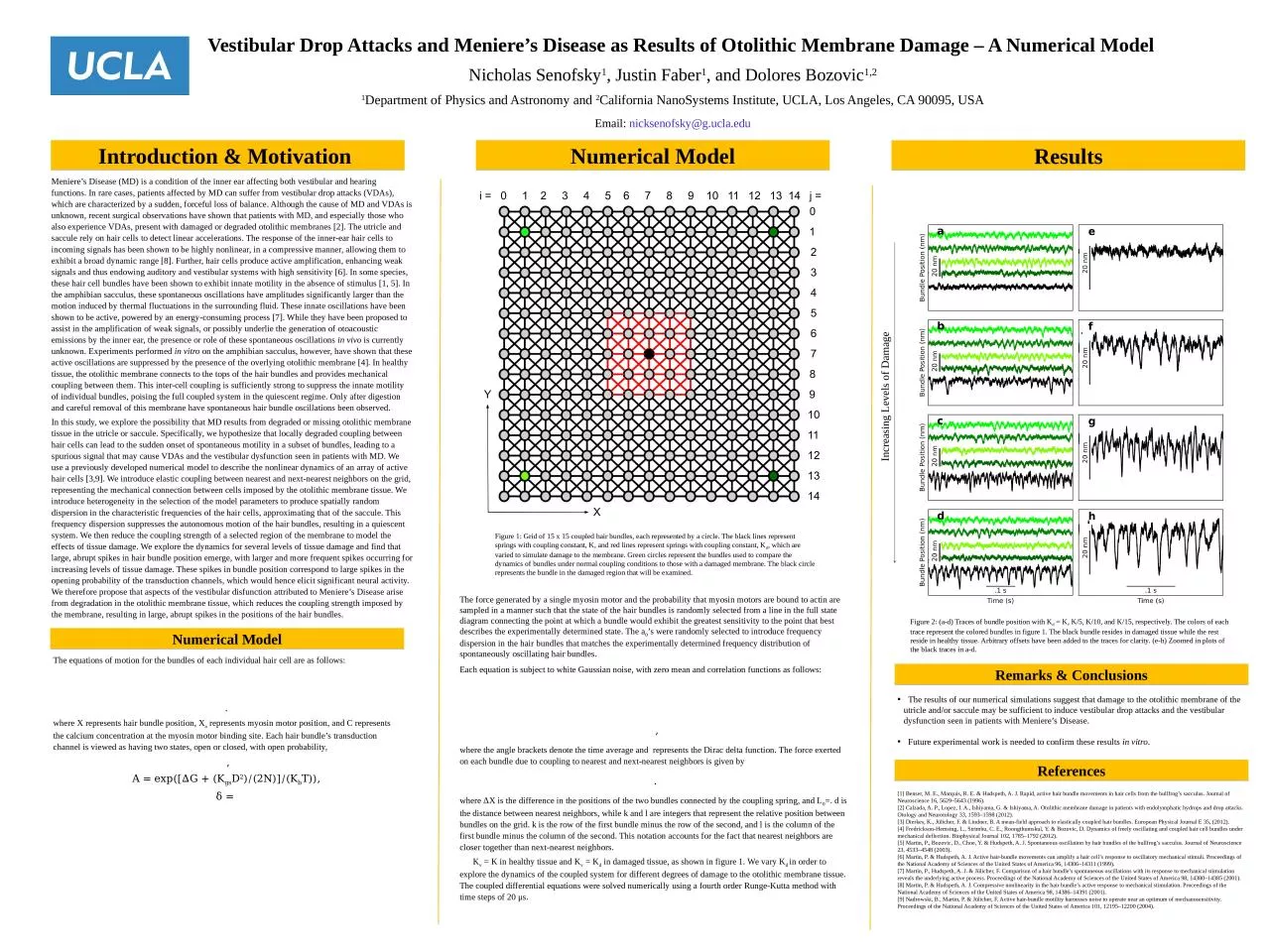
1. Vestibular Drop Attacks and Meniere’s Disease as Results of Otolithic Membrane Damage – A Numerical Model Remarks & ConclusionsNicholas Senofsky1, Justin Faber1, and Dolores Bozovic1,21Department of Physics and Astronomy and 2California NanoSystems Institute, UCLA, Los Angeles, CA 90095, USAEmail: nicksenofsky@g.ucla.edu The results of our numerical simulations suggest that damage to the otolithic membrane of the utricle and/or saccule may be sufficient to induce vestibular drop attacks and the vestibular dysfunction seen in patients with Meniere’s Disease. Future experimental work is needed to confirm these results in vitro.ReferencesIntroduction & Motivation ResultsNumerical ModelMeniere’s Disease (MD) is a condition of the inner ear affecting both vestibular and hearing functions. In rare cases, patients affected by MD can suffer from vestibular drop attacks (VDAs), which are characterized by a sudden, forceful loss of balance. Although the cause of MD and VDAs is unknown, recent surgical observations have shown that patients with MD, and especially those who also experience VDAs, present with damaged or degraded otolithic membranes [2]. The utricle and saccule rely on hair cells to detect linear accelerations. The response of the inner-ear hair cells to incoming signals has been shown to be highly nonlinear, in a compressive manner, allowing them to exhibit a broad dynamic range [8]. Further, hair cells produce active amplification, enhancing weak signals and thus endowing auditory and vestibular systems with high sensitivity [6]. In some species, these hair cell bundles have been shown to exhibit innate motility in the absence of stimulus [1, 5]. In the amphibian sacculus, these spontaneous oscillations have amplitudes significantly larger than the motion induced by thermal fluctuations in the surrounding fluid. These innate oscillations have been shown to be active, powered by an energy-consuming process [7]. While they have been proposed to assist in the amplification of weak signals, or possibly underlie the generation of otoacoustic emissions by the inner ear, the presence or role of these spontaneous oscillations in vivo is currently unknown. Experiments performed in vitro on the amphibian sacculus, however, have shown that these active oscillations are suppressed by the presence of the overlying otolithic membrane [4]. In healthy tissue, the otolithic membrane connects to the tops of the hair bundles and provides mechanical coupling between them. This inter-cell coupling is sufficiently strong to suppress the innate motility of individual bundles, poising the full coupled system in the quiescent regime. Only after digestion and careful removal of this membrane have spontaneous hair bundle oscillations been observed.In this study, we explore the possibility that MD results from degraded or missing otolithic membrane tissue in the utricle or saccule. Specifically, we hypothesize that locally degraded coupling between hair cells can lead to the sudden onset of spontaneous motility in a subset of bundles, leading to a spurious signal that may cause VDAs and the vestibular dysfunction seen in patients with MD. We use a previously developed numerical model to describe the nonlinear dynamics of an array of active hair cells [3,9]. We introduce elastic coupling between nearest and next-nearest neighbors on the grid, representing the mechanical connection between cells imposed by the otolithic membrane tissue. We introduce heterogeneity in the selection of the model parameters to produce spatially random dispersion in the characteristic frequencies of the hair cells, approximating that of the saccule. This frequency dispersion suppresses the autonomous motion of the hair bundles, resulting in a quiescent system. We then reduce the coupling strength of a selected region of the membrane to model the effects of tissue damage. We explore the dynamics for several levels of tissue damage and find that large, abrupt spikes in hair bundle position emerge, with larger and more frequent spikes occurring for increasing levels of tissue damage. These spikes in bundle position correspond to large spikes in the opening probability of the transduction channels, which would hence elicit significant neural activity. We therefore propose that aspects of the vestibular disfunction attributed to Meniere’s Disease arise from degradation in the otolithic membrane tissue, which reduces the coupling strength imposed by the membrane, resulting in large, abrupt spikes in the positions of the hair bundles.Numerical ModelFigure 2: (a-d) Traces of bundle position with Kd = K, K/5, K/10, and K/15, respectively. The colors of each trace represent the colored bundles in figure 1. The black bundle resides in damaged tissue while the rest reside in healthy tissue. Arbitrary offsets have been added to the traces for clarity. (e-h) Zoomed in plots of the black traces in a-d.Figure 1: Grid of 15 x 15 coupled hair bundles, each represented by a circle. The black lines represent springs with coupling constant, K, and red lines represent springs with coupling constant, Kd, which are varied to simulate damage to the membrane. Green circles represent the bundles used to compare the dynamics of bundles under normal coupling conditions to those with a damaged membrane. The black circle represents the bundle in the damaged region that will be examined. The equations of motion for the bundles of each individual hair cell are as follows:.where X represents hair bundle position, Xa represents myosin motor position, and C represents the calcium concentration at the myosin motor binding site. Each hair bundle’s transduction channel is viewed as having two states, open or closed, with open probability, ,A = exp([ΔG + (KgsD2)/(2N)]/(KbT)),δ = The force generated by a single myosin motor and the probability that myosin motors are bound to actin are sampled in a manner such that the state of the hair bundles is randomly selected from a line in the full state diagram connecting the point at which a bundle would exhibit the greatest sensitivity to the point that best describes the experimentally determined state. The a0’s were randomly selected to introduce frequency dispersion in the hair bundles that matches the experimentally determined frequency distribution of spontaneously oscillating hair bundles.Each equation is subject to white Gaussian noise, with zero mean and correlation functions as follows: ,where the angle brackets denote the time average and represents the Dirac delta function. The force exerted on each bundle due to coupling to nearest and next-nearest neighbors is given by.where ΔX is the difference in the positions of the two bundles connected by the coupling spring, and L0=. d is the distance between nearest neighbors, while k and l are integers that represent the relative position between bundles on the grid. k is the row of the first bundle minus the row of the second, and l is the column of the first bundle minus the column of the second. This notation accounts for the fact that nearest neighbors are closer together than next-nearest neighbors. Kv = K in healthy tissue and Kv = Kd in damaged tissue, as shown in figure 1. We vary Kd in order to explore the dynamics of the coupled system for different degrees of damage to the otolithic membrane tissue. The coupled differential equations were solved numerically using a fourth order Runge-Kutta method with time steps of 20 μs. [1] Benser, M. E., Marquis, R. E. & Hudspeth, A. J. Rapid, active hair bundle movements in hair cells from the bullfrog’s sacculus. Journal of Neuroscience 16, 5629–5643 (1996). [2] Calzada, A. P., Lopez, I. A., Ishiyama, G. & Ishiyama, A. Otolithic membrane damage in patients with endolymphatic hydrops and drop attacks. Otology and Neurotology 33, 1593–1598 (2012).[3] Dierkes, K., Jülicher, F. & Lindner, B. A mean-field approach to elastically coupled hair bundles. European Physical Journal E 35, (2012).[4] Fredrickson-Hemsing, L., Strimbu, C. E., Roongthumskul, Y. & Bozovic, D. Dynamics of freely oscillating and coupled hair cell bundles under mechanical deflection. Biophysical Journal 102, 1785–1792 (2012). [5] Martin, P., Bozovic, D., Choe, Y. & Hudspeth, A. J. Spontaneous oscillation by hair bundles of the bullfrog’s sacculus. Journal of Neuroscience 23, 4533–4548 (2003).[6] Martin, P. & Hudspeth, A. J. Active hair-bundle movements can amplify a hair cell’s response to oscillatory mechanical stimuli. Proceedings of the National Academy of Sciences of the United States of America 96, 14306–14311 (1999). [7] Martin, P., Hudspeth, A. J. & Jülicher, F. Comparison of a hair bundle’s spontaneous oscillations with its response to mechanical stimulation reveals the underlying active process. Proceedings of the National Academy of Sciences of the United States of America 98, 14380–14385 (2001). [8] Martin, P. & Hudspeth, A. J. Compressive nonlinearity in the hair bundle’s active response to mechanical stimulation. Proceedings of the National Academy of Sciences of the United States of America 98, 14386–14391 (2001). [9] Nadrowski, B., Martin, P. & Jülicher, F. Active hair-bundle motility harnesses noise to operate near an optimum of mechanosensitivity. Proceedings of the National Academy of Sciences of the United States of America 101, 12195–12200 (2004).Increasing Levels of Damage