
Viruses are the most primitive microorganisms infecting man General properties Obligate intracellular Possess either DNA deoxyribonucleic acid or RNA ribonucleic acid but never both Smaller than bacteria can be passed through the bacterial filters ID: 1010768
Download Presentation The PPT/PDF document "Viruses Smallest unicellular organisms t..." is the property of its rightful owner. Permission is granted to download and print the materials on this web site for personal, non-commercial use only, and to display it on your personal computer provided you do not modify the materials and that you retain all copyright notices contained in the materials. By downloading content from our website, you accept the terms of this agreement.


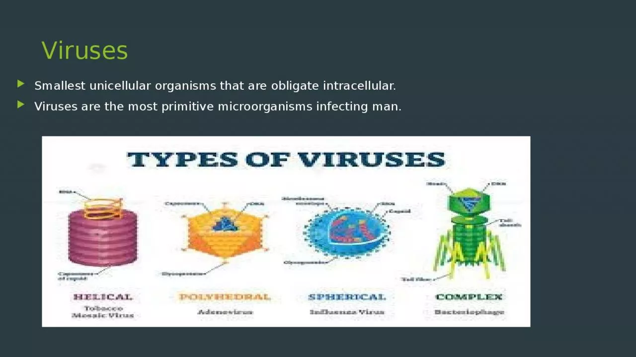
1. VirusesSmallest unicellular organisms that are obligate intracellular. Viruses are the most primitive microorganisms infecting man.
2. General propertiesObligate intracellular Possess either DNA (deoxyribonucleic acid) or RNA (ribonucleic acid), but never both Smaller than bacteria, can be passed through the bacterial filters Cannot be grown on artificial cell free media (grow in animals, embryonated eggs or tissue culture)
3. Viruses differ from bacteria :-Multiply by a complex method (not by binary fission).Do not have a proper cellular organisation. Do not have cell wall or cell membrane or cellular organelles including ribosomes Lack the enzymes necessary for protein and nucleic acid synthesis. Not susceptible to antibacterial antibiotics
4. MORPHOLOGY OF VIRUSNucleic acidNucleic acid may be single or double stranded, circular or linear, segmented or un-segmented. Viruses are classified based on nucleic acid as DNA viruses and RNA viruses.
5. CapsidComposed of a number of repeated protein subunits (polypeptides) called capsomeres. Functions: Protects the nucleic acid core from the external environmentIn non-enveloped viruses - initiates the first step of viral replication. Antigenic and specific for each virus.
6. Symmetry – Based on arrangement of capsomeres Type of symmetry ExplanationExamplesIcosahedral (cubical) symmetryCapsomeres are arranged as if they lay on the faces of an icosahedron20 triangular facets and 12 corners or vertices Rigid structure.All DNA viruses (except poxviruses)Most of the RNA viruses have icosahedral symmetry
7. Symmetry – Based on arrangement of capsomeres Type of symmetry ExplanationExamplesHelical symmetryCapsomeres are coiled surrounding the nucleic acid in the form of a helix or spiral. Flexible structure.RNA viruses such as-myxoviruses, rhabdoviruses, filoviruses, bunyaviruses, etc.
8. Symmetry – Based on arrangement of capsomeres Type of symmetry ExplanationExamplesComplex symmetry Do not have either of the above symmetry. Poxviruses
9. EnvelopeEnvelope surrounding the nucleocapsid. Lipoprotein in nature. Lipid part is derived from host cell membraneProtein part is virus coded, made up of subunits called peplomers. Peplomers - project as spikes on the surface of the envelope.
10. EnvelopeMore susceptible to heat and lipid solvents like etherAntigenic - facilitates entry of the virusMore than one kind of peplomers can be seen in some viruses, e.g. Influenza viruses possess haemagglutinin and neuraminidase peplomers.
11. EnvelopeMost viruses are enveloped except: Non-enveloped DNA viruses- parvovirus, adenovirus and papovavirusNon-enveloped RNA viruses-picornavirus, hepatitis A virus &hepatitis E virus.
12. Size of the virusesSize vary from 20-300nm in size. Smallest virus - parvovirus(20 nm)Largest - poxvirus (300nm).Because of the small size, viruses can pass through bacterial filters and they cannot be visualized under light microscope.
13. Size of the viruses – Determined byElectron microscope (best method)Ultracentrifugation- Viral particles suspended in liquid medium when subjected to ultracentrifugation, will settle down at a sedimentation rate that is proportional to their size.Passage through membrane filters of different pore sizes- The maximum pore size that prevents a virus to pass through multiplied by 0.64 yields the approximate diameter of the viral particle.
14. CLASSIFICATIONFamilyDNA typeEnvelopeSymmetrySize (nm)Representative VirusesHerpesviridaeds,linearYesIcosahedron150-200Herpes simplex virus - 1 Herpes simplex virus- 2Varicella-zoster virusEpstein-Barr virusCytomegalovirusHuman herpes virus 6,7 & 8Hepadnaviridaeds, circular,incompleteYesIcosahedron40–48Hepatitis B virusParvoviridaess, linearAbsentIcosahedron18–26Parvovirus B19Papovaviridaeds, circularAbsentIcosahedron45-55Human papillomaviruses JC virus and BK virusPoxviridaeds, linearYesComplex230 x 400Variola (smallpox) Molluscum contagiosum virusAdenoviridaeds, linearAbsentIcosahedron70–90Human adenoviruses
15. CLASSIFICATIONRNA Viruses RNA typeEnvelopeSymmetrySize(nm)Representative VirusesPicornaviridaess, +ve sense AbsentIcosahedral28–30PoliovirusCoxsackievirusEchovirus EnterovirusRhinovirus Hepatitis A virusCaliciviridaess, +ve senseAbsentIcosahedral27-40Norwalk agent Hepatitis E virusTogaviridaess, +ve senseYesIcosahedral50-70Rubella virus Eastern equine encephalitis virus Western equine encephalitis virus
16. CLASSIFICATIONRNA Viruses RNA typeEnvelopeSymmetrySize(nm)Representative VirusesFlaviviridaess, +ve senseYesIcosahedral (?)40-60Yellow fever virus Dengue virus St. Louis encephalitis virus West Nile virusHepatitis C virus Coronaviridaess, +ve senseYesHelical120-160CoronavirusesRhabdoviridaess, -ve senseYesHelical75x180Rabies virus Vesicular stomatitis virusFiloviridaess, -ve senseYesHelical80 x 1000Marburg virus Ebola virus
17. CLASSIFICATIONRNA Viruses RNA typeEnvelopeSymmetrySize(nm)Representative VirusesParamyxoviridaess, -ve senseYesHelical150–300Parainfluenza virus Mumps virus Measles virus Respiratory syncytial virus Newcastle disease virusMetapneumovirusOrthomyxoviridaess, -ve sense, 8 segmentsYesHelical80–120Influenza viruses- A, B, and CBunyaviridaess, -ve sense, 3 circular segmentsYesHelical80–120HantavirusCalifornia encephalitis virus Sandfly fever virus
18. CLASSIFICATIONRNA Viruses RNA typeEnvelopeSymmetrySize(nm)Representative VirusesBunyaviridaess, -ve sense, 3 circular segmentsYesHelical80–120HantavirusCalifornia encephalitis virus Sandfly fever virusArenaviridaess, -ve sense, RNA,2 circular segmentsYesHelical (?)50-300Lymphocytic choriomeningitis virus Lassa fever virus South American hemorrhagic fever virusReoviridaeds, 10–12 segmentsAbsentIcosahedral60-80Rotavirus ReovirusColorado tick fever virusRetroviridae2 identical copies of +ve sense ss RNAYesIcosahedral (spherical)80-110HTLV (Human T Lymphotropic virus) HIV (Human immunodeficiency virus)
19. Adsorption/attachmentFirst and most specific step.Involves receptor interactions. Viruses have attachment sites on their envelopes or capsid proteins that bind to the complementary receptor sites present on the host cell surface. HIV: Viral surface glycoprotein gp120binds toCD4molecules on the host cells.Influenza: Viral hemagglutinin (an envelope protein) binds specifically to glycoprotein receptors present on the surface of respiratory epithelium.
20. Viral Replication Viruses undergo a complex way of cell division.Replication of viruses passes through six sequential steps: 1) Adsorption/attachment2) Penetration 3) Uncoating4) Biosynthesis5) Assembly6) Maturation7) Release
21. PenetrationMechanismExplanationPhagocytosis (viropexis)Receptor mediated endocytosisMembrane fusionSome enveloped viruses (e.g. HIV) enter by fusion of their envelope proteins with the plasma membrane of the host cell so that only the nucleocapsid enters into the cytoplasm, whereas the viral envelope remains attached to the host cell membrane. Injection of nucleic acidBacteriophages cannot penetrate the rigid bacterial cell wall, hence only the nucleic acid is injected while the capsid remains attached to the cell wall.
22. UncoatingBy the action of lysosomal enzymes of the host cells, the viral capsid gets separated and the nucleic acid is released into the cytoplasm.Absent for bacteriophages.
23. BiosynthesisFollowing viral components are synthesized:Nucleic acidCapsid proteinEnzymes required for various stages of viral replicationRegulatory proteins to shut down the host cell metabolism.
24. Site of Nucleic acid replicationDNA viruses: DNA replication occurs in the nucleus except in poxviruses, which synthesize DNA in the cytoplasm.RNA viruses - RNA replication occurs in cytoplasm except in retroviruses and orthomyxoviruses, which synthesize RNA in the nucleus.
25. AssemblyViral nucleic acid and proteins are packaged together to form progeny viruses (nucleocapsids).Occurs in the host cell nucleus or cytoplasm.DNA viruses are assembled in the nucleus except hepadnaviruses and poxviruses (in cytoplasm)RNA viruses are assembled in the cytoplasm.
26. MaturationFollowing assembly, maturation of daughter virions take place either in the nucleus or cytoplasm or membranes (golgi or endoplasmic reticulum or plasma membrane)
27. ReleaseLysis of the host cells (non enveloped viruses and bacteriophages).Budding through host cell membrane (enveloped viruses) - During budding, they acquire a part of the host cell membrane to form the lipid part of their envelopes.Envelope is acquired either from plasma membrane (influenza virus) or from nuclear membrane (e.g. herpesviruses). Viral glycoproteins are then inserted into the envelopes. Excess viral glycoproteins are synthesized to saturate cell receptors so thatthe viruses will not stick to the host cell following release.
28. Eclipse phase Defined as ‘interval between the penetration of the virus into the host cell till the appearance of first infectious virus progeny particle’. During this period, the virus cannot be demonstrated inside the host cell. Duration -15 to 30 minutes for bacteriophages and 15-30 hours for most of the animal viruses.
29. ReferencesTextbook of Medical Microbiology by Ananthnarayan, PanikerTextbook of Medical Microbiology by C.P Baweja Textbook of Medical Microbiology by S. Bhat, A.S.SastryTextbook of Medical Microbiology by D.R.Arora, Brij bala Arora